
1. Endoplasmic Reticulum: Structure, Types, Functions and Cellular Importance
The endoplasmic reticulum (ER) is one of the most important membrane-bound organelles of eukaryotic cells. It forms an extensive network of interconnected membranous tubules, flattened sacs, and cisternae distributed throughout the cytoplasm. The endoplasmic reticulum is closely associated with the nucleus and plays central roles in the synthesis, processing, folding, modification, and transport of many cellular molecules.
The endoplasmic reticulum is broadly divided into two major forms:
- Rough endoplasmic reticulum (RER)
- Smooth endoplasmic reticulum (SER)
The rough endoplasmic reticulum has ribosomes attached to its cytosolic surface, giving it a rough appearance under the electron microscope. It is mainly involved in the synthesis and initial processing of proteins that are destined for secretion, membranes, lysosomes, or specific intracellular compartments.
The smooth endoplasmic reticulum lacks ribosomes on its cytosolic surface. It performs several functions, including lipid synthesis, carbohydrate metabolism, detoxification, calcium storage, and steroid production.
Although rough and smooth ER have different specialized functions, they are structurally and functionally interconnected. Together, they form an integrated system that contributes to the organization, metabolism, and homeostasis of the cell.
2. Discovery and Historical Background
2.1 Early Observations
The endoplasmic reticulum was identified through microscopic studies of cells during the development of modern cell biology.
With improvements in electron microscopy, researchers were able to observe an extensive membranous network within the cytoplasm.
2.2 Discovery of the Endoplasmic Reticulum
The term endoplasmic reticulum was introduced to describe the reticular, or network-like, membrane system found within the cytoplasm.
The word “endoplasmic” refers to its location within the cytoplasm, while “reticulum” means a network.
2.3 Relationship with Ribosomes
Researchers later recognized that certain regions of this membrane network possessed numerous ribosomes attached to their outer surface.
These ribosome-containing regions were called the rough endoplasmic reticulum.
Regions lacking attached ribosomes were recognized as the smooth endoplasmic reticulum.
This structural distinction helped explain why different regions of the ER perform different cellular functions.
3. General Characteristics of the Endoplasmic Reticulum
The endoplasmic reticulum has several characteristic features.
3.1 Membrane-Bound Organelle
The ER is surrounded by a membrane that is continuous with the outer nuclear membrane.
This continuity creates a physical connection between the nucleus and the endomembrane system.
3.2 Extensive Membrane Network
The ER is not a single isolated structure. It consists of an extensive network of:
- Tubules
- Cisternae
- Vesicles
- Membrane sheets
The organization of these structures varies according to the cell type and its physiological condition.
3.3 Presence in Eukaryotic Cells
The endoplasmic reticulum is a characteristic organelle of eukaryotic cells.
It is found in:
- Animal cells
- Plant cells
- Fungal cells
- Protist cells
Prokaryotic cells do not possess a typical membrane-bound endoplasmic reticulum.
3.4 Dynamic Nature
The ER is highly dynamic.
Its membranes continuously undergo:
- Fusion
- Fission
- Remodeling
- Expansion
- Contraction
- Interaction with other organelles
This dynamic nature allows the ER to adapt to changing cellular requirements.
4. Structure of the Endoplasmic Reticulum
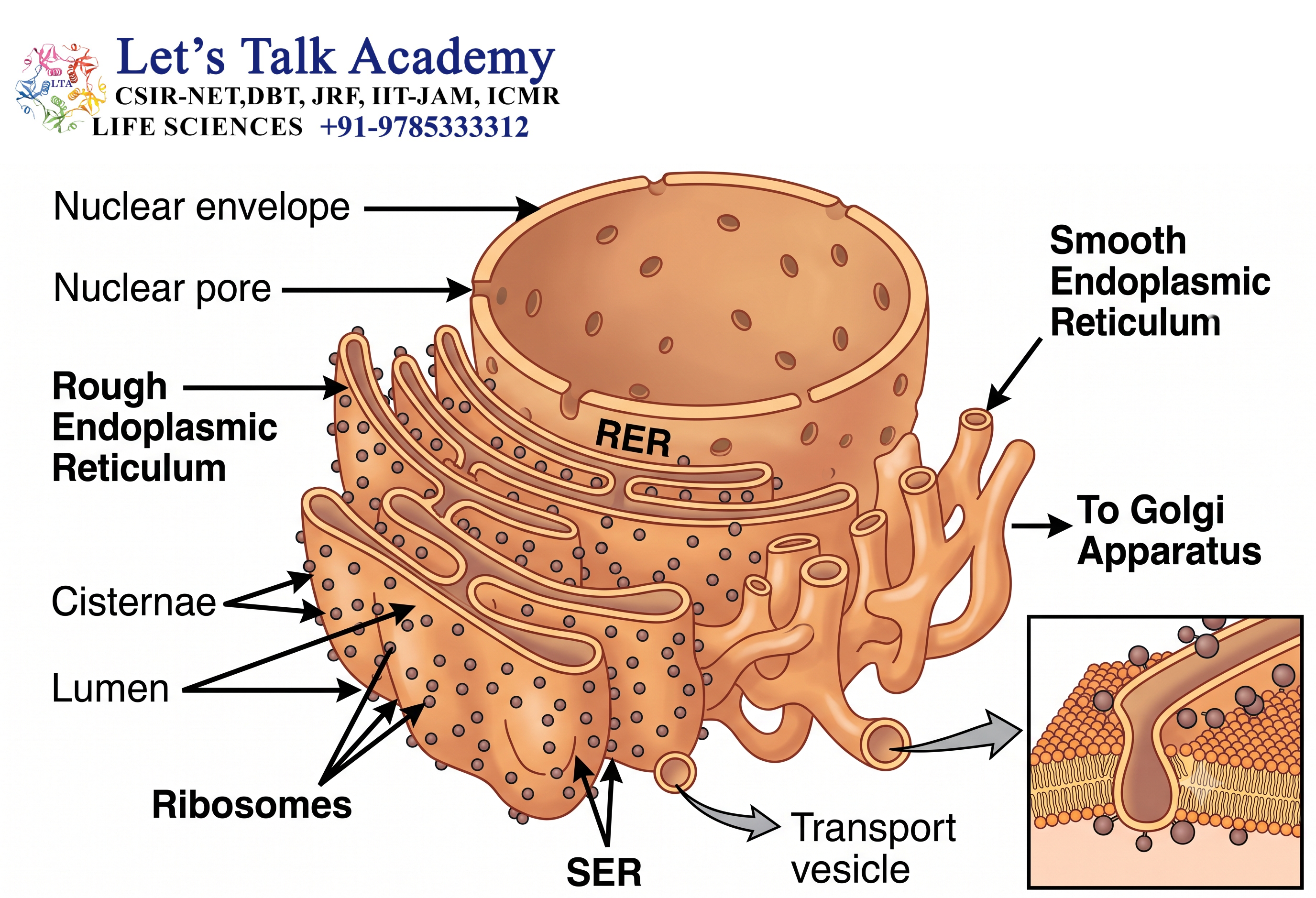
The endoplasmic reticulum consists primarily of a membrane and an internal space called the ER lumen.
4.1 ER Membrane
The ER membrane is a lipid bilayer containing numerous proteins.
These proteins perform functions such as:
- Transport
- Enzymatic reactions
- Protein folding
- Calcium movement
- Lipid synthesis
- Membrane organization
- Signal transduction
4.2 ER Lumen
The space enclosed by the ER membrane is called the ER lumen or ER cisternal space.
The lumen provides an important environment for:
- Protein folding
- Protein modification
- Calcium storage
- Assembly of certain protein complexes
- Quality control of newly synthesized proteins
4.3 Cisternae
Cisternae are flattened membrane-bound sacs that form part of the ER network.
They are particularly prominent in regions of rough ER.
4.4 Tubules
ER tubules are narrow membrane-bound structures that form an interconnected network.
Smooth ER is particularly rich in tubular structures.
4.5 Connection with the Nuclear Envelope
The ER membrane is continuous with the outer nuclear membrane.
This connection allows close coordination between nuclear activity and cytoplasmic membrane trafficking.
The perinuclear space between the inner and outer nuclear membranes is continuous with the ER lumen.
5. Types of Endoplasmic Reticulum
The endoplasmic reticulum is mainly divided into rough and smooth forms.
5.1 Rough Endoplasmic Reticulum
The rough endoplasmic reticulum (RER) contains ribosomes attached to its cytosolic surface.
The ribosomes give the RER its characteristic rough appearance.
The RER is especially well developed in cells that synthesize large quantities of proteins for secretion or membrane insertion.
Examples include:
- Pancreatic acinar cells
- Plasma cells
- Certain endocrine cells
5.1.1 Protein Synthesis
One of the major functions of the RER is the synthesis of proteins.
Ribosomes attached to the RER synthesize proteins that enter the ER lumen or become inserted into the ER membrane.
These proteins may eventually be transported to:
- Plasma membrane
- Golgi apparatus
- Lysosomes
- Secretory vesicles
- Extracellular space
5.1.2 Protein Folding
Newly synthesized proteins entering the ER must fold into their correct three-dimensional structures.
The ER contains molecular chaperones and other proteins that assist in proper protein folding.
5.1.3 Protein Modification
Several proteins undergo modifications within the ER.
These may include:
- Formation of disulfide bonds
- Addition of specific carbohydrate groups
- Protein processing
- Quality-control modifications
5.1.4 Membrane Protein Production
The RER is responsible for the synthesis of many membrane proteins.
These proteins become inserted into the ER membrane during translation.
They can subsequently be transported through the endomembrane system to their final destination.
5.2 Smooth Endoplasmic Reticulum
The smooth endoplasmic reticulum (SER) lacks ribosomes on its cytosolic surface.
It is usually composed predominantly of interconnected tubules.
The SER performs several important functions.
These include:
- Lipid synthesis
- Steroid synthesis
- Detoxification
- Carbohydrate metabolism
- Calcium storage
- Membrane production
5.3 Sarcoplasmic Reticulum
A specialized form of smooth ER found in muscle cells is called the sarcoplasmic reticulum (SR).
Its primary function is the storage and regulated release of Ca²⁺ ions.
Calcium release from the sarcoplasmic reticulum is essential for muscle contraction.
6. Rough Endoplasmic Reticulum and Ribosomes
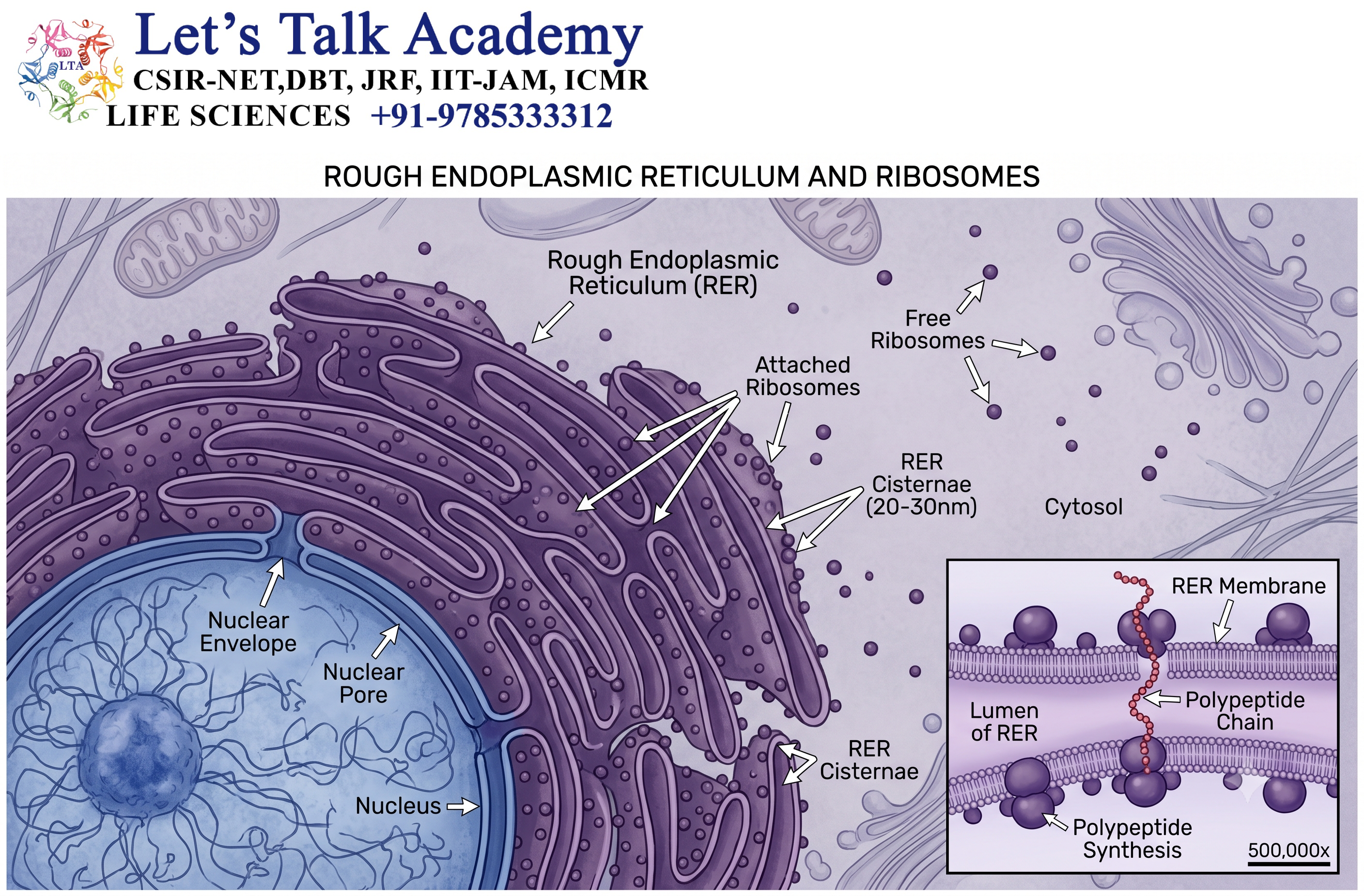
The relationship between ribosomes and the RER is central to protein synthesis.
6.1 Ribosomes on the Cytosolic Surface
Ribosomes are not permanently attached to the ER.
Instead, ribosomes become associated with the ER when they synthesize proteins containing appropriate targeting information.
6.2 Signal Sequence
Many proteins destined for the secretory pathway contain an N-terminal signal sequence.
This sequence helps direct the translating ribosome and growing protein toward the ER.
6.3 Signal Recognition Particle
The signal recognition particle (SRP) recognizes the signal sequence as it emerges from the ribosome.
SRP temporarily slows translation and directs the ribosome-nascent chain complex toward the ER membrane.
6.4 SRP Receptor
The SRP-ribosome complex interacts with the SRP receptor on the ER membrane.
The ribosome is then positioned near a protein-conducting channel.
6.5 Translocon
The Sec61 translocon is a major protein-conducting channel in the ER membrane.
It allows newly synthesized proteins to enter the ER lumen or become inserted into the ER membrane.
This process is known as co-translational translocation.
7. Protein Synthesis in the Rough ER
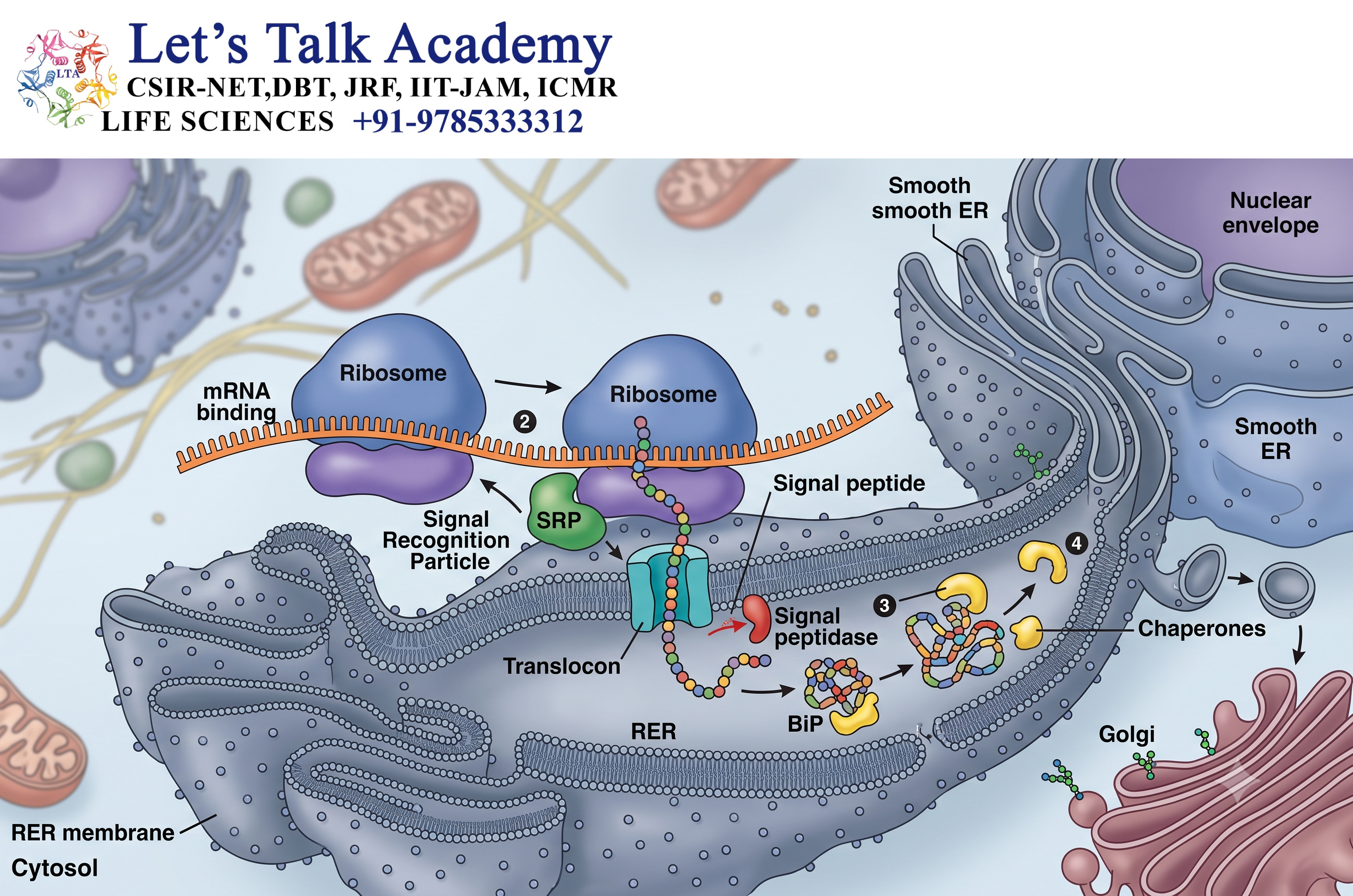
Protein synthesis at the RER follows a coordinated sequence.
7.1 Initiation of Translation
Translation begins on a free ribosome in the cytosol.
If the newly synthesized protein contains an ER-targeting signal sequence, SRP recognizes it.
7.2 Targeting to the ER
The SRP directs the ribosome toward the ER membrane.
The ribosome interacts with the SRP receptor and becomes associated with the translocon.
7.3 Protein Translocation
Translation resumes, and the growing polypeptide passes through the translocon into the ER lumen.
For membrane proteins, portions of the protein are inserted directly into the ER membrane.
7.4 Completion of Protein Synthesis
After translation is completed, the newly synthesized protein undergoes folding and processing.
Properly processed proteins can then leave the ER and travel toward the Golgi apparatus.
8. Protein Folding in the Endoplasmic Reticulum
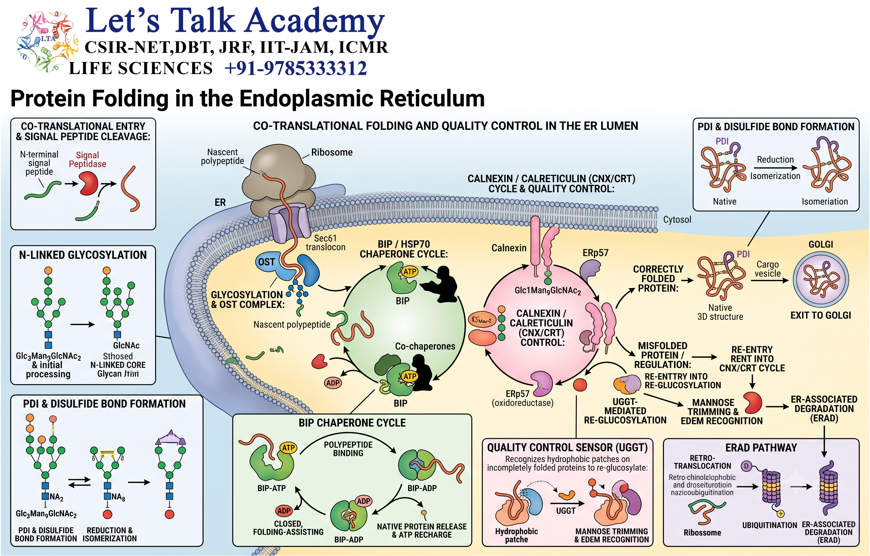
Protein folding is one of the most important functions of the ER.
8.1 Importance of Proper Folding
Proteins must adopt specific three-dimensional structures to function correctly.
Incorrectly folded proteins may lose their function or become toxic to the cell.
8.2 Molecular Chaperones
The ER contains molecular chaperones that assist proteins in achieving proper conformations.
One important ER chaperone is BiP, a member of the Hsp70 family.
8.3 Disulfide Bond Formation
The ER provides an environment suitable for the formation of disulfide bonds.
Disulfide bonds contribute to the stability of many secreted and membrane proteins.
8.4 Protein Quality Control
The ER contains quality-control systems that distinguish correctly folded proteins from misfolded proteins.
Correctly folded proteins can proceed through the secretory pathway.
Misfolded proteins may be retained and subjected to further processing or degradation.
9. Endoplasmic Reticulum Quality Control
The ER must prevent defective proteins from reaching their final destinations.
9.1 Recognition of Misfolded Proteins
Molecular chaperones and other quality-control proteins recognize improperly folded proteins.
9.2 Retention
Misfolded proteins may be temporarily retained within the ER.
This gives them an opportunity to fold correctly.
9.3 ER-Associated Degradation
Proteins that cannot be properly folded may be directed toward ER-associated degradation (ERAD).
During ERAD, defective proteins are transported back toward the cytosol and targeted for degradation, commonly through the ubiquitin-proteasome system.
9.4 Importance of Quality Control
ER quality control prevents defective proteins from accumulating in other cellular compartments.
This is essential for maintaining cellular proteostasis.
10. Unfolded Protein Response
When misfolded or unfolded proteins accumulate in the ER, cells activate a protective signaling network called the unfolded protein response (UPR).
10.1 Causes of ER Stress
ER stress can result from:
- Excessive protein synthesis
- Mutations producing unstable proteins
- Disturbance of calcium balance
- Oxidative stress
- Nutrient imbalance
- Certain toxins
- Infection or other cellular stresses
10.2 Major UPR Pathways
Three major ER stress sensors are:
- IRE1
- PERK
- ATF6
These pathways collectively attempt to restore ER homeostasis.
10.3 Reduction of Protein Synthesis
One response to ER stress is a reduction in general protein synthesis.
This decreases the amount of newly synthesized protein entering the ER.
10.4 Increased Chaperone Production
The UPR can increase the production of molecular chaperones.
This enhances the cell’s capacity to fold newly synthesized proteins.
10.5 Increased Degradation
The UPR can also enhance mechanisms for removing misfolded proteins.
10.6 Cell Death During Severe Stress
If ER stress is severe or prolonged and homeostasis cannot be restored, signaling pathways may contribute to programmed cell death.
11. Functions of the Smooth Endoplasmic Reticulum
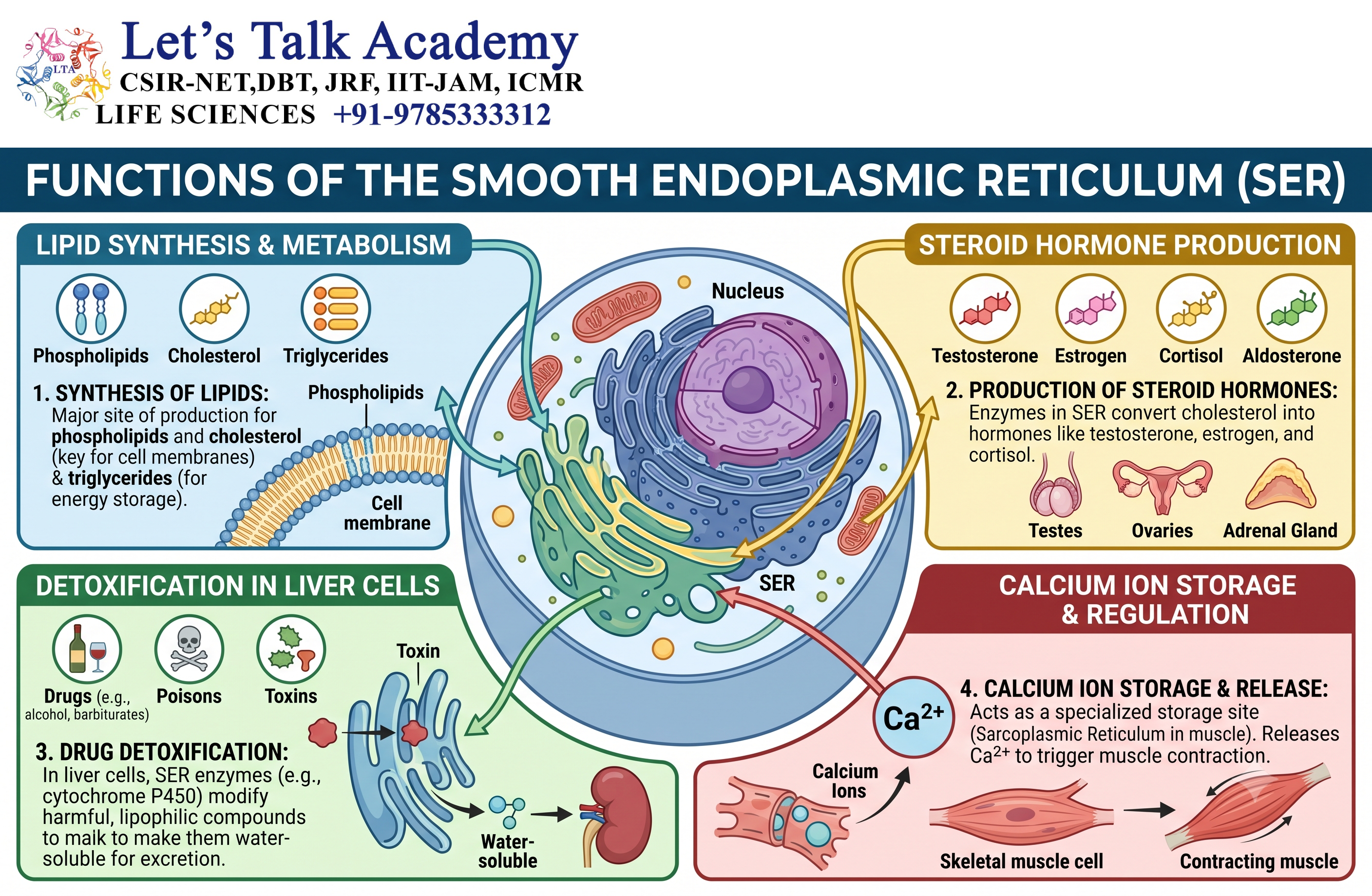
The smooth ER performs a wide range of metabolic functions.
11.1 Lipid Synthesis
The SER is a major site of synthesis of many cellular lipids.
These include:
- Phospholipids
- Cholesterol
- Triglycerides
- Other lipid molecules
11.2 Membrane Biogenesis
Newly synthesized lipids are important for producing cellular membranes.
The ER contributes significantly to the expansion and maintenance of the endomembrane system.
11.3 Steroid Hormone Synthesis
Cells that synthesize steroid hormones contain abundant smooth ER.
Steroid-producing tissues include cells of:
- Adrenal cortex
- Gonads
- Certain endocrine tissues
11.4 Detoxification
The SER is involved in detoxification reactions, especially in liver cells.
Many detoxification enzymes belong to the cytochrome P450 family.
These enzymes modify lipid-soluble compounds, making them easier to process and eliminate.
11.5 Carbohydrate Metabolism
The ER participates in several aspects of carbohydrate metabolism.
In liver cells, the ER is involved in processes associated with glucose homeostasis.
11.6 Calcium Storage
The ER serves as an important intracellular calcium reservoir.
Controlled calcium release and uptake are essential for:
- Muscle contraction
- Signal transduction
- Secretion
- Enzyme regulation
- Cellular signaling
12. Calcium Storage and Signaling
Calcium ions act as important intracellular signaling molecules.
12.1 Calcium as a Second Messenger
Changes in cytosolic Ca²⁺ concentration can regulate numerous cellular processes.
These include:
- Muscle contraction
- Neurotransmitter release
- Hormone secretion
- Enzyme activity
- Gene expression
12.2 ER Calcium Stores
The ER stores a large pool of intracellular calcium.
Calcium concentration inside the ER is maintained through specialized transport proteins.
12.3 SERCA Pump
The sarcoplasmic/endoplasmic reticulum Ca²⁺-ATPase (SERCA) uses ATP to transport calcium from the cytosol into the ER.
This allows the ER to refill its calcium stores.
12.4 Calcium Release
Calcium can be released from the ER through specialized channels.
Important calcium-release channels include:
- IP₃ receptors
- Ryanodine receptors
The released calcium can act as a rapid intracellular signal.
13. Endoplasmic Reticulum and Lipid Metabolism
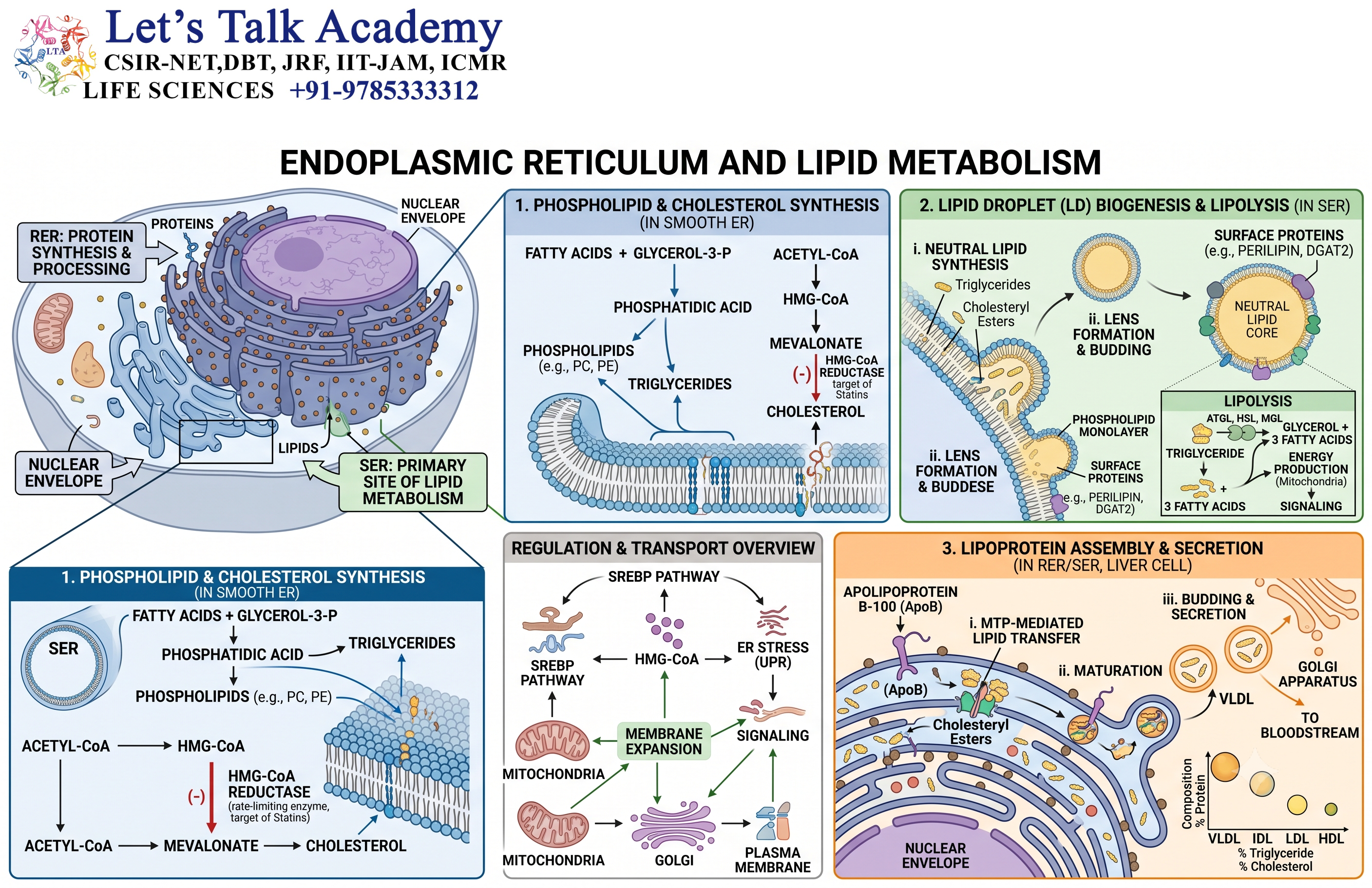
The ER is a major center for cellular lipid metabolism.
13.1 Phospholipid Synthesis
Many phospholipids are synthesized in the ER membrane.
Phospholipids are essential components of biological membranes.
13.2 Cholesterol Metabolism
The ER participates in cholesterol synthesis and regulation.
Cholesterol is essential for membrane structure and serves as a precursor for steroid hormones and other molecules.
13.3 Triglyceride Synthesis
The ER contributes to triglyceride production.
Triglycerides are important for energy storage.
13.4 Lipid Droplet Formation
Lipid droplets originate from the ER.
They store neutral lipids and provide an important reservoir for cellular energy metabolism.
14. Endoplasmic Reticulum and Vesicular Transport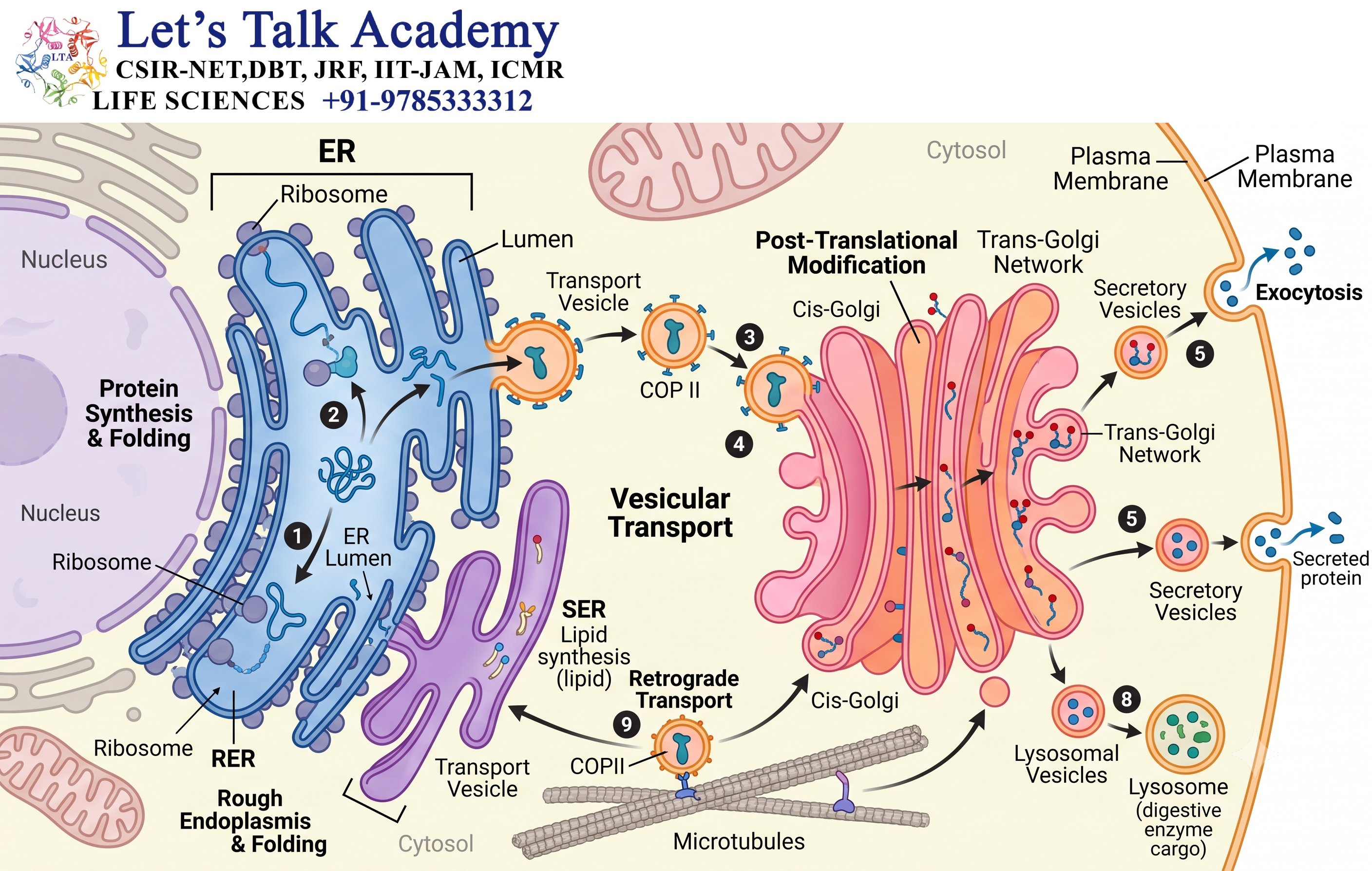
The ER is the starting point for much of the secretory pathway.
14.1 Formation of Transport Vesicles
Proteins and lipids leaving the ER are packaged into transport vesicles.
14.2 COPII-Coated Vesicles
COPII-coated vesicles transport cargo from the ER toward the Golgi apparatus.
COPII-mediated transport is an important component of anterograde trafficking.
14.3 ER-to-Golgi Transport
Newly synthesized proteins move from the ER to the Golgi apparatus for further modification, sorting, and packaging.
14.4 Retrograde Transport
Some proteins and membrane components need to return from the Golgi to the ER.
COPI-coated vesicles participate in many retrograde transport processes.
This bidirectional trafficking maintains the composition and function of the ER and Golgi compartments.
15. Endoplasmic Reticulum and the Golgi Apparatus
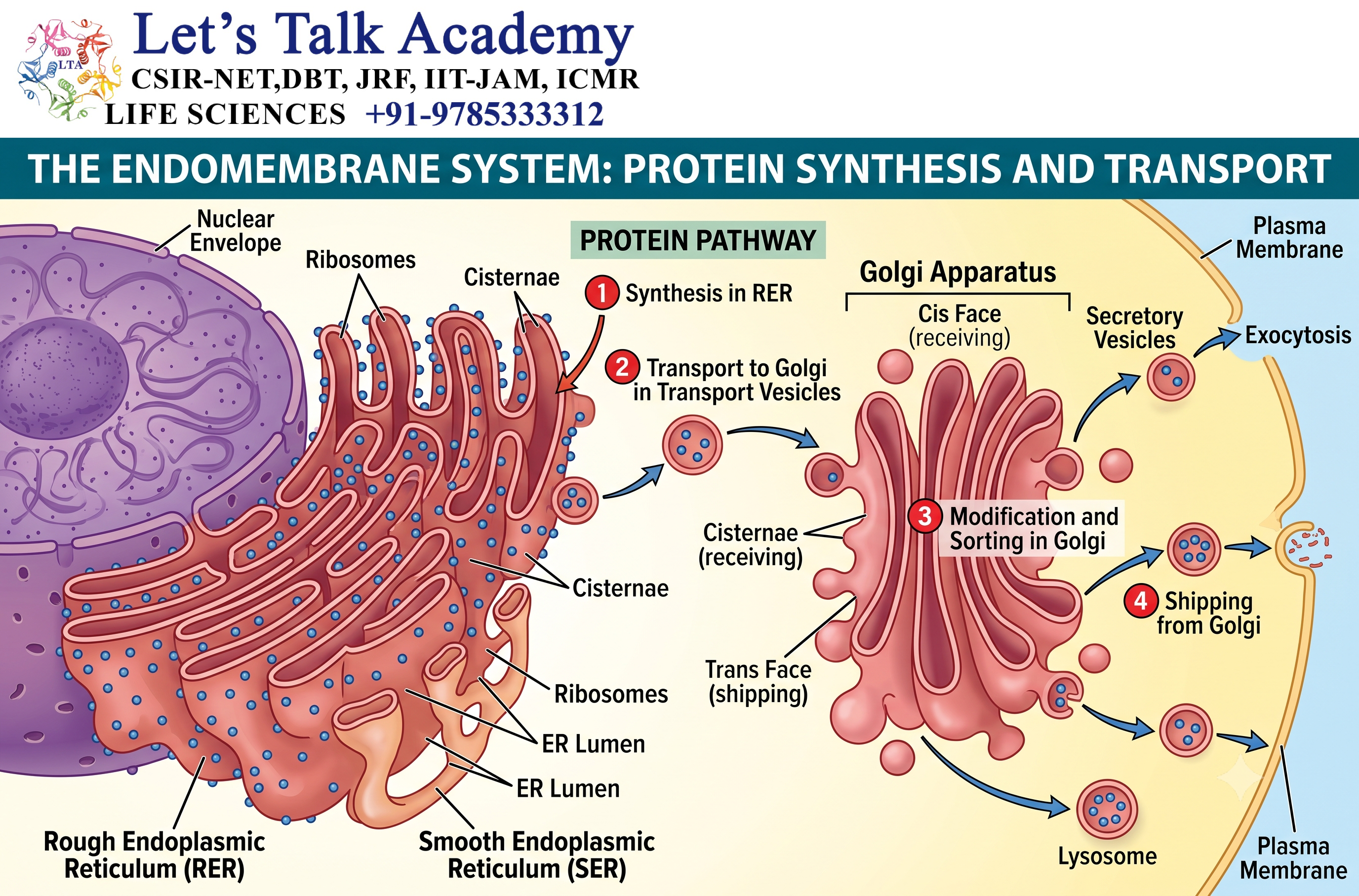
The ER and Golgi apparatus work closely together.
15.1 ER as the Entry Point
Many proteins entering the secretory pathway first enter the ER.
15.2 Transport to Golgi
Correctly folded proteins are transported from the ER to the Golgi apparatus.
15.3 Further Modification
The Golgi modifies proteins and lipids through processes such as:
- Glycosylation
- Proteolytic processing
- Sulfation
- Sorting
15.4 Final Destination
After Golgi processing, molecules may be directed toward:
- Plasma membrane
- Lysosomes
- Secretory vesicles
- Extracellular space
Thus, the ER and Golgi form an integrated manufacturing and distribution system.
16. Endoplasmic Reticulum and Secretory Pathway
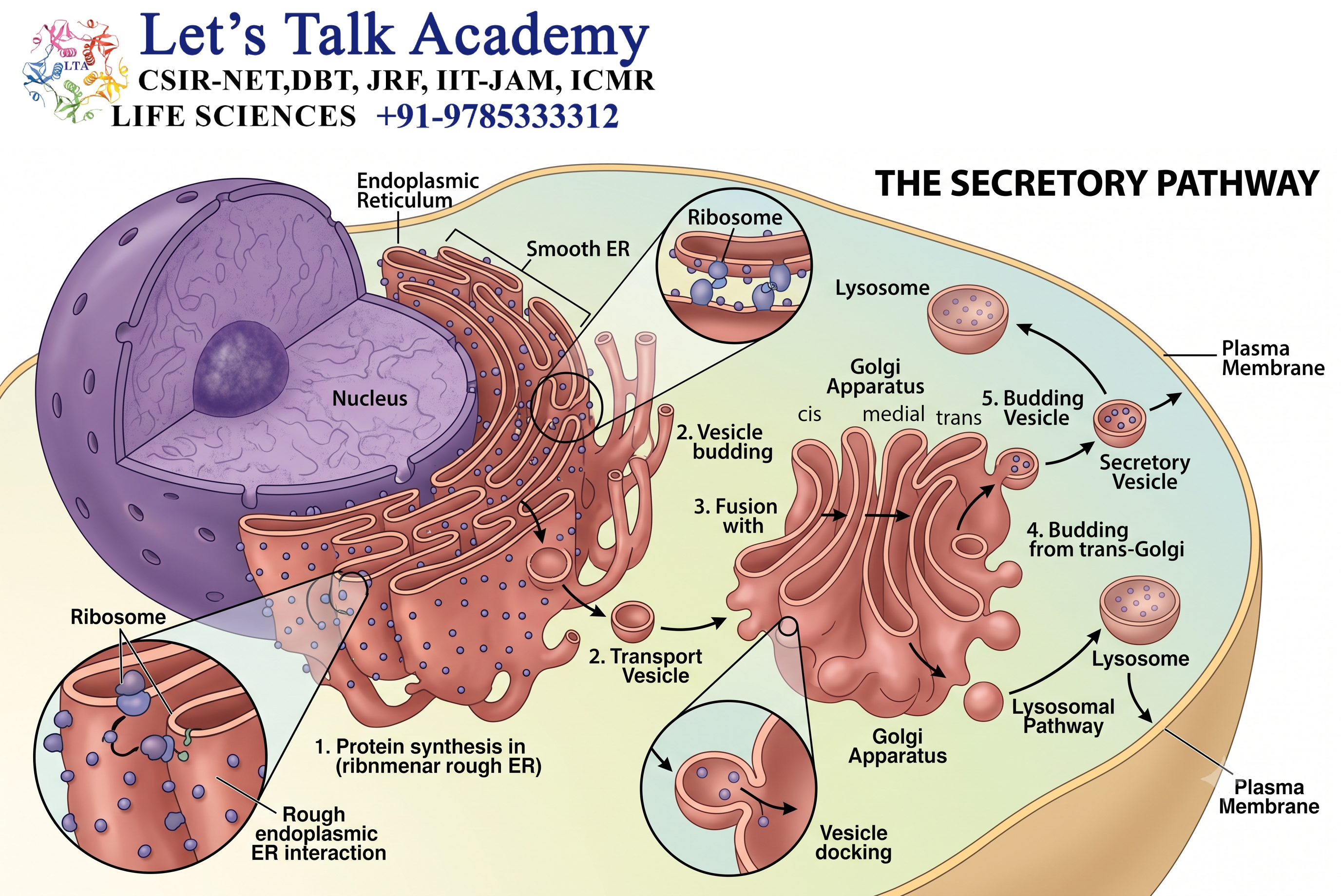
The ER is the first major organelle of the classical secretory pathway.
A simplified sequence is:
Ribosome → ER → Transport Vesicle → Golgi Apparatus → Secretory Vesicle → Final Destination
Proteins destined for secretion enter the ER during synthesis.
They undergo folding and initial modification before being transported toward the Golgi apparatus.
17. Endoplasmic Reticulum in Different Cell Types
The amount and organization of ER vary according to cellular function.
17.1 Pancreatic Cells
Protein-secreting pancreatic cells contain extensive rough ER because they synthesize large quantities of digestive enzymes.
17.2 Plasma Cells
Plasma cells produce large amounts of antibodies.
Therefore, they contain highly developed rough ER.
17.3 Liver Cells
Liver cells contain both rough and smooth ER.
The RER supports protein synthesis, while the SER participates in lipid metabolism and detoxification.
17.4 Steroid-Producing Cells
Cells that synthesize steroid hormones contain abundant smooth ER.
17.5 Muscle Cells
Muscle cells contain specialized sarcoplasmic reticulum for calcium storage and release.
18. Endoplasmic Reticulum and Autophagy
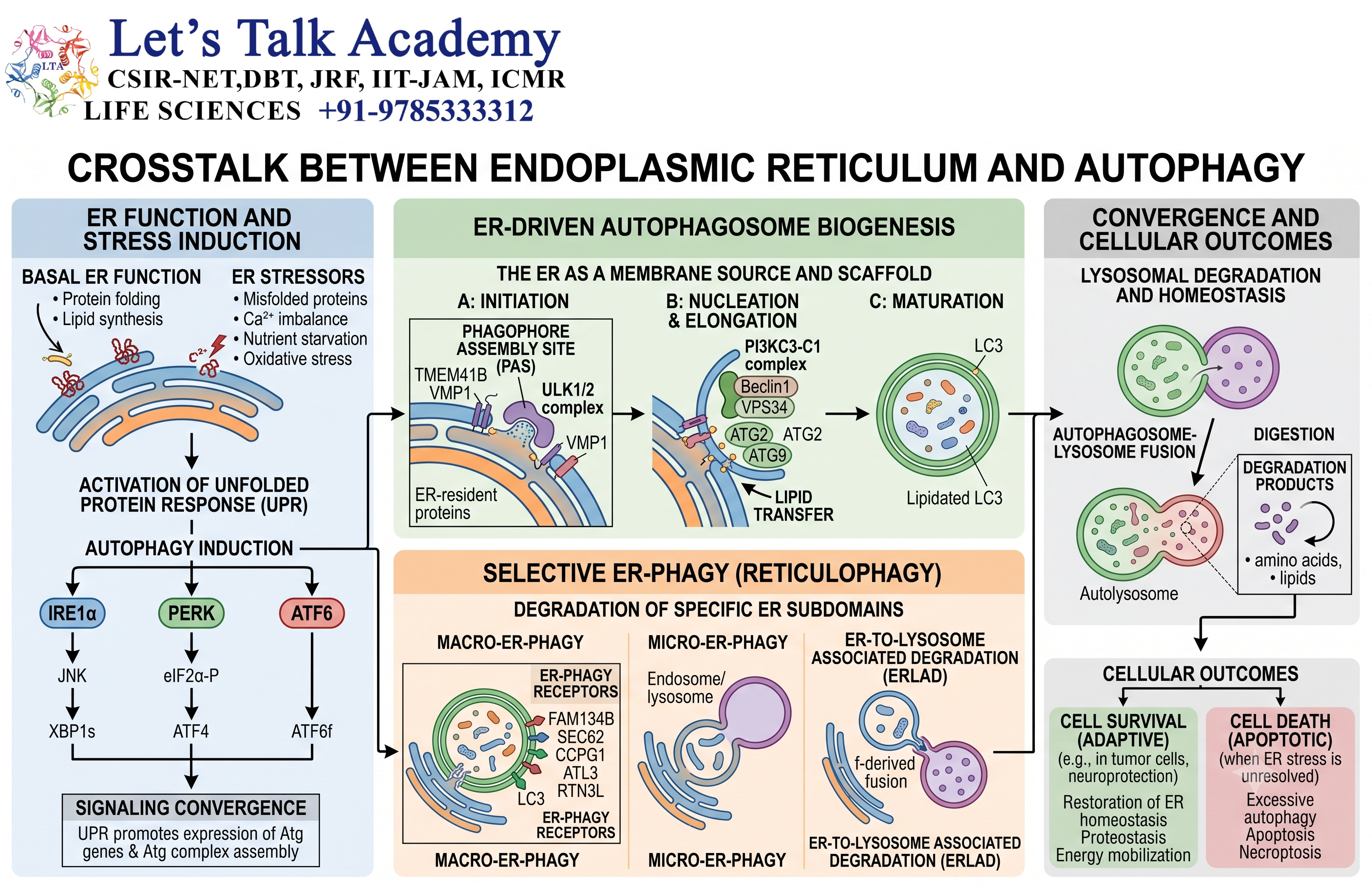
The ER also participates in autophagy.
18.1 ER as a Membrane Source
ER membranes can contribute to the formation of autophagic structures.
18.2 ER–Autophagosome Relationship
The formation of autophagosomes involves complex interactions among membranes from different cellular compartments, with the ER playing an important role.
18.3 Removal of Damaged Components
Through autophagy, cellular components can ultimately be delivered to lysosomes for degradation.
Thus, the ER is functionally connected with the lysosomal recycling system.
19. Endoplasmic Reticulum Contact Sites
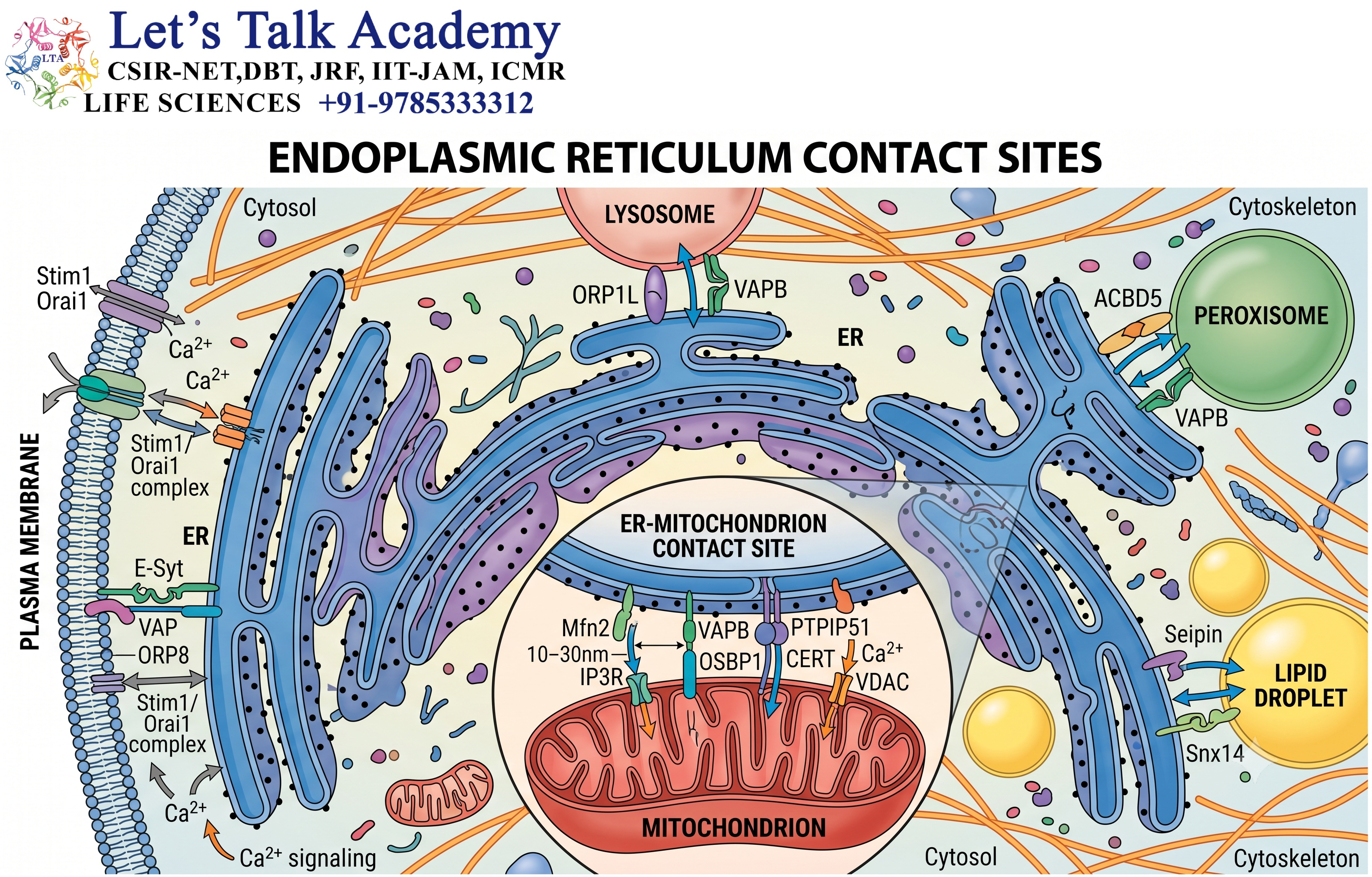
The ER forms specialized regions of close physical association with other organelles.
These regions are called membrane contact sites.
19.1 ER–Mitochondria Contact Sites
The ER can establish close contact with mitochondria.
These contacts are important for:
- Calcium transfer
- Lipid exchange
- Metabolic signaling
- Mitochondrial function
19.2 ER–Plasma Membrane Contacts
ER-plasma membrane contact sites contribute to calcium signaling and lipid transfer.
19.3 ER–Golgi Contacts
Interactions between ER and Golgi compartments facilitate efficient membrane and cargo trafficking.
These contact sites demonstrate that organelles do not function independently.
20. Endoplasmic Reticulum and Cellular Homeostasis
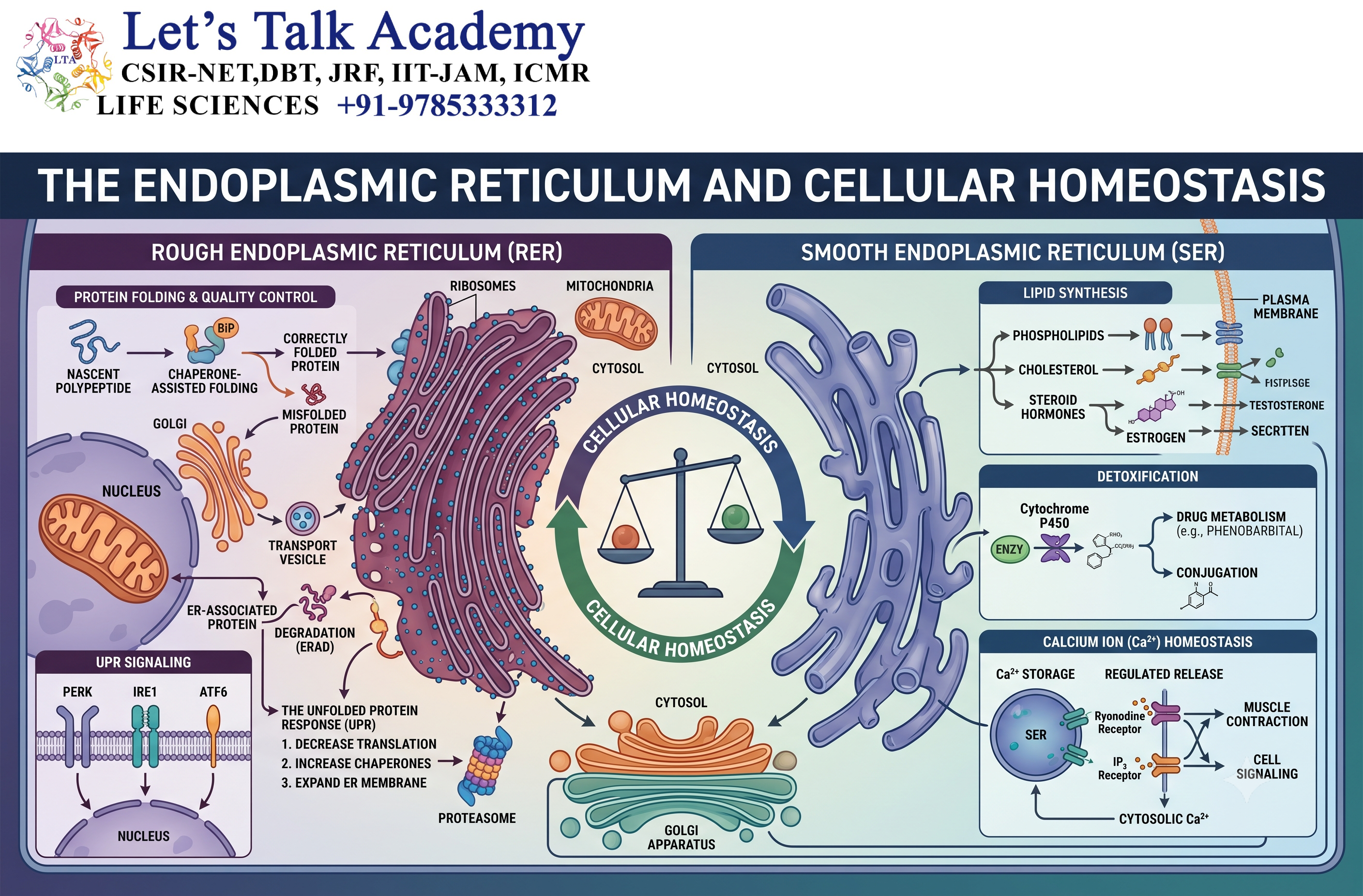
The ER contributes extensively to cellular homeostasis.
It regulates:
- Protein folding
- Lipid synthesis
- Calcium balance
- Membrane production
- Protein quality control
- Stress responses
A healthy ER is therefore essential for normal cellular function.
21. Endoplasmic Reticulum Stress
ER stress occurs when the protein-folding capacity of the ER becomes insufficient to handle the load of unfolded or misfolded proteins.
21.1 Causes of ER Stress
ER stress may occur due to:
- Excessive protein production
- Genetic mutations
- Disturbed calcium balance
- Oxidative stress
- Metabolic disturbances
- Environmental stress
21.2 Cellular Response
The cell activates the unfolded protein response to restore ER function.
The response attempts to:
- Reduce protein load
- Increase protein-folding capacity
- Increase degradation of defective proteins
- Restore ER homeostasis
21.3 Prolonged ER Stress
If ER stress cannot be resolved, prolonged activation of stress pathways may contribute to inflammation, metabolic dysfunction, or cell death.
22. Endoplasmic Reticulum and Disease
Because the ER performs essential functions, defects in ER processes can contribute to disease.
ER dysfunction has been associated with:
- Metabolic disorders
- Neurodegenerative diseases
- Diabetes
- Liver disorders
- Inflammatory conditions
- Protein-misfolding diseases
- Certain cancers
The specific relationship varies depending on the disease and affected cellular pathway.
23. ER-Associated Degradation
ER-associated degradation (ERAD) is an important protein quality-control pathway.
23.1 Recognition
Misfolded proteins are identified within the ER.
23.2 Retrotranslocation
The defective protein is transported from the ER lumen or membrane toward the cytosol.
23.3 Ubiquitination
The protein is often tagged with ubiquitin, which marks it for degradation.
23.4 Proteasomal Degradation
The ubiquitinated protein is ultimately degraded by the 26S proteasome.
This pathway prevents accumulation of defective proteins within the ER.
24. Endoplasmic Reticulum and Protein Glycosylation
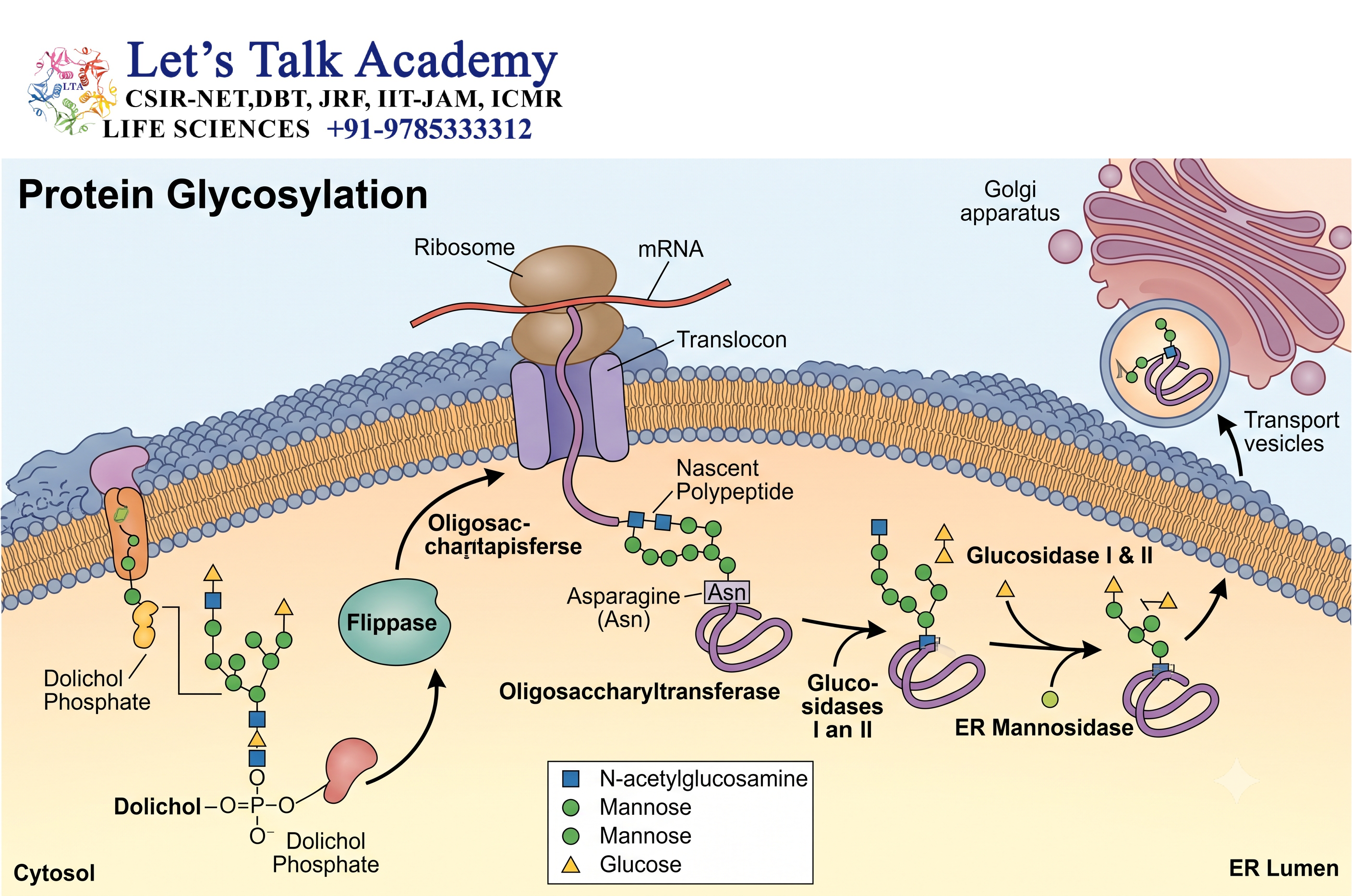
Protein glycosylation is an important ER-associated process.
24.1 N-Linked Glycosylation
Many proteins entering the ER undergo N-linked glycosylation.
A preassembled oligosaccharide is transferred to specific asparagine residues in the growing protein.
24.2 Role in Protein Folding
N-linked glycans are not merely decorative modifications.
They can participate in protein folding and quality-control mechanisms.
24.3 Calnexin and Calreticulin
Calnexin and calreticulin are ER-associated molecular chaperones that participate in the folding and quality control of glycoproteins.
25. Endoplasmic Reticulum and Membrane Biogenesis
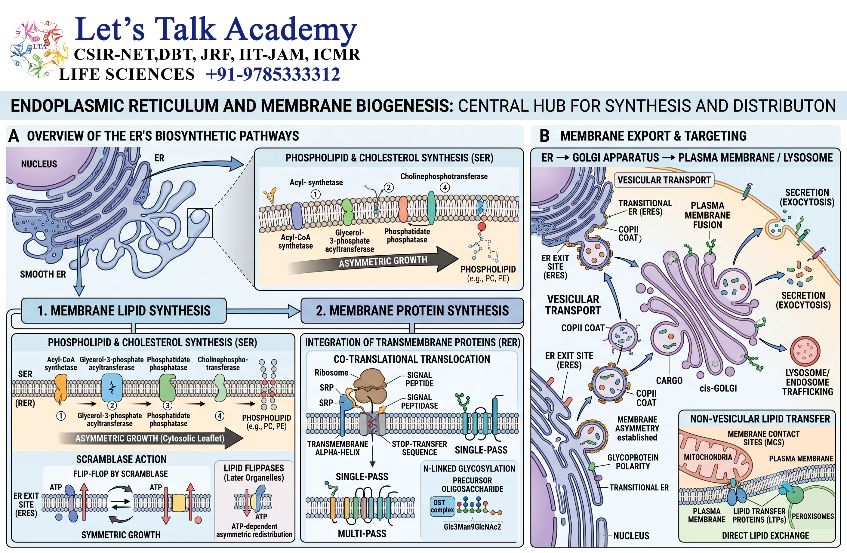
The ER is one of the major sites where new biological membranes are generated.
25.1 Lipid Production
Lipids are synthesized within the ER membrane.
25.2 Membrane Expansion
Newly synthesized lipids allow ER membranes to expand.
25.3 Distribution of Membrane Components
Lipids and membrane proteins can be transported from the ER to other organelles.
Therefore, the ER serves as an important manufacturing center for the cellular membrane system.
26. Endoplasmic Reticulum and Detoxification
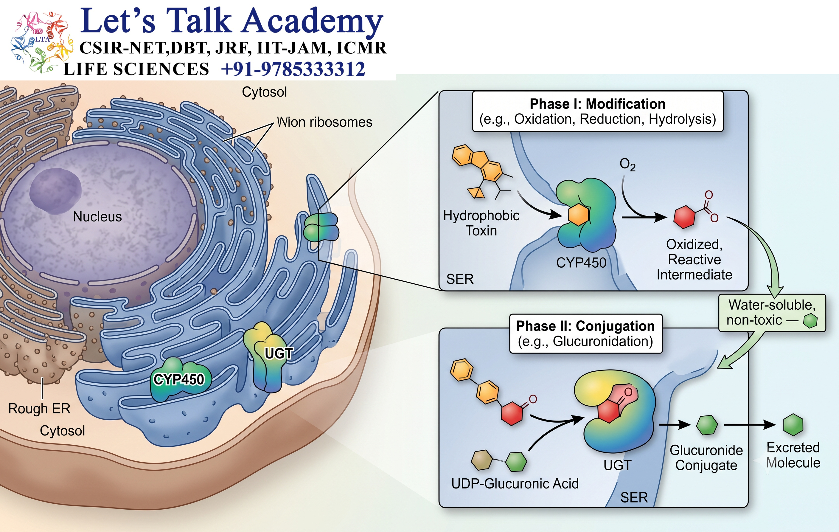
The smooth ER is particularly important in detoxification.
26.1 Liver Cells
Hepatocytes contain extensive smooth ER because the liver performs numerous metabolic and detoxification functions.
26.2 Cytochrome P450 System
Cytochrome P450 enzymes modify many lipid-soluble substances.
These include:
- Drugs
- Environmental chemicals
- Steroid-related compounds
- Other foreign substances
26.3 Increased Detoxification Demand
Exposure to certain substances can increase the amount of smooth ER and associated detoxification enzymes in some cells.
This illustrates the adaptive nature of the ER.
27. Endoplasmic Reticulum and Carbohydrate Metabolism
The ER contributes to carbohydrate metabolism, particularly in specialized metabolic tissues.
In liver cells, ER-associated enzymes participate in processes related to glucose production and homeostasis.
The ER therefore contributes not only to structural organization but also to systemic metabolic regulation.
28. Endoplasmic Reticulum and Steroid Biosynthesis
Steroid-producing cells have abundant smooth ER.
Steroid hormones are synthesized from cholesterol through a series of enzymatic reactions.
Important steroid-producing tissues include:
- Adrenal cortex
- Ovaries
- Testes
- Placenta
The ER works together with mitochondria in steroid biosynthesis.
29. Endoplasmic Reticulum and Protein Export
Proteins synthesized in the ER do not all remain there.
Correctly folded proteins are packaged into transport vesicles.
The general pathway is:
Protein synthesis → ER entry → Folding → Quality control → Vesicle formation → Golgi → Sorting → Final destination
This pathway allows cells to efficiently distribute proteins throughout the body of the cell and, when required, outside the cell.
30. Difference Between Rough and Smooth Endoplasmic Reticulum
| Feature | Rough ER | Smooth ER |
|---|---|---|
| Ribosomes | Present on cytosolic surface | Absent |
| Major structure | Flattened cisternae and sheets | Tubular network |
| Major function | Protein synthesis and processing | Lipid synthesis and metabolism |
| Protein folding | Major role | Limited |
| Calcium storage | Present but not its defining function | Important, especially in specialized cells |
| Detoxification | Limited | Major role |
| Steroid synthesis | Limited | Major role |
| Membrane protein synthesis | Major | Not a primary function |
31. Difference Between ER and Golgi Apparatus
The ER and Golgi apparatus are closely connected but perform different functions.
| Feature | Endoplasmic Reticulum | Golgi Apparatus |
|---|---|---|
| Main role | Synthesis and initial processing | Modification, sorting and packaging |
| Protein synthesis | RER performs major role | No direct protein synthesis |
| Lipid synthesis | Major site | Some lipid modification |
| Protein folding | Major role | Further processing |
| Calcium storage | Important | Not its major function |
| Vesicle formation | ER-to-Golgi transport | Multiple outgoing pathways |
32. Difference Between ER and Lysosomes
| Feature | Endoplasmic Reticulum | Lysosomes |
|---|---|---|
| Main function | Synthesis, processing and transport | Degradation |
| Membrane | Single membrane | Single membrane |
| Internal environment | ER lumen has specialized conditions for folding | Strongly acidic |
| Major enzymes | Folding and metabolic enzymes | Acid hydrolases |
| Protein synthesis | RER-associated | No |
| Autophagy | Participates in formation and regulation | Performs degradation |
| Recycling | Indirectly contributes | Major function |
33. Regulation of Endoplasmic Reticulum Function
The ER continuously adapts to the needs of the cell.
Its activity can be influenced by:
- Nutrient availability
- Protein synthesis rate
- Calcium concentration
- Lipid availability
- Cellular stress
- Hormonal signals
- Metabolic conditions
This regulation ensures that the ER does not simply function as a passive membrane network.
34. Endoplasmic Reticulum and Cellular Communication
The ER communicates with other organelles through both membrane trafficking and membrane contact sites.
Important interactions include:
ER ↔ Golgi
ER ↔ Mitochondria
ER ↔ Plasma membrane
ER ↔ Endosomes
ER ↔ Lysosomes
These interactions allow the cell to coordinate protein trafficking, lipid distribution, calcium signaling, metabolism, and stress responses.
35. Importance of the Endoplasmic Reticulum
The ER can be considered one of the major manufacturing and quality-control centers of the cell.
Its importance includes:
- Protein synthesis
- Protein folding
- Protein quality control
- Lipid synthesis
- Membrane biogenesis
- Calcium storage
- Detoxification
- Steroid synthesis
- Carbohydrate metabolism
- Vesicular transport
- ER stress signaling
- Autophagy-related functions
- Communication with other organelles
36. Key Terms Related to the Endoplasmic Reticulum
Endoplasmic Reticulum
An extensive intracellular membrane network involved in protein synthesis, lipid metabolism, calcium storage, and intracellular transport.
Rough Endoplasmic Reticulum
The ribosome-associated portion of the ER involved primarily in synthesis and processing of proteins entering the secretory pathway.
Smooth Endoplasmic Reticulum
The ribosome-free portion of the ER involved in lipid synthesis, detoxification, calcium storage, and other metabolic functions.
Sarcoplasmic Reticulum
A specialized ER in muscle cells responsible mainly for calcium storage and release.
ER Lumen
The internal space enclosed by the ER membrane.
Translocon
A membrane-associated protein-conducting channel through which newly synthesized proteins enter the ER or become inserted into its membrane.
Signal Recognition Particle
A ribonucleoprotein complex that recognizes ER-targeting signal sequences and directs translating ribosomes to the ER.
ER Stress
A cellular condition caused by accumulation of unfolded or misfolded proteins within the ER.
Unfolded Protein Response
A cellular signaling response activated by ER stress to restore protein-folding capacity and ER homeostasis.
ERAD
A quality-control pathway that removes misfolded proteins from the ER for degradation.
37. Conceptual Flowchart of Rough ER-Mediated Protein Processing
Ribosome begins translation
↓
ER signal sequence emerges
↓
Signal Recognition Particle binds
↓
Ribosome directed to ER membrane
↓
SRP receptor interaction
↓
Ribosome associates with Sec61 translocon
↓
Protein enters ER lumen or membrane
↓
Protein folding and modification
↓
Quality control
↓
Correctly folded protein
↓
Transport vesicle
↓
Golgi apparatus
↓
Sorting and further modification
↓
Final cellular destination
38. Conceptual Flowchart of Smooth ER Functions
Smooth ER
↓
Lipid synthesis
→ Phospholipids
→ Cholesterol
→ Triglycerides
↓
Steroid synthesis
→ Steroid hormones
↓
Detoxification
→ Cytochrome P450 system
↓
Calcium storage
→ Ca²⁺ uptake
→ Controlled Ca²⁺ release
↓
Carbohydrate metabolism
↓
Membrane biogenesis
39. Integrated Role of the Endoplasmic Reticulum
The ER can be understood as a central hub connecting several major cellular processes.
Protein Production
The rough ER produces proteins destined for secretion, membranes, lysosomes, and other components of the endomembrane system.
Lipid Production
The smooth ER produces many lipids required for membranes and metabolism.
Quality Control
The ER checks whether newly synthesized proteins are correctly folded.
Calcium Regulation
The ER stores and releases calcium according to cellular signals.
Transport
The ER sends correctly processed proteins and lipids toward the Golgi apparatus.
Stress Response
The ER detects disturbances in protein folding and activates adaptive signaling pathways.
This integration makes the ER essential for maintaining cellular organization.
40. Summary
The endoplasmic reticulum is a highly organized and dynamic membrane network that performs numerous essential functions within eukaryotic cells.
It exists primarily in two forms: rough ER and smooth ER. Rough ER contains ribosomes and is primarily responsible for the synthesis, folding, modification, and quality control of proteins entering the secretory pathway. Smooth ER lacks ribosomes and plays major roles in lipid synthesis, steroid production, detoxification, carbohydrate metabolism, and calcium storage.
The ER is also a central component of intracellular trafficking. Proteins and lipids produced in the ER are transported toward the Golgi apparatus through vesicular pathways. The ER also communicates directly with other organelles through specialized membrane contact sites.
Protein folding and quality control are particularly important ER functions. When unfolded or misfolded proteins accumulate, the cell activates the unfolded protein response. If the stress is successfully controlled, ER homeostasis is restored. If severe stress persists, it can contribute to cellular dysfunction and cell death.
The ER also serves as a major intracellular calcium reservoir. Calcium release from specialized ER compartments regulates muscle contraction, secretion, signal transduction, and numerous other cellular processes.
The smooth ER is especially important in metabolically active cells. It participates in lipid and steroid synthesis and contains enzymes involved in detoxification.
Therefore, the endoplasmic reticulum should not be viewed simply as a network of membranes. It is a multifunctional cellular factory, quality-control system, calcium reservoir, metabolic center, and transport hub that connects many fundamental processes required for cell survival and normal function.
41. Concluding Perspective
The endoplasmic reticulum occupies a central position in cellular organization.
From the synthesis of a newly translated protein to its folding, quality control, transport, and final delivery, the ER is involved at multiple stages. At the same time, its membranes provide the foundation for lipid synthesis and membrane biogenesis, while specialized regions regulate intracellular calcium.
Its connection with the nuclear envelope, Golgi apparatus, mitochondria, plasma membrane, endosomes, and lysosomes allows the ER to coordinate activities across the cell.
The distinction between rough and smooth ER represents functional specialization within a single interconnected system. The rough ER is optimized for protein production and processing, whereas the smooth ER supports lipid metabolism, detoxification, calcium regulation, and steroid production.
The ER is also highly responsive to cellular stress. Through quality-control mechanisms, ER-associated degradation, and the unfolded protein response, it protects the cell from the harmful consequences of protein misfolding.
Thus, the endoplasmic reticulum is best understood as a dynamic and multifunctional organelle that integrates protein biology, lipid metabolism, calcium signaling, membrane trafficking, cellular stress responses, and homeostasis.
Understanding the structure and functions of the ER provides a foundation for understanding how eukaryotic cells synthesize, process, transport, recycle, and regulate their molecular components.


