
1. Introduction to DNA Damage and Repair
DNA is the fundamental genetic material of almost all living organisms. It stores the information required for growth, development, metabolism, reproduction, and cellular regulation. Because DNA carries such essential information, maintaining its structural and chemical integrity is extremely important.
However, DNA is not chemically inert. Every cell experiences thousands of DNA lesions during its lifetime. These lesions can arise spontaneously from normal cellular metabolism or can be induced by environmental factors such as ultraviolet radiation, ionizing radiation, chemicals, and certain biological agents.
If DNA damage is not repaired correctly, it can interfere with DNA replication and transcription and may result in mutations, chromosome abnormalities, cellular dysfunction, senescence, or cell death. On the other hand, excessive or incorrectly repaired DNA damage can contribute to genomic instability and disease development.
Cells therefore possess highly organized DNA damage response and repair systems. These systems detect damaged DNA, temporarily stop cell-cycle progression when necessary, recruit appropriate repair proteins, remove or bypass the damaged region, restore the correct DNA sequence, and finally allow the cell to resume its normal activities.
DNA repair is not a single pathway. Different types of DNA damage require different repair mechanisms. For example, a damaged base may be repaired by base excision repair, bulky DNA lesions may be corrected by nucleotide excision repair, replication errors are primarily corrected by mismatch repair, and DNA double-strand breaks may be repaired by homologous recombination or non-homologous end joining.
Understanding these mechanisms is essential for understanding mutation, genome stability, aging, cancer biology, genetic disorders, and the cellular response to radiation and chemical stress.
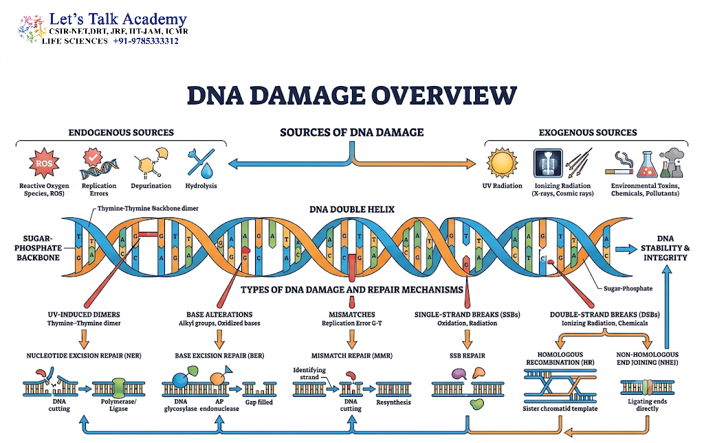
2. Concept of DNA Damage
DNA damage refers to any chemical or structural alteration in DNA that changes its normal structure or interferes with its biological functions.
DNA damage may involve:
- Modification of individual bases
- Loss of a nitrogenous base
- Formation of abnormal chemical bonds
- Single-strand breaks
- Double-strand breaks
- DNA-protein crosslinks
- Interstrand crosslinks
- Replication-associated lesions
- Bulky DNA adducts
- Errors introduced during DNA replication
A useful distinction should be made between DNA damage and mutation.
DNA damage is generally a temporary abnormality that can potentially be repaired. A mutation is a permanent alteration in the DNA sequence that remains after replication or repair. Therefore, unrepaired or incorrectly repaired DNA damage can eventually become a mutation.
3. Sources of DNA Damage
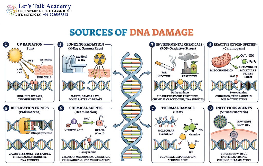
DNA damage can originate from both internal and external sources.
3.1 Endogenous Sources
Endogenous DNA damage arises from normal cellular processes.
Major endogenous sources include:
- Reactive oxygen species
- Spontaneous hydrolysis
- Errors during DNA replication
- Normal DNA metabolism
- Spontaneous deamination
- Replication stress
- Metabolic by-products
3.2 Reactive Oxygen Species
Reactive oxygen species, commonly abbreviated as ROS, are generated during normal cellular metabolism, particularly during mitochondrial respiration.
Important ROS include:
- Superoxide radicals
- Hydrogen peroxide
- Hydroxyl radicals
ROS can oxidize DNA bases and the deoxyribose sugar. One of the most important oxidative DNA lesions is 8-oxoguanine (8-oxoG).
8-oxoG is particularly important because it can mispair with adenine during DNA replication, potentially producing a G:C to T:A transversion mutation.
Cells use antioxidant systems as the first line of defense, while DNA repair mechanisms remove oxidative lesions that escape these protective systems.
3.3 Spontaneous Depurination
DNA bases can spontaneously detach from the sugar-phosphate backbone.
Loss of a purine base, either adenine or guanine, produces an AP site, also called an abasic site.
An AP site contains the sugar-phosphate backbone but lacks a nitrogenous base.
AP sites are primarily repaired through the base excision repair pathway.
3.4 Spontaneous Deamination
Deamination involves removal of an amino group from a DNA base.
Examples include:
Cytosine → Uracil
5-methylcytosine → Thymine
The conversion of cytosine into uracil is particularly important because uracil normally does not belong in DNA.
Deamination of 5-methylcytosine is also biologically significant because it can generate C→T transition mutations.
3.5 Replication Errors
DNA polymerases have high fidelity, but they are not absolutely error-free.
During replication, a nucleotide may occasionally be inserted incorrectly. For example, a polymerase may insert G opposite T instead of A.
Many of these errors are corrected immediately by the proofreading activity of replicative DNA polymerases. Remaining mismatches are corrected primarily by the mismatch repair pathway.
4. Exogenous Sources of DNA Damage
External environmental agents can also damage DNA.
Important exogenous sources include:
- Ultraviolet radiation
- Ionizing radiation
- Chemical mutagens
- Tobacco smoke components
- Certain industrial chemicals
- Some therapeutic agents
- Environmental pollutants
4.1 Ultraviolet Radiation
Ultraviolet radiation can produce covalent bonds between adjacent pyrimidines, particularly thymine residues.
The two major lesions are:
- Cyclobutane pyrimidine dimers
- 6-4 photoproducts
These lesions distort the DNA helix and interfere with replication and transcription.
In humans, such lesions are primarily repaired by nucleotide excision repair.
4.2 Ionizing Radiation
Ionizing radiation has sufficient energy to remove electrons from molecules and can generate highly reactive species.
It can produce:
- Single-strand breaks
- Double-strand breaks
- Base modifications
- Sugar damage
- DNA-protein crosslinks
Among these lesions, DNA double-strand breaks are particularly dangerous because both strands of the DNA molecule are disrupted.
4.3 Chemical Mutagens
Chemical agents can modify DNA bases or interfere with DNA replication.
Examples include:
- Alkylating agents
- Base analogues
- Intercalating agents
- Crosslinking agents
- Oxidizing chemicals
Different chemicals produce different lesions and consequently activate different repair pathways.
5. Major Types of DNA Damage
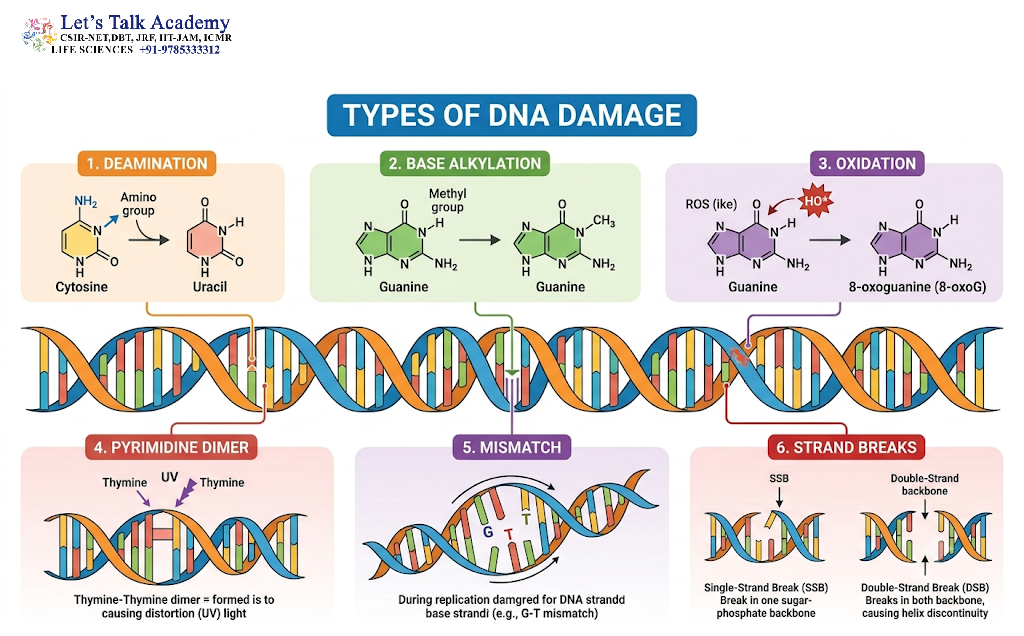
DNA damage can be broadly classified according to its structural nature.
5.1 Base Modification
A DNA base may undergo chemical modification without breaking the DNA backbone.
Examples include:
- Oxidation
- Alkylation
- Deamination
Modified bases can interfere with correct base pairing.
5.2 Depurination
Removal of adenine or guanine creates an abasic or AP site.
If left unrepaired, AP sites can cause replication errors and DNA strand breaks.
5.3 Pyrimidine Dimers
UV radiation can cause covalent linkage between adjacent pyrimidines.
Thymine dimers are classic examples.
5.4 Single-Strand Breaks
A single-strand break occurs when one strand of the DNA backbone is broken.
These lesions are generally less dangerous than double-strand breaks because the complementary strand remains available as a template.
5.5 Double-Strand Breaks
A double-strand break occurs when both DNA strands are broken in close proximity.
Double-strand breaks can result in:
- Deletions
- Insertions
- Chromosomal rearrangements
- Translocations
- Loss of genetic information
They are therefore among the most serious forms of DNA damage.
5.6 DNA Crosslinks
Crosslinks form abnormal covalent bonds involving DNA strands or DNA and proteins.
An interstrand crosslink connects the two DNA strands and prevents their separation.
Such lesions are particularly problematic because replication and transcription require separation of the DNA strands.
6. DNA Damage Response
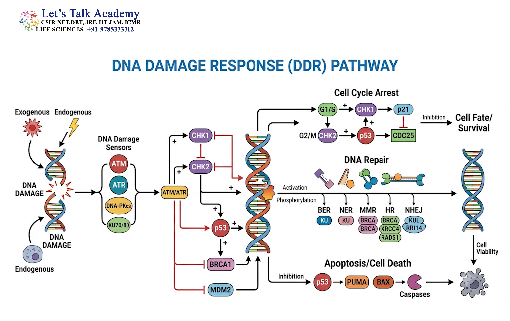
DNA repair is part of a broader cellular process known as the DNA damage response (DDR).
The DNA damage response includes several coordinated steps:
- Damage detection
- Signal generation
- Cell-cycle checkpoint activation
- Recruitment of repair proteins
- DNA repair
- Recovery or elimination of severely damaged cells
The DNA damage response prevents damaged DNA from being copied or transmitted to daughter cells.
6.1 Damage Sensors
Specialized proteins recognize abnormal DNA structures.
For example, proteins involved in sensing double-strand breaks help recruit signaling proteins to the damaged region.
6.2 Signal Transduction
DNA damage activates protein kinases that modify downstream proteins.
Important signaling proteins include:
- ATM
- ATR
- DNA-PK
These proteins coordinate DNA repair with cell-cycle control.
6.3 Cell-Cycle Checkpoints
Checkpoints temporarily halt cell-cycle progression to provide time for DNA repair.
Major checkpoints include:
- G1/S checkpoint
- Intra-S checkpoint
- G2/M checkpoint
The tumor suppressor protein p53 is an important regulator of the cellular response to DNA damage.
When damage is severe, p53 can promote cell-cycle arrest, cellular senescence, or apoptosis.
7. Direct Reversal of DNA Damage
Some forms of DNA damage can be corrected without removing the damaged nucleotide.
This process is known as direct repair or direct reversal.
7.1 Photoreactivation
Photoreactivation uses light energy to reverse pyrimidine dimers.
The enzyme responsible is photolyase.
Photolyase binds to the damaged DNA and uses energy from visible or near-ultraviolet light to break the abnormal bonds.
Although photoreactivation is found in many organisms, it is not the primary pathway for repairing UV-induced pyrimidine dimers in placental mammals.
7.2 O6-Methylguanine Repair
The DNA repair protein O6-methylguanine-DNA methyltransferase (MGMT) can remove an alkyl group from the O6 position of guanine.
MGMT transfers the alkyl group to itself and becomes inactive in the process.
Therefore, MGMT is often described as a suicide repair protein.
8. Base Excision Repair
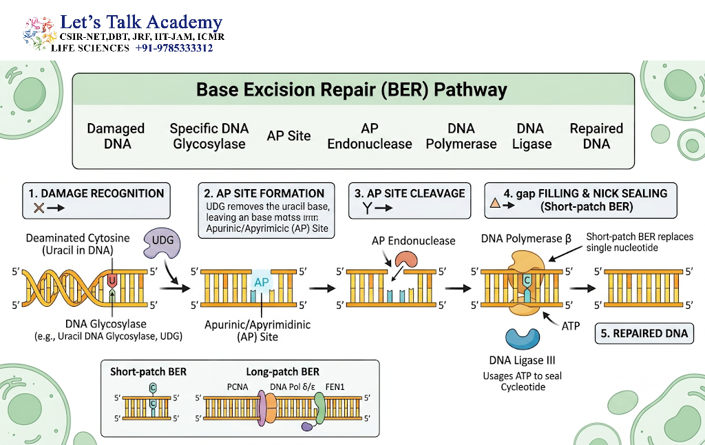
Base excision repair (BER) primarily repairs small, non-helix-distorting lesions.
Examples include:
- Oxidized bases
- Deaminated bases
- Alkylated bases
- Abasic sites
The pathway generally involves the following steps:
Damaged base → Base removal → AP site processing → Gap filling → Ligation
8.1 DNA Glycosylase
The first major enzyme in BER is a DNA glycosylase.
DNA glycosylases recognize specific damaged bases and cleave the N-glycosidic bond connecting the base to the sugar.
This produces an AP site.
Different glycosylases recognize different lesions.
For example:
- Uracil-DNA glycosylase removes uracil
- OGG1 recognizes 8-oxoguanine lesions
- Alkylpurine DNA glycosylases recognize certain alkylated bases
8.2 AP Endonuclease
The resulting AP site is processed by AP endonuclease.
The enzyme cleaves the DNA backbone near the AP site, creating a suitable substrate for subsequent repair reactions.
8.3 DNA Polymerase
A DNA polymerase inserts the correct nucleotide using the undamaged strand as a template.
8.4 DNA Ligase
Finally, DNA ligase seals the remaining nick in the DNA backbone.
9. Nucleotide Excision Repair
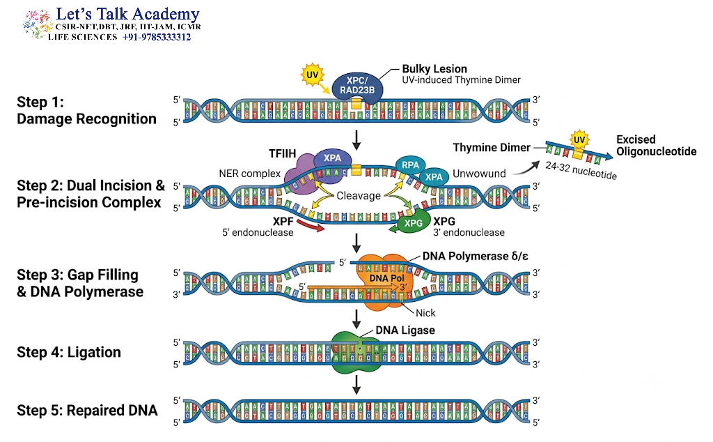
Nucleotide excision repair (NER) removes relatively large, helix-distorting DNA lesions.
Major lesions repaired by NER include:
- UV-induced pyrimidine dimers
- 6-4 photoproducts
- Bulky chemical adducts
Unlike BER, which removes an individual damaged base, NER removes a short stretch of nucleotides containing the lesion.
9.1 Global Genome Nucleotide Excision Repair
Global genome NER continuously surveys the entire genome for DNA distortions.
Proteins recognize abnormalities in DNA structure and initiate repair.
9.2 Transcription-Coupled Nucleotide Excision Repair
Transcription-coupled NER is activated when RNA polymerase encounters a DNA lesion on a transcriptionally active gene.
The stalled transcription machinery acts as a signal that directs repair proteins to the damaged region.
9.3 General Steps of NER
NER can be summarized as:
Damage recognition → DNA unwinding → Dual incision → Removal of damaged oligonucleotide → DNA synthesis → Ligation
Proteins from the TFIIH complex, including helicases such as XPB and XPD, help unwind DNA around the lesion.
Endonucleases then cut the damaged strand on both sides of the lesion.
The damaged oligonucleotide is removed, and the resulting gap is filled by DNA polymerase and sealed by DNA ligase.
9.4 Disorders Associated with NER Defects
Defects in nucleotide excision repair can produce severe human disorders.
A classic example is xeroderma pigmentosum, characterized by extreme sensitivity to sunlight and a greatly increased risk of skin cancer.
Other NER-related disorders include:
- Cockayne syndrome
- Trichothiodystrophy
These disorders demonstrate the importance of efficient DNA repair for genome stability and normal development.
10. Mismatch Repair
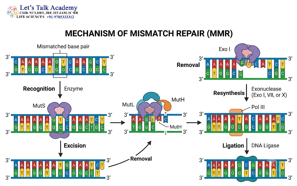
Mismatch repair (MMR) corrects errors that escape the proofreading activity of DNA polymerases.
Common targets include:
- Base-base mismatches
- Small insertion loops
- Small deletion loops
Mismatch repair is particularly important immediately after DNA replication.
10.1 Recognition of the Mismatch
In bacteria, proteins such as MutS recognize mismatched bases.
MutL participates in coordinating subsequent repair events.
In eukaryotes, homologous proteins perform similar functions, including the MutS homolog (MSH) and MutL homolog (MLH) protein families.
10.2 Removal of the Incorrect Strand
The repair machinery identifies the newly synthesized strand containing the replication error.
The section containing the mismatch is removed.
10.3 Resynthesis
DNA polymerase synthesizes the correct sequence using the parental strand as the template.
DNA ligase then seals the remaining nick.
10.4 Biological Importance of Mismatch Repair
Defects in mismatch repair increase mutation rates and genomic instability.
In humans, inherited defects in MMR genes are associated with Lynch syndrome, which increases the risk of several cancers, particularly colorectal and endometrial cancers.
11. Repair of Single-Strand Breaks
Single-strand breaks can arise from oxidative stress, radiation, enzymatic reactions, or incomplete DNA repair.
Proteins such as PARP1 can recognize single-strand DNA breaks and recruit repair factors.
The damaged DNA end may require processing before DNA synthesis and ligation can occur.
DNA polymerases fill the missing nucleotides, while DNA ligases seal the final nick.
Single-strand break repair is closely connected to BER because some BER intermediates temporarily contain single-strand breaks.
12. Double-Strand Break Repair
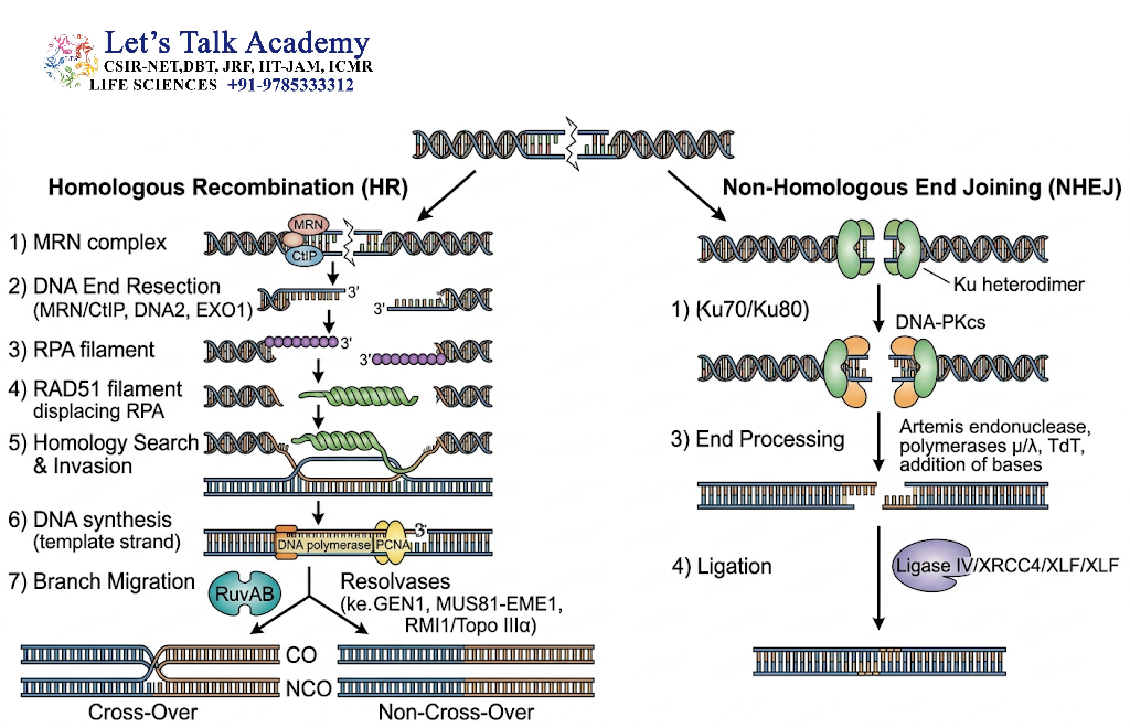
DNA double-strand breaks are particularly dangerous because no intact complementary strand remains directly connected across the break.
Two major pathways repair double-strand breaks:
- Homologous recombination
- Non-homologous end joining
The choice of pathway depends on factors such as cell-cycle stage, DNA-end structure, and availability of a homologous template.
13. Homologous Recombination
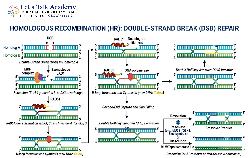
Homologous recombination (HR) is a highly accurate mechanism for repairing double-strand breaks because it uses a homologous DNA sequence as a template.
In proliferating eukaryotic cells, HR is particularly important during the S and G2 phases, when a sister chromatid is available.
13.1 Major Steps of Homologous Recombination
The major stages include:
- Break recognition
- DNA-end resection
- Formation of single-stranded DNA
- Strand invasion
- DNA synthesis
- Resolution or dissolution of recombination intermediates
13.2 Role of RAD51
RAD51 is a central protein in homologous recombination.
RAD51 forms a nucleoprotein filament on single-stranded DNA and facilitates the search for a homologous DNA sequence.
13.3 Role of BRCA Proteins
BRCA1 and BRCA2 are important components of homologous recombination-mediated DNA repair.
BRCA2 helps load RAD51 onto single-stranded DNA.
Defects in BRCA1 or BRCA2 can compromise homologous recombination and increase genomic instability.
14. Non-Homologous End Joining
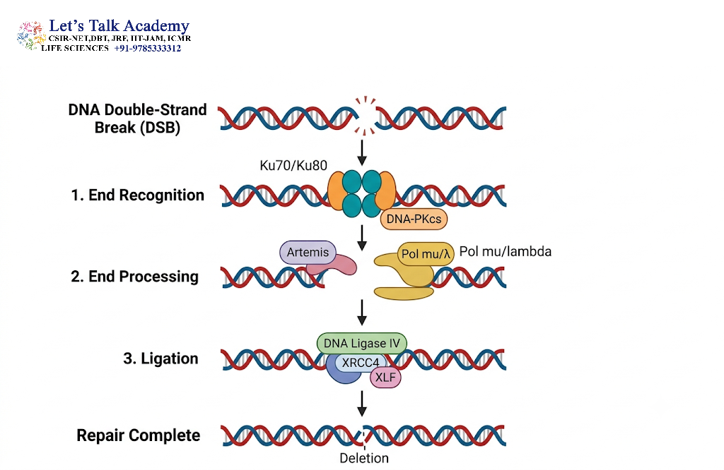
Non-homologous end joining (NHEJ) repairs double-strand breaks by directly joining broken DNA ends.
Unlike homologous recombination, NHEJ does not require a long homologous DNA template.
Important proteins include:
- Ku70
- Ku80
- DNA-PKcs
- Artemis
- XRCC4
- DNA ligase IV
- XLF
14.1 Recognition of DNA Ends
The Ku70/Ku80 heterodimer binds to broken DNA ends.
It helps protect and organize the DNA ends for subsequent repair.
14.2 End Processing
If the broken ends are not directly compatible, they may need to be processed.
Nucleases and DNA polymerases can modify the ends before joining.
14.3 DNA Ligation
DNA ligase IV, together with XRCC4 and associated factors, joins the DNA ends.
Because NHEJ can involve processing or nucleotide loss, it may occasionally introduce small insertions or deletions.
15. Alternative End Joining and Microhomology-Mediated Repair
Cells also possess alternative pathways for repairing double-strand breaks.
One important mechanism is microhomology-mediated end joining (MMEJ).
This pathway uses short homologous sequences called microhomologies near the broken DNA ends.
Because DNA sequences between the microhomologies may be removed, MMEJ is generally more error-prone than classical NHEJ and homologous recombination.
It can contribute to:
- Small deletions
- Chromosomal rearrangements
- Genome instability
16. Interstrand Crosslink Repair
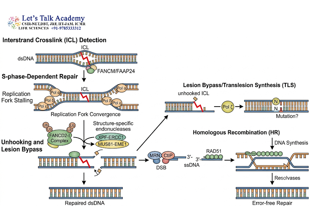
Interstrand crosslinks prevent the two DNA strands from separating.
This creates a major problem because replication and transcription require strand separation.
Repair of interstrand crosslinks involves coordinated activity of multiple repair pathways.
The Fanconi anemia pathway plays a central role in detecting and processing these lesions.
Several Fanconi anemia proteins cooperate with:
- Nucleotide excision repair proteins
- Translesion synthesis machinery
- Homologous recombination proteins
Defects in this pathway can cause Fanconi anemia, a disorder associated with chromosome instability, bone marrow failure, developmental abnormalities, and increased cancer susceptibility.
17. Translesion DNA Synthesis
Sometimes DNA replication encounters a lesion that cannot be immediately removed.
In such situations, specialized DNA polymerases can replicate across the damaged region.
This process is called translesion synthesis (TLS).
TLS prevents replication forks from becoming permanently stalled, but many TLS polymerases have lower fidelity than normal replicative DNA polymerases.
Therefore, TLS can allow replication to continue but may increase the probability of mutation.
A key regulatory protein in this process is PCNA, whose modification can influence the recruitment of specialized polymerases.
18. DNA Damage Tolerance
DNA damage tolerance mechanisms allow cells to continue DNA replication even when lesions remain temporarily unrepaired.
These mechanisms do not necessarily remove the original lesion immediately.
Instead, they help the replication machinery bypass or tolerate the damaged region.
DNA damage tolerance is particularly important when immediate repair is impossible.
19. DNA Repair and Cell-Cycle Regulation
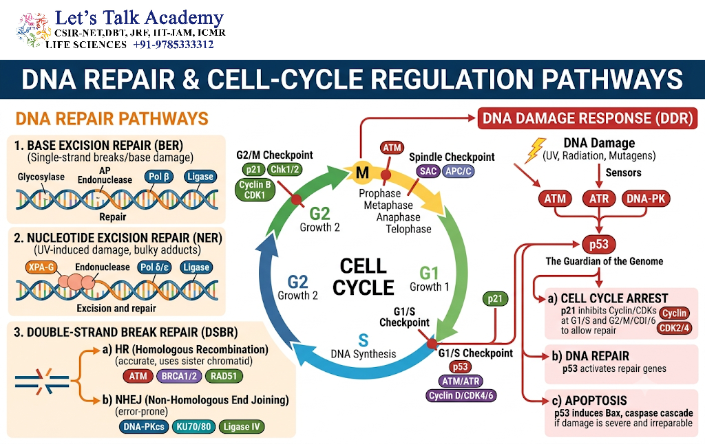
DNA repair and cell-cycle progression are tightly connected.
Cells should not proceed into DNA replication or mitosis when their genome contains dangerous levels of damage.
19.1 G1/S Checkpoint
The G1/S checkpoint prevents damaged DNA from being replicated.
p53 can induce expression of p21, a cyclin-dependent kinase inhibitor.
p21 suppresses cell-cycle progression and provides additional time for DNA repair.
19.2 Intra-S Checkpoint
The intra-S checkpoint slows DNA replication in response to replication stress and DNA damage.
ATR signaling is particularly important in this response.
19.3 G2/M Checkpoint
The G2/M checkpoint prevents entry into mitosis when DNA replication is incomplete or DNA damage remains unresolved.
This checkpoint provides an additional opportunity for repair before chromosome segregation.
20. Apoptosis in Response to Severe DNA Damage
Not all DNA damage can or should be repaired.
When DNA damage is extensive and irreparable, the cell may activate apoptosis, a controlled form of programmed cell death.
This prevents severely damaged cells from continuing to divide and potentially transmitting harmful mutations.
p53 is an important regulator of this decision.
Depending on the type and severity of damage, p53 may promote:
- Cell-cycle arrest
- DNA repair
- Senescence
- Apoptosis
Thus, DNA repair and programmed cell death work together to protect the organism from genomic instability.
21. DNA Damage and Cancer
Genomic stability is essential for preventing cancer.
Cancer can develop when DNA damage accumulates because of:
- Increased exposure to DNA-damaging agents
- Defective DNA repair
- Mutations in checkpoint proteins
- Abnormal replication
- Failure of apoptosis
Mutations in DNA repair genes can increase the accumulation of additional mutations.
This creates a cycle in which defective genome maintenance promotes genomic instability, which in turn increases the likelihood of further mutations.
Important cancer-associated genes involved in DNA damage responses include:
- TP53
- BRCA1
- BRCA2
- ATM
- ATR
- MSH2
- MLH1
- MSH6
- PMS2
22. DNA Repair Defects and Human Diseases
Defects in DNA repair pathways can produce characteristic genetic disorders.
22.1 Xeroderma Pigmentosum
Defects in nucleotide excision repair cause extreme sensitivity to UV radiation.
Affected individuals have a greatly increased risk of skin cancers.
22.2 Lynch Syndrome
Lynch syndrome is associated with inherited defects in mismatch repair genes.
It increases the risk of colorectal and several other cancers.
22.3 Ataxia-Telangiectasia
Mutations affecting ATM can cause ataxia-telangiectasia.
The disorder is associated with impaired responses to DNA double-strand breaks, neurological abnormalities, immune dysfunction, and increased cancer susceptibility.
22.4 Fanconi Anemia
Fanconi anemia results from defects in proteins involved in interstrand crosslink repair.
It is associated with chromosome instability and increased susceptibility to malignancy.
23. DNA Repair in Mitochondria
Mitochondrial DNA is also exposed to damage, particularly because mitochondria are major sites of reactive oxygen species generation.
Mitochondrial DNA repair mechanisms include:
- Base excision repair
- Single-strand break repair
- Limited forms of other repair processes
Because mitochondrial DNA encodes components essential for oxidative phosphorylation, damage to mitochondrial genomes can affect cellular energy metabolism.
24. DNA Repair in Prokaryotes and Eukaryotes
DNA repair mechanisms are conserved across organisms, although the proteins and regulatory systems can differ.
Bacteria often provide experimentally useful models for understanding DNA repair.
For example, the bacterial UvrABC system is a classical model for nucleotide excision repair, while MutS and MutL are central components of bacterial mismatch repair.
Eukaryotic organisms possess more complex repair systems that are closely integrated with chromatin organization, cell-cycle regulation, and checkpoint signaling.
25. Important DNA Repair Enzymes and Proteins
Several proteins are particularly important in DNA repair.
| Protein/Enzyme | Major Function |
|---|---|
| DNA glycosylase | Removes damaged bases |
| AP endonuclease | Cleaves DNA at AP sites |
| DNA polymerase | Fills DNA gaps |
| DNA ligase | Seals DNA nicks |
| MGMT | Directly reverses O6-alkylguanine damage |
| MutS/MSH proteins | Recognize mismatches |
| MutL/MLH proteins | Coordinate mismatch repair |
| XPA–XPG proteins | Participate in NER |
| ATM | Responds to DNA double-strand breaks |
| ATR | Responds strongly to replication stress and single-stranded DNA |
| RAD51 | Central protein in homologous recombination |
| BRCA1/BRCA2 | Promote homologous recombination |
| Ku70/Ku80 | Recognize DNA ends during NHEJ |
| DNA-PKcs | NHEJ signaling and end processing |
| DNA ligase IV | Joins DNA ends during NHEJ |
| PARP1 | Detects and coordinates repair of single-strand breaks |
26. Comparison of Major DNA Repair Pathways
| Repair Pathway | Major Damage Repaired | Important Features |
|---|---|---|
| Direct repair | Certain chemically modified bases | Reverses damage directly |
| BER | Small base lesions | Removes individual damaged bases |
| NER | Bulky, helix-distorting lesions | Removes a short DNA segment |
| MMR | Replication mismatches | Corrects newly synthesized DNA errors |
| HR | Double-strand breaks | Uses homologous template |
| NHEJ | Double-strand breaks | Directly joins DNA ends |
| MMEJ | Some double-strand breaks | Uses short microhomologies |
| TLS | Replication-blocking lesions | Bypasses DNA damage |
27. Fidelity of DNA Repair
DNA repair itself must be highly accurate.
The objective is not simply to remove damaged DNA but to restore the correct genetic sequence.
Repair accuracy depends on:
- Recognition of the correct lesion
- Selection of the correct DNA strand
- Use of the undamaged strand as a template
- Accurate DNA synthesis
- Proper ligation
- Coordination with DNA replication and cell-cycle progression
When repair is inaccurate, mutations or chromosomal rearrangements can arise.
28. DNA Damage, Mutation, and Genetic Variation
DNA damage can contribute to mutations when it is incorrectly repaired or replicated across.
Mutations can include:
- Base substitutions
- Insertions
- Deletions
- Inversions
- Duplications
- Translocations
Not all mutations are harmful. Some have little or no effect, while others may alter protein function or gene regulation.
However, accumulation of damaging mutations can compromise cellular function and contribute to disease.
29. DNA Repair and Aging
DNA damage accumulates throughout life.
Several factors may contribute to this accumulation, including:
- Continuous metabolic activity
- Oxidative stress
- Replication errors
- Environmental exposure
- Declining efficiency of some repair processes
Persistent DNA damage can activate cellular senescence or apoptosis.
Therefore, DNA repair systems are important components of cellular maintenance and long-term genome stability.
30. DNA Repair as a Therapeutic Target
The dependence of cancer cells on DNA repair pathways has created important opportunities for targeted therapy.
A well-known example involves PARP inhibitors.
Cells with defective homologous recombination, particularly those carrying certain BRCA1 or BRCA2 mutations, can become unusually dependent on other DNA repair mechanisms.
Blocking PARP-mediated repair can therefore selectively increase DNA damage in susceptible cancer cells.
This principle is an example of synthetic lethality, in which loss of either one of two pathways may be tolerated, but simultaneous loss of both becomes lethal.
31. Integration of DNA Repair Pathways
DNA repair pathways do not operate as completely independent systems.
A single DNA lesion may pass through several repair stages.
For example, oxidative damage can initially be recognized by a DNA glycosylase, processed through BER, and finally repaired by DNA synthesis and ligation.
Similarly, double-strand break repair can involve:
Damage sensing → End processing → Homologous recombination or end joining → DNA synthesis → Ligation → Checkpoint recovery
The choice of pathway depends on lesion type, chromatin environment, cell-cycle stage, and availability of repair factors.
32. Key Molecular Principles of DNA Repair
Several general principles are common to almost all DNA repair pathways.
32.1 Damage Recognition
The repair machinery must first distinguish damaged DNA from normal DNA.
32.2 Damage Removal or Reversal
The lesion is either chemically reversed or physically removed.
32.3 DNA Synthesis
When nucleotides are removed, DNA polymerase restores the missing sequence using an intact template.
32.4 DNA Ligation
DNA ligase restores continuity of the sugar-phosphate backbone.
32.5 Checkpoint Control
Cell-cycle progression is regulated to prevent damaged DNA from being replicated or transmitted.
32.6 Recovery
Once repair is complete, signaling pathways are turned off and the cell resumes normal activity.
33. Flowchart of DNA Damage and Repair
A simplified overview can be represented as:
DNA damage
↓
Damage recognition
↓
DNA damage signaling
↓
Cell-cycle checkpoint activation
↓
Selection of appropriate repair pathway
↓
Damage removal or reversal
↓
DNA synthesis
↓
DNA ligation
↓
Genome restoration
↓
Cell-cycle recovery
If the damage is irreparable:
Persistent DNA damage → Senescence or apoptosis


