
1. Introduction
The adaptive immune system depends on highly specific receptors that allow lymphocytes to recognize foreign and abnormal molecular structures. The two major antigen-recognition receptors of adaptive immunity are the B-cell receptor (BCR) and the T-cell receptor (TCR).
The B-cell receptor is expressed on the surface of B lymphocytes and is closely related to membrane-bound immunoglobulin. It recognizes specific molecular structures on antigens and initiates signaling pathways that regulate B-cell activation, proliferation, differentiation, and antibody production.
The T-cell receptor is expressed on T lymphocytes and recognizes antigen-derived peptide fragments presented by major histocompatibility complex (MHC) molecules. TCR signaling regulates T-cell activation, differentiation, cytokine production, cytotoxicity, and immune memory.
Although both receptors provide antigen specificity, they differ significantly in their structure, antigen-recognition mechanisms, and signaling systems.
The basic principle is:
Antigen recognition → Receptor signaling → Lymphocyte activation → Differentiation → Effector and memory responses
2. Definition of B-Cell Receptor
The B-cell receptor (BCR) is a membrane-associated antigen-recognition complex present on B lymphocytes.
The BCR consists primarily of:
- Membrane-bound immunoglobulin
- CD79a
- CD79b
The membrane immunoglobulin provides antigen-binding specificity, whereas CD79a and CD79b are mainly responsible for transmitting intracellular activation signals.
3. Definition of T-Cell Receptor
The T-cell receptor (TCR) is a membrane-bound receptor expressed on T lymphocytes that recognizes antigen-derived peptides in association with MHC molecules.
The conventional TCR is usually composed of:
- α chain
- β chain
The TCR is associated with the CD3 signaling complex, which transmits activation signals into the T cell.
4. Major Functions of BCR and TCR
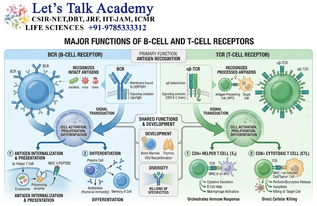
Both receptors are essential for adaptive immunity.
BCR Functions
- Antigen recognition
- B-cell activation
- Antigen internalization
- Antigen presentation through MHC II
- Clonal expansion
- Plasma-cell differentiation
- Memory B-cell formation
TCR Functions
- Recognition of peptide–MHC complexes
- T-cell activation
- Clonal expansion
- T-cell differentiation
- Cytokine production
- Cytotoxic responses
- Immune regulation
- Memory T-cell formation
5. General Structure of BCR
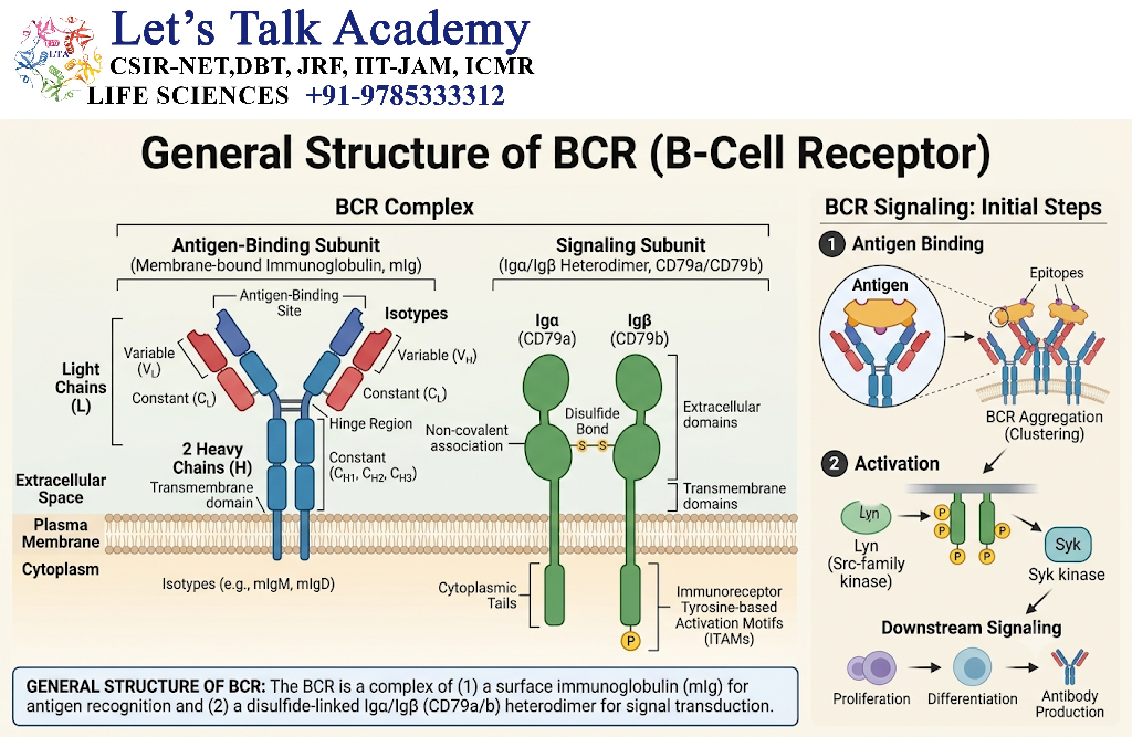
The BCR contains membrane-bound immunoglobulin molecules.
A typical membrane immunoglobulin contains:
- Two heavy chains
- Two light chains
- Variable regions
- Constant regions
- Antigen-binding sites
- Membrane-spanning regions
- Cytoplasmic tails
However, the cytoplasmic tails of membrane immunoglobulins are short and are not sufficient for strong intracellular signaling.
Therefore, CD79a and CD79b provide the major signaling functions.
6. BCR Structure
A simplified representation is:
             Antigen
                ↓
        ┌──────────────┐
        │ Membrane Ig  │
        │   Fab region │
        └──────┬───────┘
               │
        Cell membrane
          │         │
       CD79a     CD79b
          │         │
         ITAM      ITAM
          │         │
       Signaling into cell
The antigen-binding portion is formed by the variable regions of the immunoglobulin.
7. Membrane Immunoglobulin and Secreted Antibody
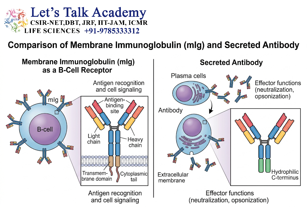
B cells can express immunoglobulin in two major forms.
Membrane-Bound Form
The immunoglobulin remains attached to the B-cell membrane and functions as part of the BCR.
Secreted Form
After differentiation into plasma cells, immunoglobulin can be secreted as soluble antibody.
The antigen-binding specificity of the membrane and secreted forms from the same B-cell clone is generally related because they are generated from the same rearranged immunoglobulin genes, although their RNA processing differs.
8. BCR Antigen Recognition
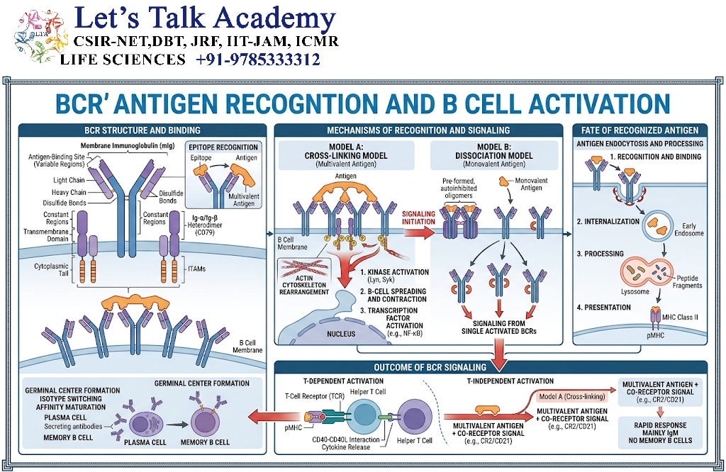
BCRs can recognize antigens in their relatively intact molecular form.
They may recognize:
- Proteins
- Polysaccharides
- Lipids
- Glycoproteins
- Other molecular structures
Recognition depends on the three-dimensional complementarity between the antigenic epitope and the antibody-binding site.
Therefore:
Antigen epitope ↔ BCR antigen-binding site
9. BCR and Epitope Recognition
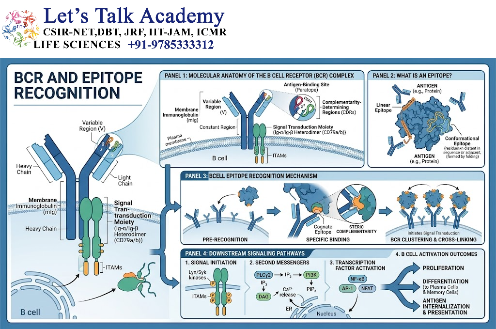
BCRs can recognize both:
Linear Epitopes
Continuous amino acid sequences or molecular regions.
Conformational Epitopes
Three-dimensional structures created by protein folding.
This allows B cells to recognize structural features that do not need to be processed into peptides before initial BCR binding.
10. BCR Signaling Proteins
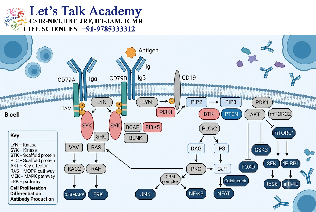
The major signaling components associated with BCR include:
- CD79a
- CD79b
- Src-family kinases
- Syk
- BLNK
- PLCγ2
- PI3K
- BTK
- NF-κB
- NFAT
- AP-1
These components convert extracellular antigen recognition into intracellular biochemical signals.
11. BCR Signaling Mechanism
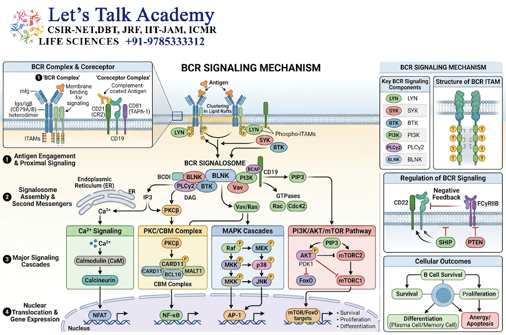
The simplified pathway is:
Antigen
   ↓
BCR cross-linking
   ↓
Src-family kinase activation
   ↓
CD79a/CD79b ITAM phosphorylation
   ↓
Syk activation
   ↓
BLNK signaling complex
   ↓
PLCγ2 / PI3K / MAPK pathways
   ↓
Ca²⁺ / NFAT / NF-κB / AP-1
   ↓
Gene expression
   ↓
B-cell activation
12. Role of ITAMs in BCR Signaling
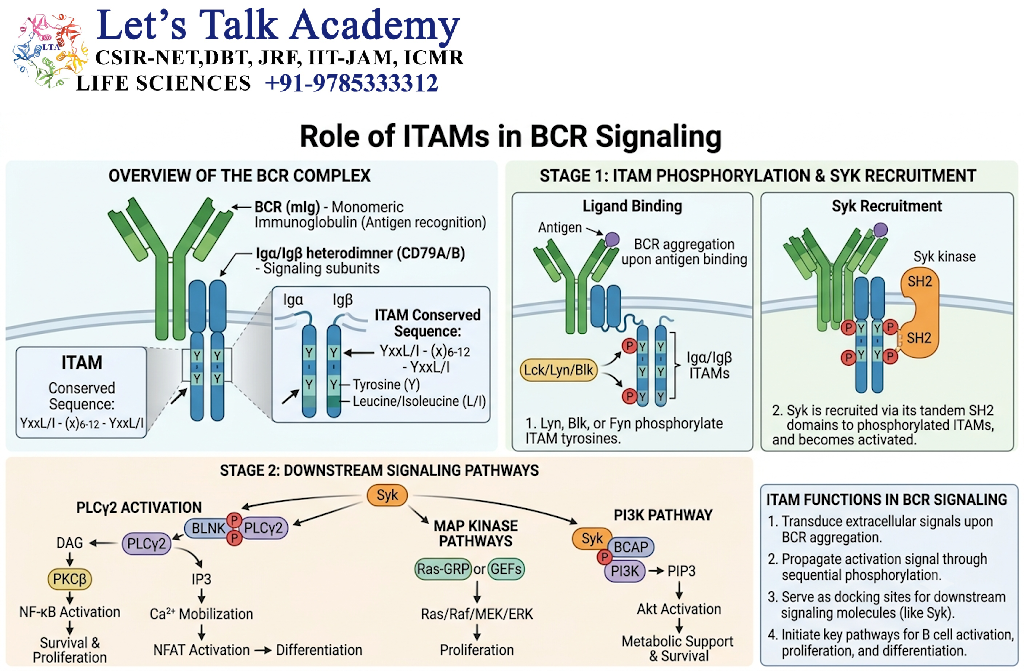
ITAM stands for Immunoreceptor Tyrosine-based Activation Motif.
CD79a and CD79b contain ITAM sequences in their cytoplasmic regions.
Following antigen-induced BCR engagement:
ITAM phosphorylation → Syk recruitment/activation → Downstream signaling
ITAMs therefore function as important molecular switches for receptor activation.
13. Role of Syk
Spleen tyrosine kinase (Syk) is a major signaling enzyme downstream of the BCR.
After phosphorylation of CD79a/CD79b ITAMs, Syk is recruited and activated.
Syk then helps initiate signaling complexes that activate multiple downstream pathways.
These pathways regulate:
- Calcium signaling
- Cytoskeletal changes
- Gene transcription
- Cell survival
- Proliferation
- Differentiation
14. BCR and Calcium Signaling
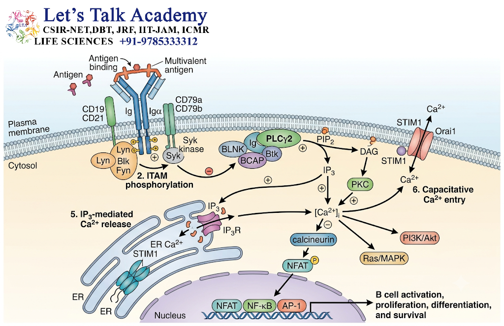
BCR stimulation activates PLCγ2.
PLCγ2 hydrolyzes membrane phospholipid PIP₂ to produce:
- IP₃
- DAG
IP₃ promotes calcium release from intracellular stores.
DAG contributes to activation of protein kinase C pathways.
Thus:
BCR → PLCγ2 → PIP₂ cleavage → IP₃ + DAG → Ca²⁺ + PKC signaling
These pathways activate transcription factors such as NFAT and NF-κB.
15. BCR and PI3K–AKT Signaling
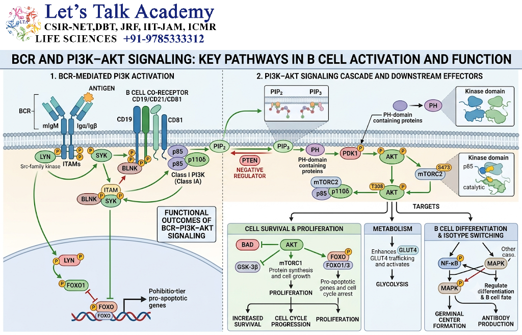
BCR signaling can also activate the PI3K–AKT pathway.
This pathway contributes to:
- Cell survival
- Metabolism
- Growth
- Proliferation
- Differentiation
The strength and duration of PI3K signaling are tightly regulated.
16. BCR Co-Receptor Complex
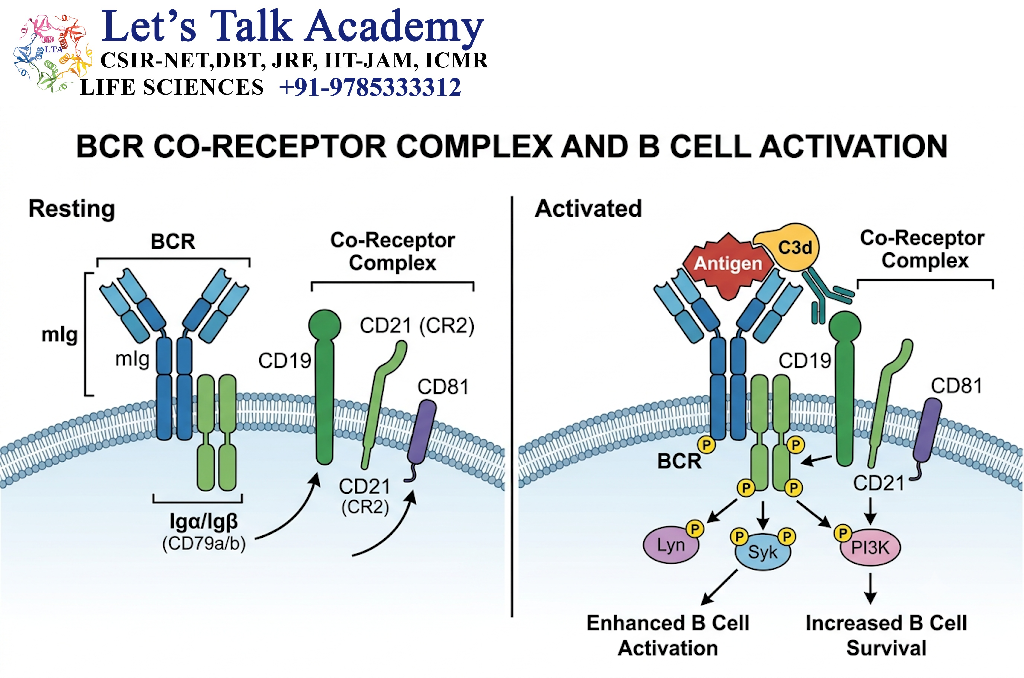
BCR signaling can be enhanced by a co-receptor complex involving:
- CD19
- CD21
- CD81
CD21 can recognize complement fragments attached to antigen.
Co-engagement of this complex with the BCR can amplify intracellular signaling and lower the threshold required for B-cell activation.
17. BCR Internalization
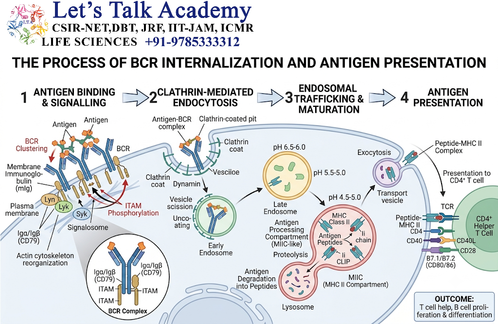
After binding antigen, B cells can internalize the antigen–BCR complex.
The antigen is then processed into peptides.
These peptides can be loaded onto MHC Class II molecules.
The B cell subsequently displays:
Peptide–MHC II
for recognition by CD4⁺ helper T cells.
This creates an important connection between BCR-mediated antigen recognition and T-cell-dependent antibody responses.
18. BCR and B-Cell Activation
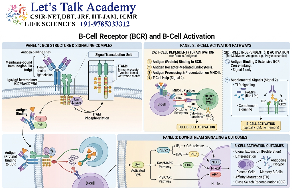
The overall sequence is:
Antigen
   ↓
BCR recognition
   ↓
BCR signaling
   ↓
Antigen internalization
   ↓
MHC II presentation
   ↓
T-cell help
   ↓
B-cell proliferation
   ↓
Differentiation
   ↓
Plasma cells + Memory B cells
19. Definition of TCR
The T-cell receptor (TCR) is a highly specific antigen receptor expressed on T lymphocytes.
The most common TCR is the αβ TCR, consisting of:
- TCR α chain
- TCR β chain
A smaller population of T cells expresses γδ TCRs.
The TCR provides antigen specificity, but the associated CD3 complex is essential for signaling.
20. Structure of the αβ TCR
Each TCR α and β chain contains:
- Variable domain
- Constant domain
- Transmembrane region
- Short cytoplasmic region
The variable domains contain complementarity-determining regions (CDRs) involved in antigen recognition.
The CDR loops create the molecular surface that interacts with peptide–MHC complexes.
21. TCR Structure
A simplified representation is:
              Peptide
                 ↓
            ┌─────────┐
            │   MHC   │
            └────┬────┘
                 │
          ┌─────────────┐
          │   TCR       │
          │ α       β   │
          └─┬─────────┬─┘
            │         │
         CD3 complex
            │
        ITAM-containing
        signaling chains
            │
        Inside the cell
The TCR itself has a very short cytoplasmic tail and therefore depends on associated signaling proteins.
22. TCR and Peptide–MHC Recognition
Unlike BCRs, conventional TCRs generally do not recognize free soluble antigen directly.
Instead, they recognize:
Peptide + MHC
This is a fundamental property of conventional T-cell recognition.
For example:
CD8⁺ T cell → peptide–MHC I
CD4⁺ T cell → peptide–MHC II
23. TCR Complementarity-Determining Regions
TCR variable domains contain six major CDR loops:
- CDR1α
- CDR2α
- CDR3α
- CDR1β
- CDR2β
- CDR3β
CDR3 regions are particularly important because they are highly variable and often make extensive contacts with the antigenic peptide.
CDR1 and CDR2 also contribute strongly to interactions with MHC.
24. TCR Signaling Complex
The TCR associates with the CD3 complex.
Major CD3 components include:
- CD3γ
- CD3δ
- CD3ε
- CD3ζ
The CD3 chains contain ITAMs that transmit activation signals.
Thus:
TCR = antigen-recognition component
CD3 = major signaling component
25. TCR Signaling Mechanism
TCR signaling can be summarized as:
Peptide–MHC → TCR engagement → CD4/CD8 involvement → Lck activation → CD3 ITAM phosphorylation → ZAP-70 activation → downstream signaling → gene expression
Important pathways include:
- Calcium–NFAT
- Ras–MAPK–AP-1
- PKCθ–NF-κB
26. Role of Lck
Lck is a Src-family tyrosine kinase associated mainly with CD4 and CD8 co-receptors.
Following TCR engagement, Lck phosphorylates ITAMs within CD3-associated signaling chains.
This promotes recruitment and activation of ZAP-70.
27. Role of ZAP-70
ZAP-70 is a tyrosine kinase activated downstream of phosphorylated CD3 ITAMs.
It helps activate adaptor proteins such as:
- LAT
- SLP-76
These proteins organize signaling complexes that connect TCR activation to multiple downstream pathways.
28. TCR and Calcium Signaling
TCR activation stimulates PLCγ1.
PLCγ1 hydrolyzes PIP₂ to produce:
- IP₃
- DAG
IP₃ increases intracellular calcium.
Calcium activates calcineurin, which promotes activation and nuclear entry of NFAT transcription factors.
Therefore:
TCR → PLCγ1 → IP₃ → Ca²⁺ → Calcineurin → NFAT
29. TCR and MAPK Signaling
DAG contributes to activation of signaling pathways involving:
Ras → RAF → MEK → ERK
ERK contributes to activation of transcription factors such as AP-1.
This pathway regulates:
- Proliferation
- Differentiation
- Cytokine production
- Gene expression
30. TCR and NF-κB
TCR signaling can activate protein kinase C pathways, including PKCθ, leading to activation of NF-κB.
NF-κB regulates expression of genes involved in:
- Survival
- Activation
- Cytokine production
- Proliferation
31. Three Major Signals in T-Cell Activation
TCR signaling forms part of a larger activation system.
Signal 1
TCR ↔ peptide–MHC
Signal 2
CD28 ↔ CD80/CD86
Signal 3
Cytokines
Together:
Signal 1 + Signal 2 + Signal 3 → Effective T-cell activation
32. CD4 and CD8 Co-Receptors
CD4 and CD8 are co-receptors that stabilize interactions between T cells and MHC molecules.
CD4
Interacts mainly with MHC Class II.
CD8
Interacts mainly with MHC Class I.
They also help bring Lck into proximity with the TCR–CD3 complex.
33. TCR Recognition and MHC Restriction
T-cell recognition is MHC restricted.
This means conventional T cells recognize antigen in the context of an appropriate MHC molecule.
Therefore:
TCR recognition = Peptide recognition + MHC recognition
This is fundamentally different from antibody recognition of intact antigen.
34. BCR vs TCR Antigen Recognition
| Feature | BCR | TCR |
|---|---|---|
| Antigen recognized | Can recognize intact antigen | Usually recognizes processed peptide |
| MHC requirement | Not required for initial antigen binding | Required for conventional αβ TCR recognition |
| Recognition target | Epitope | Peptide–MHC complex |
| Antigen forms | Proteins, carbohydrates, lipids, etc. | Primarily peptide antigens in classical MHC pathways |
| Secreted form | Antibody can be secreted | Not secreted as antibody-like molecule |
| Main role | Humoral immunity | Cellular immunity |
35. Generation of BCR Diversity
BCR diversity is generated primarily through V(D)J recombination during B-cell development.
Immunoglobulin genes contain variable gene segments.
For heavy chains:
V + D + J → Variable region
For light chains:
V + J → Variable region
The rearrangement process is mediated by:
- RAG1
- RAG2
Additional diversity arises from junctional changes and heavy/light-chain pairing.
36. Generation of TCR Diversity
TCR diversity is also generated through V(D)J recombination.
For the TCR β chain:
V + D + J → Variable region
For the TCR α chain:
V + J → Variable region
RAG1 and RAG2 are also essential for TCR gene rearrangement.
Additional diversity arises through:
- Junctional diversity
- Different α–β chain combinations
37. Junctional Diversity
During V(D)J recombination, nucleotide addition and deletion can occur at gene-segment junctions.
Important mechanisms include:
- Nucleotide deletion
- P-nucleotide addition
- N-nucleotide addition
These changes create enormous receptor diversity.
38. BCR and TCR Diversity Comparison
| Source of Diversity | BCR | TCR |
|---|---|---|
| V(D)J recombination | Yes | Yes |
| Junctional diversity | Yes | Yes |
| Chain pairing | Heavy + light | α + β |
| Somatic hypermutation | Yes, after activation | No for conventional TCRs |
| Affinity maturation | Yes | No equivalent process |
A major distinction is that activated B cells can undergo somatic hypermutation and affinity maturation, whereas conventional TCR genes are not subjected to an equivalent affinity-maturation process after activation.
39. Allelic Exclusion
During lymphocyte development, mechanisms of allelic exclusion help ensure that an individual B or T cell expresses a single dominant antigen-receptor specificity.
In B cells, this helps establish one productive heavy-chain and one light-chain combination.
In T cells, productive rearrangement of receptor chains is regulated to establish a defined TCR specificity.
This supports clonal selection and specificity.
40. BCR Signaling vs TCR Signaling
| Feature | BCR | TCR |
|---|---|---|
| Recognition molecule | Membrane immunoglobulin | TCR αβ or γδ |
| Signaling complex | CD79a/CD79b | CD3 complex |
| Important kinase | Syk | ZAP-70 |
| Major Src-family kinase | Lyn and related kinases | Lck |
| Major PLC | PLCγ2 | PLCγ1 |
| ITAM-bearing proteins | CD79a/CD79b | CD3 chains |
| Calcium pathway | Important | Important |
| MAPK | Important | Important |
| NF-κB | Important | Important |
| Main outcome | B-cell activation/differentiation | T-cell activation/differentiation |
41. Co-Receptors and Accessory Molecules
Lymphocyte activation involves more than the antigen receptor itself.
B-Cell Molecules
- CD19
- CD21
- CD81
- CD40
T-Cell Molecules
- CD4
- CD8
- CD28
- CD40L
- CTLA-4
- PD-1
These molecules modify receptor signaling and help regulate the intensity and outcome of immune responses.
42. BCR and TCR Signaling Thresholds
Lymphocytes must distinguish between weak, strong, persistent, and contextually appropriate receptor signals.
The outcome depends on:
- Receptor affinity
- Antigen concentration
- Duration of receptor engagement
- Co-receptor activity
- Co-stimulation
- Inhibitory signals
- Cytokines
- Cellular environment
Therefore:
Receptor engagement ≠ automatically complete activation
The surrounding signaling environment determines the final response.
43. Positive and Negative Regulation
Receptor signaling must be tightly controlled.
Positive Regulation
Examples include:
- CD19-mediated amplification
- CD28 co-stimulation
- Cytokine signaling
Negative Regulation
Examples include:
- CTLA-4
- PD-1
- Protein tyrosine phosphatases
- Receptor internalization
- Ubiquitination
- Inhibitory adaptor proteins
This balance prevents excessive immune activation.
44. Receptor Internalization and Downregulation
After activation, BCR and TCR signaling can be reduced through mechanisms such as:
- Receptor internalization
- Dephosphorylation
- Ubiquitination
- Signal-protein degradation
- Reduced receptor availability
These mechanisms help prevent continuous signaling.
45. BCR and TCR in Immune Memory
Both receptors contribute to the formation of immunological memory.
BCR
Activated B cells generate:
Plasma cells + Memory B cells
TCR
Activated T cells generate:
Effector T cells + Memory T cells
Memory cells retain antigen-receptor specificity and can respond more rapidly upon re-exposure.
46. BCR and TCR in Immune Tolerance
Receptor specificity also creates a potential risk of recognizing self-antigens.
Therefore, lymphocytes undergo tolerance mechanisms during development and after activation.
B-cell tolerance mechanisms include:
- Receptor editing
- Deletion
- Anergy
T-cell tolerance mechanisms include:
- Positive selection
- Negative selection
- Regulatory T-cell development
- Peripheral tolerance
These processes help reduce harmful self-reactivity.
47. Biological Significance of BCR
BCR-mediated recognition allows B cells to:
- Detect specific antigens
- Internalize antigen
- Present antigen through MHC II
- Receive T-cell help
- Proliferate
- Differentiate into plasma cells
- Produce antibodies
- Generate memory B cells
48. Biological Significance of TCR
TCR-mediated recognition allows T cells to:
- Detect peptide–MHC complexes
- Become activated
- Produce cytokines
- Coordinate immune responses
- Kill infected or abnormal cells
- Regulate immune activity
- Form memory populations


