
1. Introduction
Antibodies are highly specialized proteins produced by B lymphocytes and their differentiated descendants, called plasma cells. Their primary function is to recognize and bind specific molecular structures known as antigens. Antibodies participate in neutralization, opsonization, complement activation, and other immune-defense mechanisms.
The human body can produce an enormous variety of antibodies, potentially recognizing millions of different molecular structures. However, the human genome does not contain a separate, complete gene for every possible antibody. Instead, antibody diversity is generated through a combination of gene rearrangement, DNA modification, chain pairing, and antigen-driven selection.
The term antibody diversity refers to the range of different antigen-binding specificities present within the B-cell and antibody repertoire.
This diversity allows the immune system to respond to:
-
Microorganisms that have never previously entered the body
-
Different strains of the same pathogen
-
Toxins and other soluble foreign molecules
-
Altered or mutated antigens
-
Complex antigens containing multiple epitopes
Antibody diversity is generated in two broad stages:
-
Antigen-independent diversity, which develops during B-cell formation.
-
Antigen-dependent diversification, which occurs after B-cell activation.
The main mechanisms are:
-
V(D)J recombination
-
Combinatorial diversity
-
Junctional diversity
-
Heavy-chain and light-chain pairing
-
Somatic hypermutation
-
Affinity maturation
-
Class-switch recombination
-
Receptor editing and selection during B-cell development
2. Basic Structure of Immunoglobulins
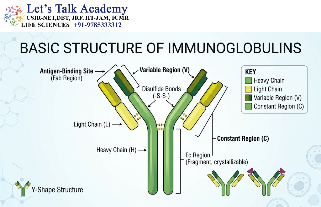
Understanding antibody diversity requires an understanding of antibody structure because the mechanisms of diversity primarily modify the region responsible for antigen recognition.
2.1 Basic Antibody Structure
A typical antibody molecule consists of four polypeptide chains:
-
Two identical heavy chains
-
Two identical light chains
The chains are connected by disulfide bonds. Each chain contains variable and constant regions.
The antibody has a characteristic Y-shaped structure with two major functional portions:
-
Fab region: responsible for antigen binding
-
Fc region: responsible for many biological effector functions
The Fab region contains the variable domains of one heavy chain and one light chain. The Fc region is formed mainly by the constant domains of the heavy chains.
2.2 Variable Regions
The variable regions are located at the amino-terminal ends of the heavy and light chains. Their amino acid sequences differ considerably between antibodies.
The variable regions form the antigen-binding sites. Each antibody generally has two antigen-binding sites with the same basic specificity because it contains two identical heavy-light-chain pairs.
Within each variable domain are three hypervariable regions called complementarity-determining regions, or CDRs.
There are:
-
CDR1 in the heavy chain and light chain
-
CDR2 in the heavy chain and light chain
-
CDR3 in the heavy chain and light chain
Thus, each antigen-binding site is formed by six CDRs.
2.3 Framework Regions
The CDRs are supported by relatively conserved sequences called framework regions.
The framework regions:
-
Maintain the three-dimensional structure of the variable domain
-
Position the CDRs correctly
-
Provide structural stability
-
Influence the final shape of the antigen-binding site
Although CDRs contribute directly to antigen recognition, changes in framework regions can also affect antibody folding and antigen binding.
2.4 The Importance of CDR3
Among the six CDRs, CDR3—particularly heavy-chain CDR3—is often highly variable.
This is because it is formed near the junctions of rearranged gene segments. During gene rearrangement, nucleotides may be added or removed at these junctions, producing extensive sequence variation.
CDR3 frequently makes an important contribution to:
-
Antigen specificity
-
Antigen-binding affinity
-
Recognition of unusual or deeply recessed epitopes
-
Recognition of conformational antigenic structures
3. Organization of Immunoglobulin Genes
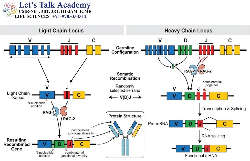
Antibody diversity is possible because immunoglobulin genes are organized into multiple gene segments rather than as single, uninterrupted variable-region genes.
The three major gene-segment categories are:
-
V — Variable
-
D — Diversity
-
J — Joining
The arrangement differs between heavy-chain and light-chain loci.
3.1 Heavy-Chain Gene Locus
The immunoglobulin heavy-chain locus contains multiple copies of:
-
V segments
-
D segments
-
J segments
-
Constant-region genes
A functional heavy-chain variable region is produced by joining one V segment, one D segment, and one J segment.
The general arrangement is:
V + D + J → Rearranged heavy-chain variable region
The heavy-chain constant region is located downstream from the rearranged variable-region sequence.
3.2 Light-Chain Gene Loci
Light-chain genes do not contain D segments. Their variable regions are created by joining:
V + J → Rearranged light-chain variable region
There are two light-chain types:
-
Kappa, or κ
-
Lambda, or λ
The kappa and lambda loci are separate genetic regions. A mature B cell generally expresses one light-chain type as part of its functional antibody receptor.
3.3 Constant-Region Genes
The constant region of the heavy chain determines the antibody class and many of its biological functions.
The major heavy-chain constant regions correspond to:
-
μ — IgM
-
δ — IgD
-
γ — IgG
-
α — IgA
-
ε — IgE
The variable-region gene determines antigen recognition, whereas the heavy-chain constant region influences the antibody’s effector function.
4. B-Cell Development and the Formation of the Primary Repertoire
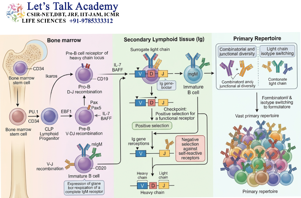
Antibody diversity begins during the development of B cells, primarily in the bone marrow.
Developing B cells undergo a series of genetic and cellular stages that generate and test their antigen receptors.
A simplified sequence is:
-
Hematopoietic stem cell
-
Lymphoid progenitor
-
Early B-cell precursor
-
Pro-B cell
-
Pre-B cell
-
Immature B cell
-
Mature naïve B cell
During these stages, immunoglobulin genes undergo rearrangement.
4.1 Heavy-Chain Rearrangement
Heavy-chain rearrangement generally begins before light-chain rearrangement.
At the pro-B-cell stage:
-
A D segment joins a J segment.
-
A V segment joins the newly formed DJ segment.
-
The rearranged VDJ region is expressed with the μ constant region.
-
The resulting μ heavy chain is tested for functionality.
If the heavy chain is functional, it can participate in the formation of the pre-B-cell receptor.
4.2 Formation of the Pre-B-Cell Receptor
The pre-B-cell receptor contains:
-
A rearranged μ heavy chain
-
A temporary surrogate light chain
-
Signaling proteins associated with the B-cell receptor
The surrogate light chain is not the final light chain. It temporarily tests whether the newly produced heavy chain can form a functional receptor.
Successful signaling indicates that the heavy chain is functional and promotes further B-cell development.
4.3 Light-Chain Rearrangement
After successful heavy-chain rearrangement, the developing B cell begins light-chain gene rearrangement.
A V segment joins a J segment in either the kappa or lambda light-chain locus.
If a functional light chain is produced, it associates with the μ heavy chain to form surface-bound IgM.
The immature B cell can then undergo tolerance testing.
4.4 Expression of the B-Cell Receptor
The B-cell receptor, or BCR, is a membrane-bound immunoglobulin associated with signaling proteins.
In a mature naïve B cell, the BCR commonly contains both:
-
IgM
-
IgD
These antibodies generally have the same antigen-binding specificity because they use the same rearranged variable-region genes but different constant regions.
5. V(D)J Recombination
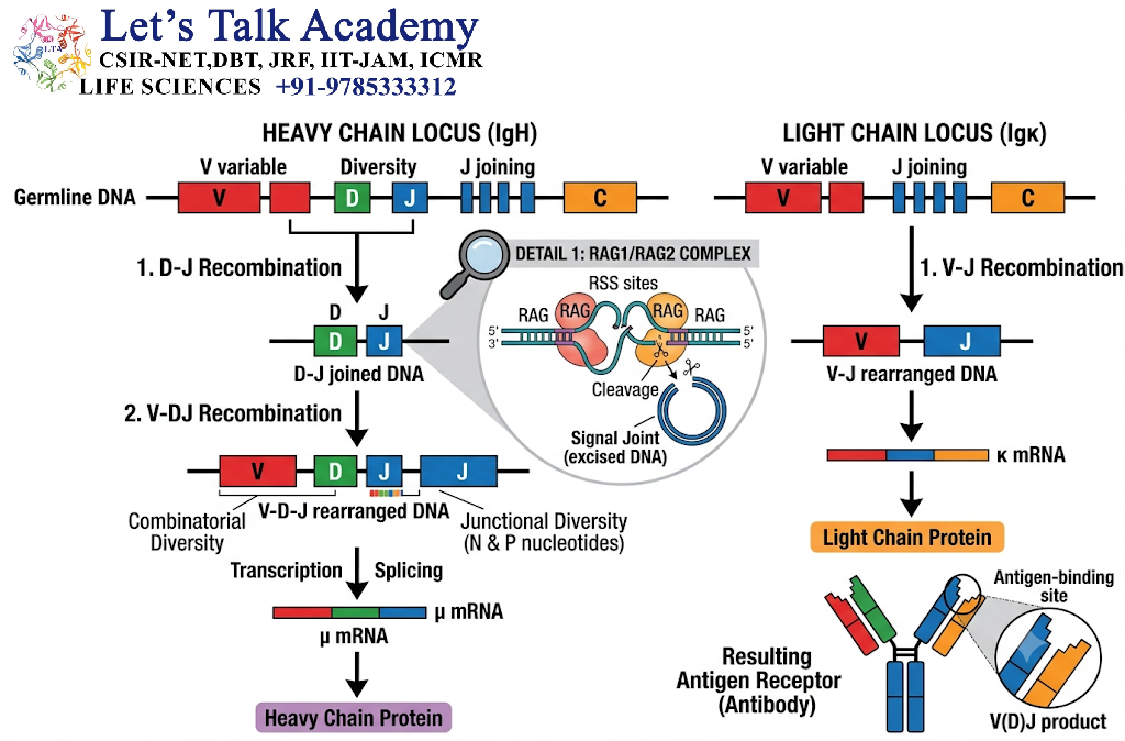
V(D)J recombination is the central mechanism by which antibody variable-region genes are assembled.
It is called V(D)J recombination because:
-
Heavy-chain genes use V, D, and J segments.
-
Light-chain genes use V and J segments.
This process takes place during B-cell development and is mediated by specialized DNA-rearrangement machinery.
5.1 Recombination-Activating Genes
The proteins RAG1 and RAG2 initiate V(D)J recombination.
They recognize DNA sequences called recombination signal sequences, or RSSs, which flank the relevant gene segments.
RAG1 and RAG2 bring selected gene segments together and create DNA breaks at appropriate locations.
The DNA ends are then processed and joined by cellular DNA-repair machinery.
5.2 Recombination Signal Sequences
Recombination signal sequences contain:
-
A conserved heptamer
-
A spacer of either 12 or 23 base pairs
-
A conserved nonamer
The 12/23 rule helps ensure that gene segments are joined in an appropriate order.
In simplified terms, a gene segment flanked by a 12-base-pair spacer is generally joined to a segment flanked by a 23-base-pair spacer.
This rule helps prevent inappropriate rearrangements.
5.3 DNA Cleavage and Hairpin Formation
RAG proteins introduce breaks between the coding regions and the recombination signal sequences.
The coding ends form hairpin-shaped DNA structures. These hairpins must be opened before the coding sequences can be joined.
The DNA-repair protein complex involved in this process includes components of the nonhomologous end-joining pathway, such as:
-
Ku70 and Ku80
-
DNA-PKcs
-
Artemis
-
XRCC4
-
DNA ligase IV
-
XLF
The precise processing of these DNA ends contributes to the generation of junctional diversity.
5.4 Formation of a Functional Rearrangement
If the rearrangement maintains the correct reading frame, a functional immunoglobulin chain may be produced.
If the rearrangement is nonproductive because of an incorrect reading frame or a disruptive mutation, the cell may attempt another rearrangement on the other allele or at another light-chain locus.
Cells that cannot produce a functional receptor generally fail to progress through B-cell development.
6. Combinatorial Diversity
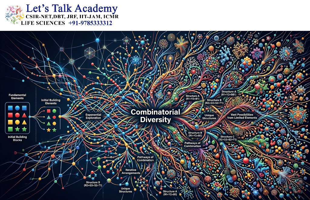
Combinatorial diversity is the variation generated by selecting different combinations of immunoglobulin gene segments during V(D)J recombination.
The immunoglobulin gene loci contain numerous V, D, and J segments. During B-cell development, one segment from each required group is selected and joined to form a variable-region gene.
Because different B cells select different combinations, they produce antibodies with different variable-region sequences.
6.1 Combinatorial Diversity in the Heavy Chain
For the heavy chain, one V segment, one D segment, and one J segment are selected.
For example, a developing B cell might produce:
-
V1–D2–J3
-
V4–D1–J2
-
V2–D5–J1
Each arrangement can encode a different heavy-chain variable region.
The exact number of possible combinations depends on the number of functional gene segments available at a particular immunoglobulin locus.
6.2 Combinatorial Diversity in the Light Chain
Light-chain diversity is generated by selecting:
-
One V segment
-
One J segment
For example, different light-chain arrangements may include:
-
V1–J2
-
V3–J4
-
V5–J1
These arrangements produce light chains with different variable-region sequences.
6.3 Combined Effect of Heavy- and Light-Chain Rearrangement
The diversity generated by heavy-chain rearrangement is multiplied when different heavy chains associate with different light chains.
For example, if four distinct heavy chains can pair with five distinct light chains, as many as twenty theoretical heavy-light-chain combinations may be possible.
This is a simplified illustration. In the body, the actual number of possible combinations is much larger and is also influenced by receptor-selection mechanisms and biological restrictions.
6.4 Importance of Combinatorial Diversity
Combinatorial diversity allows the immune system to generate a broad antibody repertoire without requiring a separate gene for every antibody.
It provides the basic structural foundation for antigen recognition before the body encounters a particular antigen.
7. Junctional Diversity
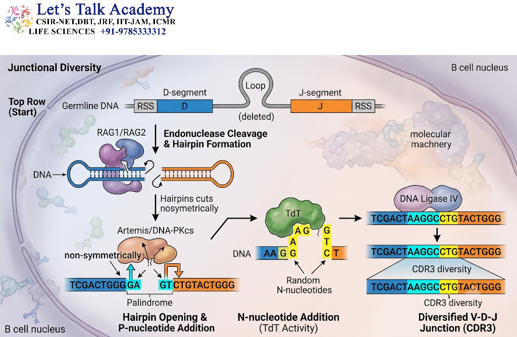
Junctional diversity is the variation produced at the points where V, D, and J gene segments are joined.
It is one of the most important contributors to antibody diversity because it can generate a very large number of different nucleotide sequences from the same selected gene segments.
For example, two B cells may select the same V, D, and J segments but join them differently. As a result, the B cells may produce antibodies with different antigen-binding properties.
7.1 Addition and Removal of Nucleotides
During V(D)J recombination, the ends of gene segments may be modified before they are joined.
The process may involve:
-
Removal of nucleotides from coding ends
-
Addition of non-templated nucleotides
-
Formation of short palindromic sequences
-
Variation in the precise position of DNA joining
These changes alter the nucleotide sequence at the junctions.
Since the nucleotide sequence determines the amino acid sequence of the antibody variable region, junctional changes can alter the structure of the antigen-binding site.
7.2 P Nucleotides
When coding-end hairpins are opened asymmetrically, short palindromic DNA sequences may be generated. These are called P nucleotides.
P nucleotides are produced from the sequence contained within the hairpin structure.
Their addition increases the number of possible sequences at the junctions between gene segments.
7.3 N Nucleotides
N nucleotides are nucleotides added randomly to the junctions without being copied from a DNA template.
The enzyme responsible for adding many of these nucleotides is terminal deoxynucleotidyl transferase, or TdT.
TdT is particularly active during the development of many B-cell populations and contributes significantly to heavy-chain diversity.
The number and sequence of N nucleotides vary from one rearrangement to another.
7.4 Exonuclease-Mediated Nucleotide Removal
Enzymes known as exonucleases may remove nucleotides from the ends of rearranging gene segments.
The number of nucleotides removed is not always the same. Therefore, even when the same V, D, and J segments are selected, different B cells may generate different junctional sequences.
Excessive nucleotide removal can sometimes make a rearrangement nonproductive by disrupting the reading frame.
7.5 Junctional Diversity and CDR3
Junctional diversity is especially important in the formation of the third complementarity-determining region, or CDR3.
The heavy-chain CDR3 region is formed at the junction of V, D, and J segments. It can vary greatly in:
-
Length
-
Amino acid composition
-
Charge
-
Flexibility
-
Three-dimensional shape
Because CDR3 is often positioned at the center of the antigen-binding site, changes in this region can substantially alter antigen recognition.
7.6 Biological Significance
Junctional diversity allows the immune system to produce antibodies that recognize:
-
Small molecular differences between antigens
-
Complex protein surfaces
-
Carbohydrate structures
-
Conformational epitopes
-
Unusual or recessed antigenic sites
However, junctional diversity can also generate nonfunctional or self-reactive receptors. Therefore, B-cell development includes checkpoints that remove or modify potentially harmful cells.
8. Heavy-Chain and Light-Chain Pairing
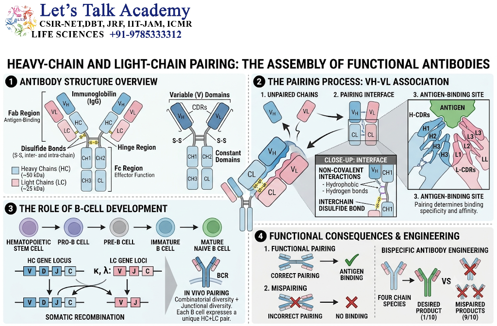
A complete antibody-binding site is formed by the combined variable regions of one heavy chain and one light chain.
The heavy chain alone does not usually determine the complete antigen specificity. The light chain contributes directly to the shape and chemical characteristics of the antigen-binding site.
8.1 Formation of the Antigen-Binding Site
Each antigen-binding site contains:
-
Three heavy-chain CDRs
-
Three light-chain CDRs
The six CDRs work together to recognize an epitope.
The heavy-chain CDRs may contribute strongly to antigen recognition, but the light-chain CDRs can also be essential for:
-
Hydrogen bonding
-
Electrostatic interactions
-
Hydrophobic interactions
-
Shape complementarity
-
Antigen-binding specificity
8.2 Random Association of Chains
Different heavy chains can potentially associate with different light chains.
For example:
|
Heavy chain |
Light chain |
Result |
|---|---|---|
|
H1 |
L1 |
Antibody A |
|
H1 |
L2 |
Antibody B |
|
H2 |
L1 |
Antibody C |
|
H2 |
L2 |
Antibody D |
The resulting antibodies may recognize different epitopes or bind the same epitope with different affinities.
8.3 Restrictions on Chain Pairing
Although heavy- and light-chain pairing contributes to diversity, pairing is not completely unrestricted.
The chains must:
-
Fold correctly
-
Associate structurally
-
Form a stable antigen-binding site
-
Pass B-cell developmental checkpoints
Some heavy-light-chain combinations are unstable or do not produce a functional receptor.
8.4 Importance of Chain Pairing in the Antibody Repertoire
Heavy- and light-chain pairing greatly increases the number of possible antibody-binding sites.
It also explains why antibodies with similar heavy-chain sequences may have different antigen specificities when paired with different light chains.
9. Allelic Exclusion and Isotype Exclusion
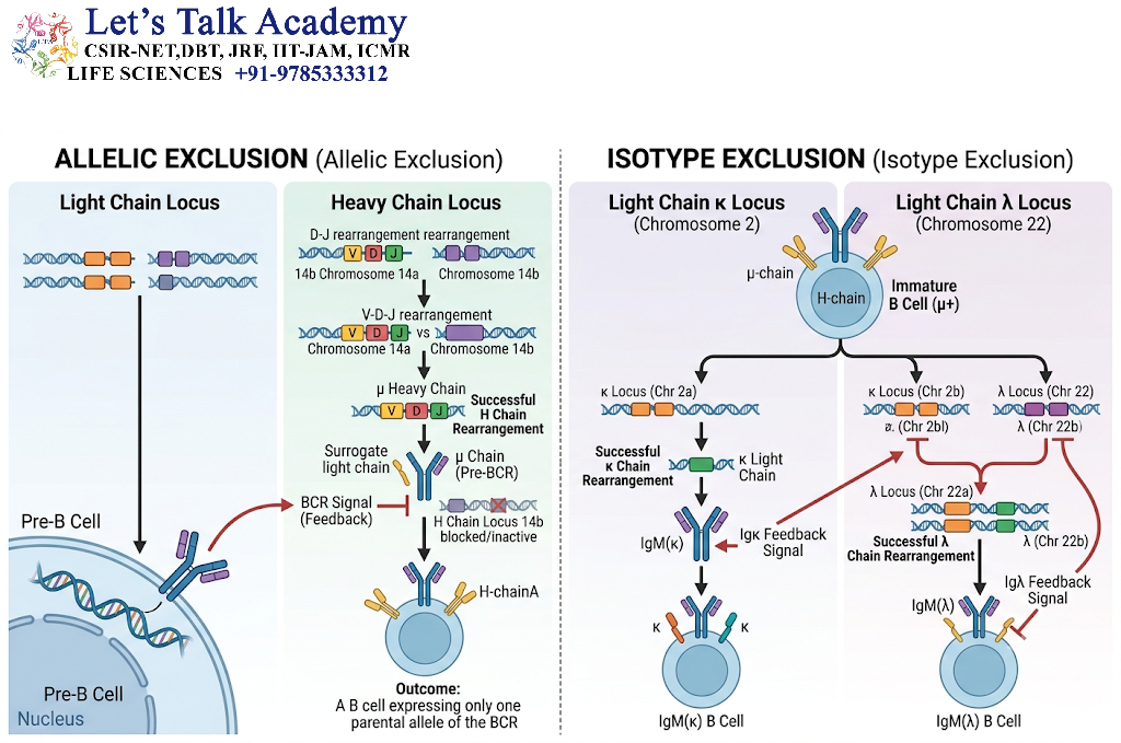
The immune system must generate diverse antibodies while ensuring that each mature B cell generally expresses one principal antigen specificity.
Two important mechanisms involved in this process are allelic exclusion and isotype exclusion.
9.1 Allelic Exclusion
Humans possess two copies of most autosomal genes, one inherited from each parent. The two copies are called alleles.
During B-cell development, rearrangement of immunoglobulin genes is regulated so that a B cell generally expresses one productive heavy-chain allele and one productive light-chain allele.
Once a functional receptor is produced, additional rearrangement is usually suppressed.
This process is called allelic exclusion.
9.2 Importance of Allelic Exclusion
Allelic exclusion ensures that:
-
Each B cell generally expresses one principal BCR specificity.
-
Antigen activation selects a relatively specific B-cell clone.
-
Clonal expansion produces cells with related antigen receptors.
-
Antibody responses can be traced to individual B-cell populations.
Allelic exclusion is not absolute in every biological circumstance, but it is a major organizing principle of B-cell biology.
9.3 Isotype Exclusion
Isotype exclusion refers to the general tendency of a B cell to express either a kappa or a lambda light chain rather than both as its principal light-chain type.
This helps maintain a relatively consistent antigen-binding specificity within an individual B-cell clone.
10. Receptor Editing and B-Cell Selection
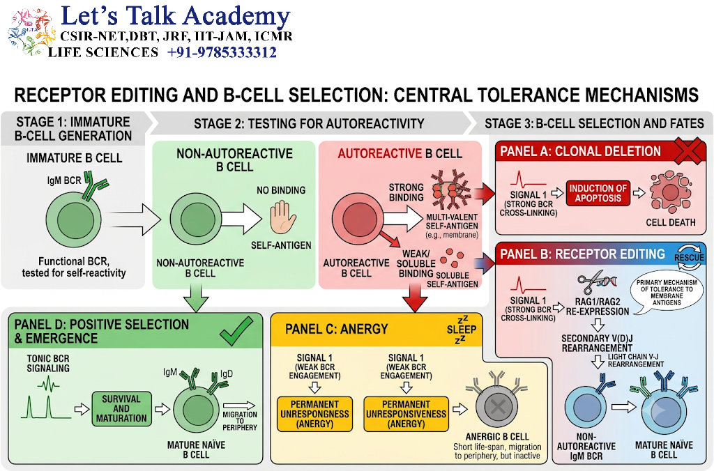
The random generation of antibody receptors can produce receptors that recognize the body’s own molecules.
To reduce the risk of autoimmunity, developing B cells undergo mechanisms of central tolerance.
These mechanisms include:
-
Receptor editing
-
Clonal deletion
-
Anergy
-
Changes in B-cell developmental fate
10.1 Recognition of Self-Antigens
An immature B cell in the bone marrow is tested for its ability to recognize self-antigens.
If its B-cell receptor binds strongly to certain self-antigens, the cell may be considered potentially dangerous.
The response depends partly on:
-
The strength of receptor binding
-
Whether the self-antigen is present in a membrane-bound or soluble form
-
The availability of the antigen
-
The developmental stage of the B cell
10.2 Receptor Editing
Receptor editing allows an immature B cell to replace a potentially self-reactive light chain with another light chain.
The cell may initiate additional light-chain gene rearrangements, particularly at the kappa locus. If suitable rearrangements are unavailable or unsuccessful, rearrangement at the lambda locus may occur.
The newly produced light chain combines with the existing heavy chain.
If the new receptor no longer strongly recognizes self-antigens, the B cell may continue developing.
10.3 Clonal Deletion
If a developing B cell remains strongly self-reactive and receptor editing is unsuccessful, it may undergo apoptosis.
This process is called clonal deletion.
It removes some potentially harmful B-cell clones from the developing repertoire.
10.4 Anergy
Some self-reactive B cells are not immediately destroyed but become functionally unresponsive.
This state is called anergy.
Anergic B cells may survive for a period of time but respond poorly to antigenic stimulation.
10.5 Balance Between Diversity and Tolerance
B-cell development must achieve two goals:
-
Generate a sufficiently diverse antibody repertoire.
-
Limit the number of strongly self-reactive B cells.
This balance is essential for effective immunity without excessive autoimmune damage.
11. Somatic Hypermutation
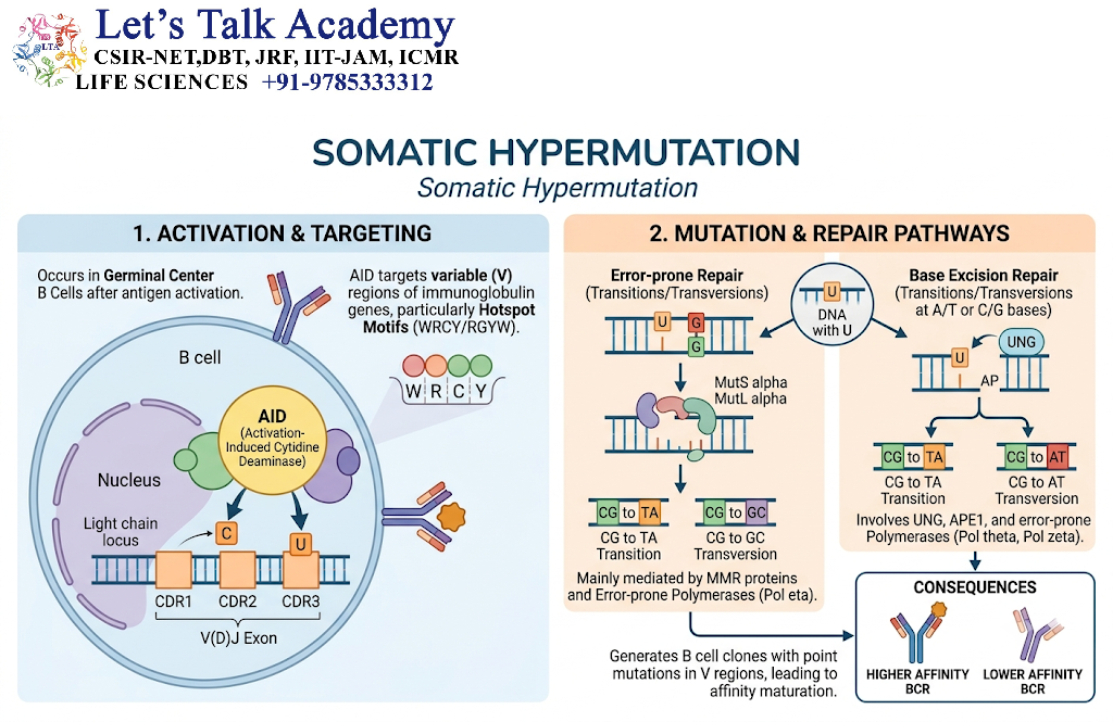
Somatic hypermutation is an antigen-dependent mechanism that introduces mutations into the variable-region genes of activated B cells.
It occurs primarily during immune responses in specialized structures called germinal centers.
Unlike V(D)J recombination, which occurs during early B-cell development, somatic hypermutation occurs after a mature B cell has been activated by an antigen.
11.1 Nature of Somatic Hypermutation
Somatic hypermutation produces a high frequency of single-nucleotide substitutions in immunoglobulin variable-region genes.
These mutations are concentrated in the DNA encoding the antigen-binding regions, although mutations can occur in framework regions as well.
The mutations may alter:
-
Amino acid sequence
-
Antigen-binding-site shape
-
Charge distribution
-
Flexibility
-
Binding affinity
-
Antigen specificity
11.2 Activation of B Cells
Somatic hypermutation usually occurs after B cells have encountered antigen and received appropriate activation signals.
In many responses, especially those involving protein antigens, B cells receive help from T follicular helper cells, or Tfh cells.
Important activation signals include:
-
Binding of antigen to the B-cell receptor
-
Antigen presentation by the B cell
-
Recognition by helper T cells
-
CD40–CD40L interaction
-
Cytokine signaling
Activated B cells may then enter germinal centers and undergo rapid proliferation and mutation.
11.3 Role of Activation-Induced Cytidine Deaminase
The enzyme activation-induced cytidine deaminase, or AID, is essential for somatic hypermutation.
AID converts cytosine bases in DNA into uracil. This creates mismatched or abnormal DNA structures that are processed by DNA-repair pathways.
The repair process can introduce mutations into the immunoglobulin variable-region genes.
AID therefore initiates the molecular events that allow the antibody variable region to evolve during an immune response.
11.4 Types of Mutations
Somatic hypermutation mainly produces point mutations, including:
-
Silent mutations, which do not change the encoded amino acid
-
Missense mutations, which change one amino acid into another
-
Occasionally, mutations that introduce a stop codon or disrupt protein function
Missense mutations in CDRs may alter antigen binding significantly.
Mutations in framework regions may affect the stability or folding of the antibody.
11.5 Possible Outcomes
A mutation may produce an antibody that:
-
Binds antigen more strongly
-
Binds antigen more weakly
-
Recognizes a related epitope
-
Loses antigen-binding ability
-
Retains approximately the same affinity
Somatic hypermutation therefore creates variation rather than automatically improving every antibody.
12. Germinal-Center Reactions
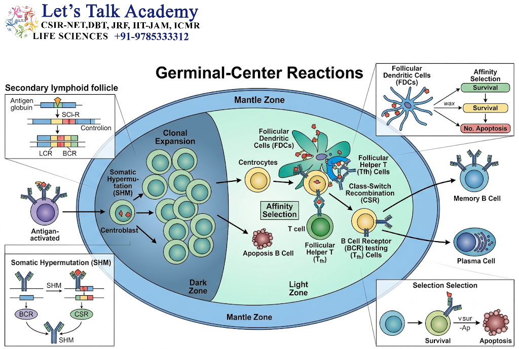
Germinal centers are specialized microenvironments that develop within secondary lymphoid organs during many immune responses.
They are commonly found in:
-
Lymph nodes
-
Spleen
-
Mucosa-associated lymphoid tissues
Germinal centers provide a site where activated B cells proliferate, mutate their antibody genes, and undergo selection.
12.1 Formation of Germinal Centers
After activation, antigen-specific B cells may migrate into lymphoid follicles and participate in germinal-center formation.
These B cells proliferate rapidly and generate populations of daughter cells with different antibody variable-region sequences.
12.2 Dark Zone
The dark zone contains rapidly dividing B cells called centroblasts.
In this region, B cells undergo:
-
Extensive proliferation
-
Somatic hypermutation
-
Generation of multiple antibody variants
The cells then migrate toward the light zone.
12.3 Light Zone
The light zone contains B cells called centrocytes, along with:
-
Follicular dendritic cells
-
T follicular helper cells
-
Antigen displayed in immune complexes
In the light zone, B cells compete for access to antigen and T-cell help.
12.4 Selection of B Cells
B cells with receptors that bind antigen effectively may capture more antigen and present more antigen-derived peptides to Tfh cells.
These B cells can receive survival and proliferation signals.
B cells with weak or nonfunctional receptors may fail to receive sufficient signals and may undergo apoptosis.
12.5 Repeated Cycles of Mutation and Selection
B cells may move repeatedly between dark and light zones.
A simplified cycle is:
-
B-cell proliferation
-
Somatic hypermutation
-
Antigen binding
-
Competition for T-cell help
-
Survival or elimination
-
Further proliferation and mutation
Repeated cycles can enrich the response for B cells producing antibodies with improved antigen-binding properties.
13. Affinity Maturation
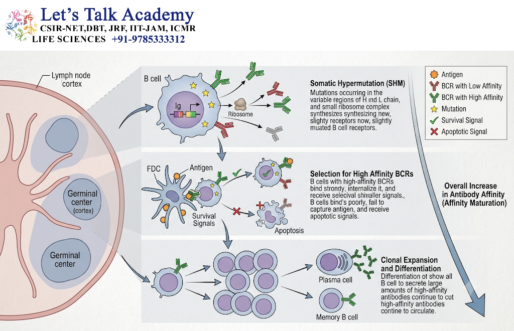
Affinity maturation is the gradual increase in the average antigen-binding affinity of antibodies during an immune response.
It is closely associated with somatic hypermutation and selection within germinal centers.
13.1 Affinity and Avidity
Affinity refers to the strength of interaction between one antigen-binding site and one epitope.
Avidity refers to the combined strength of multiple interactions between a multivalent antibody and a multivalent antigen.
Affinity maturation primarily concerns changes in the binding properties of individual antibody-binding sites, although these changes may also influence overall avidity.
13.2 Mechanism of Affinity Maturation
Affinity maturation generally involves the following stages:
-
Antigen activates a B cell.
-
The B cell proliferates.
-
Somatic hypermutation creates antibody variants.
-
The variants display different antigen-binding properties.
-
B cells compete for antigen and T-cell help.
-
B cells with favorable binding properties receive survival signals.
-
Selected cells proliferate and may undergo additional mutation.
-
High-affinity plasma cells and memory B cells are generated.
13.3 Role of Follicular Dendritic Cells
Follicular dendritic cells display antigen or antigen-containing immune complexes within germinal centers.
They help maintain antigen for recognition by B cells.
B cells must capture antigen through their BCRs and then obtain appropriate survival signals, often from Tfh cells.
This process allows B cells with effective antigen recognition to compete successfully.
13.4 Role of T Follicular Helper Cells
Tfh cells provide essential signals to activated B cells.
These signals include:
-
CD40L binding to CD40
-
Cytokine secretion
-
Promotion of B-cell survival
-
Support for proliferation
-
Support for differentiation
-
Assistance in class-switch recombination
Tfh-cell help is an important part of the selection process in many germinal-center responses.
13.5 Results of Affinity Maturation
Affinity maturation can lead to:
-
More efficient neutralization of toxins and viruses
-
Improved binding to microbial antigens
-
More effective opsonization
-
Better protection during repeated exposure
-
Formation of long-lived plasma cells and memory B cells
Affinity maturation does not necessarily occur in every antibody response, and the degree of maturation varies with the antigen and the type of immune response.
14. Class-Switch Recombination
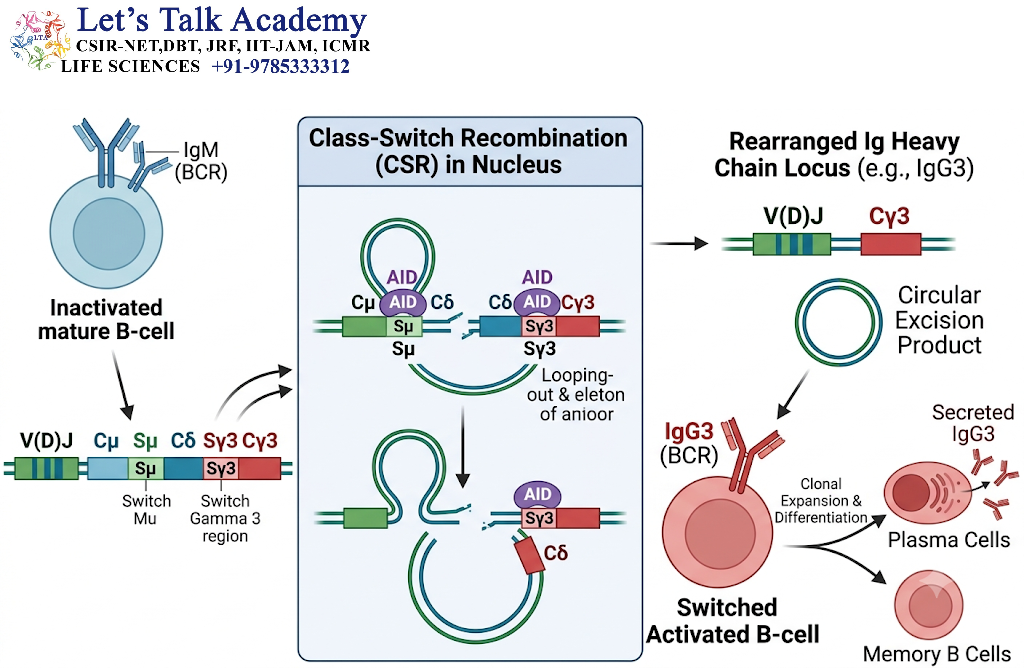
Class-switch recombination, also called isotype switching, changes the antibody heavy-chain constant region while preserving the rearranged variable region.
This process allows a B cell to produce different antibody classes with the same basic antigen-binding specificity but different biological functions.
14.1 Initial Expression of IgM and IgD
Newly mature naïve B cells commonly express both IgM and IgD on their surfaces.
These two immunoglobulins generally possess the same variable-region sequence but different constant regions.
Their expression results mainly from alternative RNA processing rather than from a new V(D)J rearrangement.
14.2 Need for Class Switching
Different tissues and types of infection require different antibody functions.
For example:
-
IgG is important in many systemic immune responses.
-
IgA is specialized for mucosal protection.
-
IgE participates in responses involving mast cells and certain parasites.
-
IgM is particularly important during early antibody responses.
Class switching allows the same antigen recognition system to be connected to different effector mechanisms.
14.3 Mechanism of Class-Switch Recombination
The heavy-chain constant-region genes are arranged in a particular order downstream of the variable-region gene.
A B cell initially expresses the μ constant region, which produces IgM.
During class switching, DNA recombination removes the intervening constant-region DNA and places a different constant-region gene next to the rearranged VDJ sequence.
For example, a B cell may switch from:
VDJ–Cμ → IgM
to:
VDJ–Cγ → IgG
The variable-region sequence remains unchanged in the basic switching process, while the constant region changes.
14.4 Switch Regions
Class-switch recombination occurs at repetitive DNA sequences called switch regions.
The μ constant-region gene is associated with the Sμ switch region. Other switch regions are located before many downstream heavy-chain constant-region genes.
DNA breaks occur in appropriate switch regions, and the intervening DNA is removed in a loop-like structure.
The remaining DNA is joined so that the existing VDJ region is positioned next to a new constant-region gene.
14.5 Role of AID
AID is essential for class-switch recombination.
As in somatic hypermutation, AID initiates DNA changes involving cytosine deamination.
However, the two processes have different outcomes:
-
Somatic hypermutation introduces mutations into variable-region genes.
-
Class-switch recombination produces DNA rearrangements involving constant-region genes.
14.6 Cytokine Influence
Cytokines and signals from T cells influence which antibody class a B cell produces.
The pattern of cytokines varies according to:
-
The type of pathogen
-
The tissue involved
-
The type of helper T-cell response
-
The surrounding inflammatory environment
The precise signals promoting each switch can differ between species and biological contexts.
14.7 Antigen Specificity and Effector Function
Class switching generally preserves the antibody’s antigen-binding specificity because the variable region remains the same.
However, the antibody’s biological activity changes because the Fc region changes.
For example, switching from IgM to IgG may alter:
-
Interaction with Fc receptors
-
Tissue distribution
-
Placental transfer
-
Complement activation
-
Opsonization capacity
-
Persistence in circulation
Thus, class switching changes how an antibody acts, not usually what antigen it recognizes.
15. Main Antibody Classes and Their Functions
15.1 Immunoglobulin M
IgM is usually the first major antibody class produced during a primary immune response.
Characteristics include:
-
Expression on naïve B cells
-
Secretion as a pentamer in many circumstances
-
High overall avidity when binding multivalent antigens
-
Strong ability to activate the classical complement pathway
IgM is important during early defense against many infections.
15.2 Immunoglobulin G
IgG is the most abundant antibody class in normal human serum.
Its functions include:
-
Neutralization of toxins and viruses
-
Opsonization of microorganisms
-
Activation of certain complement pathways
-
Interaction with Fc receptors on immune cells
-
Transfer across the placenta through specific transport mechanisms
There are several IgG subclasses, each with somewhat different biological properties.
15.3 Immunoglobulin A
IgA is especially important at mucosal surfaces.
It is found in:
-
Saliva
-
Tears
-
Mucus
-
Intestinal secretions
-
Respiratory secretions
-
Breast milk
Secretory IgA helps prevent pathogens from attaching to epithelial surfaces.
15.4 Immunoglobulin E
IgE participates in:
-
Immediate hypersensitivity reactions
-
Activation of mast cells and basophils
-
Defense against certain parasitic organisms
IgE binds strongly to high-affinity Fc receptors on certain immune cells.
Excessive or inappropriate IgE responses may contribute to allergic disease.
15.5 Immunoglobulin D
IgD is expressed mainly on the surface of naïve B cells together with IgM.
It contributes to B-cell receptor function, although its precise roles in different tissues and immune contexts continue to be investigated.
16. Primary and Secondary Antibody Responses
Antibody diversity is not limited to the generation of new antibody specificities. The immune system also changes the quality and speed of responses after repeated antigen exposure.
16.1 Primary Immune Response
The primary response occurs when an antigen is encountered for the first time.
It generally involves:
-
Activation of naïve B cells
-
Clonal expansion
-
Differentiation into plasma cells
-
Production of early antibodies, often including IgM
-
Formation of memory B cells
-
Development of germinal-center responses in many cases
Somatic hypermutation, affinity maturation, and class switching may occur during the primary response.
16.2 Secondary Immune Response
The secondary response occurs when the same or a related antigen is encountered again.
Memory B cells may respond more rapidly than naïve B cells.
The response may include:
-
Faster antibody production
-
Greater numbers of antibody-producing cells
-
Increased production of class-switched antibodies
-
Antibodies with improved average affinity
-
Expansion of selected memory B-cell populations
The secondary response is not always identical to the primary response because the antigen may have changed or because different memory B-cell populations may be recruited.
17. Clonal Selection Theory and Antibody Diversity
The clonal selection theory explains how antigen exposure leads to the expansion of particular B-cell populations.
According to this theory, the body contains many B-cell clones before antigen exposure. Each clone carries a BCR with a particular antigen-binding specificity.
17.1 Main Principles
The theory includes the following principles:
-
Each B cell carries receptors with a particular specificity.
-
An antigen binds to B cells with complementary receptors.
-
Appropriate signals activate selected B cells.
-
Activated B cells proliferate.
-
The resulting daughter cells retain the basic receptor specificity of the original clone.
-
Some daughter cells become plasma cells.
-
Others become memory B cells.
17.2 Relationship Between Diversity and Selection
Antibody diversity creates the initial range of possible specificities.
Clonal selection then identifies and expands B cells capable of recognizing the antigen.
Somatic hypermutation and affinity maturation subsequently refine the response in many types of immune reactions.
Thus, antibody production involves both:
-
Generation of receptor diversity
-
Selection of suitable receptor-bearing cells
18. Mechanisms of Antibody Diversity: A Detailed Comparison
|
Mechanism |
Time of occurrence |
Molecular event |
Main contribution |
|---|---|---|---|
|
V(D)J recombination |
During B-cell development |
Joining V, D, and J segments |
Initial variable-region diversity |
|
Combinatorial diversity |
During receptor formation |
Selection of different gene segments |
Multiple variable-region arrangements |
|
Junctional diversity |
During gene-segment joining |
Addition and removal of nucleotides |
Extensive sequence variation at junctions |
|
Heavy-light-chain pairing |
During receptor assembly |
Association of different chains |
Additional antigen-binding combinations |
|
Receptor editing |
During B-cell development |
Additional light-chain rearrangement |
Reduction of self-reactivity |
|
Somatic hypermutation |
After B-cell activation |
Point mutations in variable-region genes |
Generation of antibody variants |
|
Affinity maturation |
During germinal-center responses |
Selection of favorable variants |
Increased average binding affinity |
|
Class-switch recombination |
After B-cell activation |
Replacement of heavy-chain constant-region DNA |
Altered effector function |
19. Antigen-Independent and Antigen-Dependent Diversity
19.1 Antigen-Independent Diversity
Antigen-independent diversity is generated before a B cell encounters its specific antigen.
It includes:
-
V(D)J recombination
-
Junctional diversity
-
Heavy- and light-chain pairing
-
Receptor editing
-
Selection of functional B-cell receptors
These processes produce the naïve antibody repertoire.
19.2 Antigen-Dependent Diversification
Antigen-dependent diversification occurs after B-cell activation.
It includes:
-
Somatic hypermutation
-
Affinity maturation
-
Class-switch recombination
-
Selection and expansion of memory B-cell populations
These mechanisms alter the properties of an existing immune response rather than simply creating the initial naïve repertoire.
20. Factors That Influence the Antibody Repertoire
The antibody repertoire is dynamic and can change throughout life.
20.1 Genetic Factors
Genetic differences may influence:
-
The number and sequence of immunoglobulin gene segments
-
The availability of particular alleles
-
B-cell development
-
Receptor-selection mechanisms
-
Immune-response patterns
20.2 Age
The composition of the B-cell repertoire changes with age.
Newly generated naïve B cells, antigen-experienced memory B cells, and specialized B-cell populations may occur in different proportions at different stages of life.
20.3 Infection
Infections stimulate the expansion of B-cell clones that recognize microbial antigens.
Repeated or chronic infections may influence the composition of the memory B-cell compartment.
20.4 Vaccination
Vaccination can promote:
-
Expansion of antigen-specific B cells
-
Formation of memory B cells
-
Germinal-center reactions
-
Somatic hypermutation
-
Affinity maturation
-
Long-lived plasma-cell formation
The strength and duration of these effects depend on the vaccine formulation, antigen, vaccination schedule, and individual immune response.
20.5 Microbial Antigenic Variation
Some pathogens change their surface antigens through mutation, recombination, or other mechanisms.
Antibody diversity helps the immune system recognize changing antigens, although rapid pathogen evolution can reduce the effectiveness of pre-existing antibodies.
20.6 Immunodeficiency
Disorders affecting B-cell development or antibody gene rearrangement may reduce the diversity of the antibody repertoire.
Defects in RAG proteins, for example, can interfere with antigen-receptor formation.
20.7 Autoimmune Disease
Abnormalities in B-cell tolerance may allow self-reactive B-cell clones to persist or become activated.
These cells can produce antibodies directed against the body’s own molecules or tissues.
21. Clinical Disorders Associated with Antibody-Generation Mechanisms
21.1 Defects in V(D)J Recombination
Mutations affecting RAG1, RAG2, or other proteins involved in antigen-receptor formation may impair B-cell and T-cell development.
Depending on the severity of the defect, affected individuals may have:
-
Very few mature lymphocytes
-
Severe impairment of adaptive immunity
-
Increased susceptibility to infections
-
Abnormal immune regulation
21.2 Defects in DNA Repair
V(D)J recombination and class-switch recombination require controlled DNA breaks and repair.
Defects in DNA-repair pathways may impair immune-cell development or antibody class switching.
Some DNA-repair disorders are also associated with increased sensitivity to radiation or increased cancer risk.
21.3 Defects in AID
AID is required for somatic hypermutation and class-switch recombination.
AID deficiency can lead to:
-
Impaired affinity maturation
-
Reduced class-switched antibody production
-
Abnormally high or persistent IgM levels in some cases
-
Increased susceptibility to certain infections
The exact clinical features depend on the underlying genetic defect and other immune factors.
21.4 Autoantibody Production
If self-reactive B cells escape tolerance and become activated, they may produce autoantibodies.
Autoantibodies may recognize:
-
Nuclear components
-
Cell-surface receptors
-
Enzymes
-
Structural proteins
-
Components of the extracellular matrix
The clinical consequences depend on the target antigen and the mechanisms through which the antibodies act.
21.5 B-Cell Malignancies
The DNA rearrangements required for antibody production create opportunities for abnormal DNA repair.
Mistakes during V(D)J recombination or class-switch recombination may contribute to chromosomal rearrangements in some B-cell malignancies.
These changes can activate oncogenes or disrupt genes involved in cellular growth and survival.
22. Antibody Diversity in Laboratory and Medical Applications
Understanding antibody diversity has important applications in research, diagnosis, and treatment.
22.1 Monoclonal Antibodies
A monoclonal antibody is produced by a single B-cell clone or by cells derived from that clone.
Because it originates from one clone, it generally recognizes one particular epitope or a defined antigenic structure.
Monoclonal antibodies are used in:
-
Diagnostic tests
-
Research assays
-
Imaging
-
Cancer treatment
-
Autoimmune disease treatment
-
Infectious disease management
22.2 Polyclonal Antibodies
A polyclonal antibody preparation contains antibodies produced by multiple B-cell clones.
These antibodies may recognize several epitopes on the same antigen.
Polyclonal preparations can provide broader recognition but may vary between batches.
22.3 Recombinant Antibody Libraries
Scientists can create collections of antibody variable-region genes known as antibody libraries.
These libraries may be generated using:
-
Phage display
-
Yeast display
-
Mammalian-cell display
-
Ribosome display
-
Synthetic gene libraries
The libraries can be screened to identify antibodies that bind a particular target.
22.4 Therapeutic Antibody Engineering
Knowledge of antibody structure and diversity allows scientists to modify antibodies for therapeutic use.
Engineered changes may influence:
-
Antigen-binding affinity
-
Specificity
-
Stability
-
Tissue penetration
-
Fc-receptor interactions
-
Serum half-life
-
Immunogenicity
22.5 Diagnostic Immunoassays
Antibody diversity makes it possible to develop highly specific diagnostic reagents.
Antibodies may be used to detect:
-
Hormones
-
Microbial antigens
-
Tumor-associated markers
-
Drugs
-
Proteins
-
Autoantibodies
-
Environmental contaminants
23. Antibody Diversity and Immune Memory
Immune memory is one of the major outcomes of B-cell activation and selection.
23.1 Memory B Cells
Memory B cells are long-lived cells that retain antigen-receptor information from previous immune responses.
They may be reactivated during subsequent exposure to the same or related antigen.
Memory B-cell populations are not always uniform. They may differ in:
-
Antibody isotype
-
Antigen-binding affinity
-
Tissue distribution
-
Activation requirements
-
Differentiation potential
23.2 Long-Lived Plasma Cells
Some activated B cells differentiate into long-lived plasma cells.
These cells may reside in specialized survival niches, including areas of the bone marrow, and continuously secrete antibodies for extended periods.
Long-lived plasma cells help maintain protective antibody concentrations after infection or vaccination.
23.3 Evolution of the Memory Repertoire
The memory B-cell repertoire can change over time through:
-
Additional antigen exposure
-
Repeated vaccination
-
Cross-reactive recognition
-
Further somatic hypermutation
-
Selection of B cells against related antigens
This allows the immune system to adapt to recurring or changing threats.


