
1. Introduction to Antigens
1.1 Overview
The immune system is a highly coordinated biological defense network that protects the body against infectious organisms, harmful substances, and abnormal cells. To perform this function, immune cells must distinguish between different molecular structures. One of the most important concepts in immunology is the antigen.
An antigen is a molecule or molecular structure that can be specifically recognized by components of the immune system, particularly antibodies, B-cell receptors, or T-cell receptors. Antigens may be present on the surfaces of microorganisms, within infected cells, in body fluids, or on the surface of normal and abnormal host cells.
The term antigen is commonly associated with foreign substances that stimulate immune responses. However, a scientifically important distinction exists between an antigen and an immunogen. An antigen can be recognized by a specific immune receptor, whereas an immunogen is an antigen capable of inducing an immune response under appropriate biological conditions. Therefore, all immunogens are antigens, but not all antigens are necessarily immunogens.
Understanding antigens requires more than memorizing their definition. It involves studying their chemical composition, structural organization, antigenic determinants, recognition by lymphocytes, processing by antigen-presenting cells, and role in both protective and harmful immune reactions.
1.2 Historical Background
The study of antigens developed alongside the discovery of antibodies and the emergence of experimental immunology. Early immunological research demonstrated that exposure to certain substances could produce specific serum factors capable of reacting with those substances.
With advances in molecular biology, researchers established that immune recognition depends on precise interactions between molecular structures. The immune system does not generally recognize an entire complex microorganism or protein as a single indivisible unit. Instead, antibodies and lymphocyte receptors recognize particular regions or molecular features known as epitopes.
Modern immunology therefore examines antigens at multiple levels, including molecular structure, three-dimensional conformation, cellular localization, and interaction with antigen receptors.
1.3 Importance of Antigens in Biology
Antigens are central to several biological processes:
-
Recognition of infectious agents by the immune system.
-
Activation of antigen-specific B and T lymphocytes.
-
Production of antibodies against specific molecular targets.
-
Development of immunological memory.
-
Recognition of altered or abnormal cells.
-
Compatibility testing in blood transfusion and transplantation.
-
Vaccine development and immune monitoring.
-
Diagnosis of infectious and immune-mediated diseases.
The biological effect of an antigen depends on its properties and the context in which it is encountered. A particular antigen may stimulate protective immunity, promote immune tolerance, trigger an allergic reaction, or participate in an autoimmune process.
2. Fundamental Properties of Antigens
2.1 Definition of an Antigen
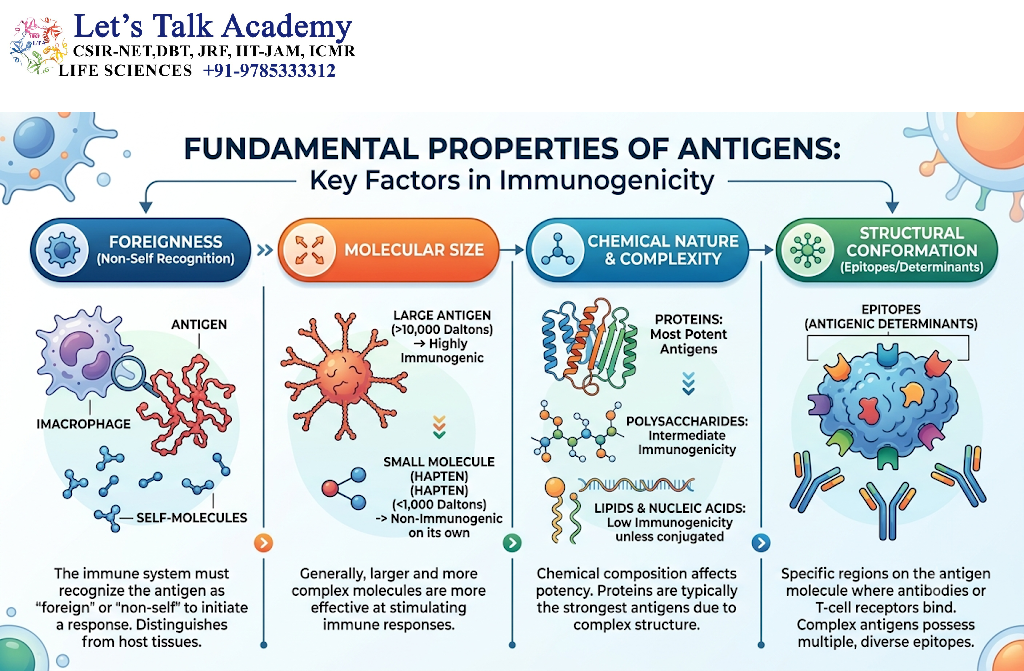
An antigen is a substance or molecular structure that binds specifically to an antibody, B-cell receptor, or T-cell receptor. The term is derived from the historical expression “antibody generator,” although the modern definition emphasizes immune recognition rather than antibody production alone.
Antigens can include proteins, polysaccharides, lipids, nucleic acids, and small chemical compounds. Their recognition depends on the molecular complementarity between the antigen and the binding site of an immune receptor.
For example, a bacterial surface protein may be recognized by an antibody. Similarly, a short peptide derived from a viral protein may be displayed by a major histocompatibility complex molecule and recognized by a T-cell receptor.
An important point is that antigen recognition does not always result in immune activation. The immune system may recognize a molecule without mounting a detectable response against it.
2.2 Antigenicity
Antigenicity refers to the ability of a substance to bind specifically to an antibody or antigen receptor.
A molecule with antigenicity contains structural features that can interact with a complementary binding site. This interaction depends on noncovalent forces, including:
-
Hydrogen bonding.
-
Electrostatic interactions.
-
Hydrophobic interactions.
-
Van der Waals forces.
-
Shape and chemical complementarity.
Antigenicity is influenced by the accessibility, shape, charge, and chemical characteristics of the recognized region.
A substance may have strong antigenicity but weak immunogenicity. For example, a small hapten can bind to an antibody even though it cannot independently induce a strong antibody response.
2.3 Immunogenicity
Immunogenicity is the ability of a substance to induce an immune response. The response may involve antibody production, activation of T lymphocytes, development of immunological memory, or other immune processes.
Immunogenicity is not an inherent property determined solely by molecular size. It depends on multiple variables, including:
-
Molecular structure and chemical complexity.
-
Degree of foreignness.
-
Molecular size and physical form.
-
Route of entry or administration.
-
Dose and frequency of exposure.
-
Presence of innate immune stimulation.
-
Genetic characteristics of the host.
-
Availability of antigen-presenting cells.
-
Previous exposure to the same or related antigen.
For example, a purified protein may be recognized by the immune system but produce only a limited response in the absence of appropriate innate signals or adjuvants.
2.4 Antigen Versus Immunogen
The distinction between antigen and immunogen is essential for understanding immune responses.
|
Feature |
Antigen |
Immunogen |
|---|---|---|
|
Basic meaning |
A molecule recognized by a specific immune receptor |
A substance capable of inducing an immune response |
|
Receptor binding |
Can bind a specific antibody or antigen receptor |
Can bind immune receptors |
|
Ability to stimulate immunity |
Not necessarily capable of inducing a response |
Capable of inducing a response under suitable conditions |
|
Example |
A hapten that binds a specific antibody |
A carrier protein that induces antibody production |
An antigen becomes immunogenic when it possesses the necessary properties to activate an appropriate immune response in a particular host and biological context.
2.5 Foreignness and Self-Recognition
The immune system is generally more responsive to molecular structures perceived as foreign or dangerous than to the body’s own molecules. This property is known as foreignness.
However, foreignness alone does not guarantee immunogenicity. Some foreign molecules are poorly immunogenic, while certain self-components may become targets of an immune response under abnormal conditions.
During lymphocyte development, mechanisms of central and peripheral tolerance help prevent strong immune responses against many self-antigens. Failure of these mechanisms may contribute to autoimmune disease.
2.6 Specificity of Antigen Recognition
Antigen recognition is highly specific but not always absolutely exclusive. An antibody or T-cell receptor generally recognizes a particular molecular structure, yet related molecules may sometimes bind to the same receptor.
This phenomenon is known as cross-reactivity. It occurs when different antigens possess identical or sufficiently similar epitopes.
Cross-reactivity can be beneficial when immune responses recognize related pathogens. It may also contribute to unwanted reactions when an immune response against one antigen interacts with a structurally similar molecule in the host.
3. Chemical Nature and Classification of Antigens
3.1 Protein Antigens
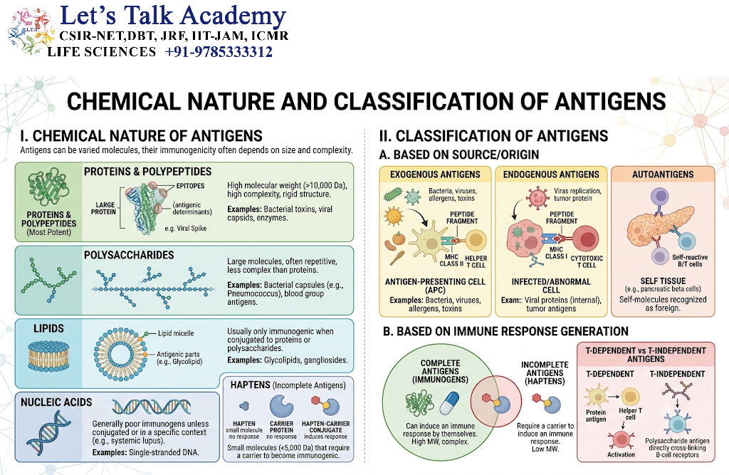
Proteins are among the most important and commonly studied antigens because they possess complex three-dimensional structures and can contain numerous antigenic determinants.
Protein antigens may be found in:
-
Bacterial toxins.
-
Viral envelope proteins.
-
Bacterial surface proteins.
-
Parasite proteins.
-
Fungal proteins.
-
Allergens.
-
Transplantation-associated molecules.
-
Tumor-associated proteins.
Protein structure influences the accessibility and nature of epitopes. Changes in folding, denaturation, or chemical modification may alter the epitopes recognized by antibodies.
Protein antigens also play a central role in T-cell-mediated immunity because proteins can be broken down into peptides and presented by major histocompatibility complex molecules.
3.2 Polysaccharide Antigens
Polysaccharides are carbohydrates composed of repeating sugar units. They are frequently present in bacterial capsules, cell walls, and other extracellular structures.
Examples include:
-
Capsular polysaccharides of bacteria.
-
Lipopolysaccharide-associated carbohydrate structures.
-
Certain fungal cell-wall carbohydrates.
-
Blood group carbohydrate determinants.
Some polysaccharides can activate B cells without requiring conventional peptide presentation to T cells. Such antigens are called T-independent antigens when they stimulate antibody responses through mechanisms that do not require conventional T-cell help.
Polysaccharide antigens are important in vaccine design. Conjugating a polysaccharide to a carrier protein can improve the quality and durability of the immune response, particularly in young children.
3.3 Lipid Antigens
Lipids can function as antigens when they are recognized by specialized immune receptors. Many lipid antigens are presented to lymphocytes through molecules other than conventional MHC class I and class II proteins.
For example, CD1 molecules can present certain lipid and glycolipid antigens to specialized T cells.
Lipid antigens are important in the study of:
-
Microbial infections.
-
Mycobacterial immunity.
-
Inflammation.
-
Metabolic disorders.
-
Immune recognition of glycolipids.
The immune recognition of lipids demonstrates that antigen recognition is not restricted to conventional protein-based mechanisms.
3.4 Nucleic Acid Antigens
DNA and RNA can participate in immune recognition, particularly when they are associated with microorganisms or released from damaged cells.
Nucleic acids may be detected by innate immune receptors such as certain Toll-like receptors and cytosolic nucleic acid sensors. In some pathological conditions, self-derived nucleic acids may activate immune pathways and contribute to autoimmune inflammation.
Although nucleic acids can act as immune-stimulating molecules, their antigenic behavior depends on their molecular form, associated proteins, delivery route, and the receptors involved.
3.5 Glycoprotein and Lipoprotein Antigens
Glycoproteins contain carbohydrate groups attached to protein molecules, whereas lipoproteins contain lipid components associated with proteins.
These molecules are common on the surfaces of cells and microorganisms. Their carbohydrate or lipid components may influence immune recognition, stability, and antigen processing.
Many viral surface proteins are glycoproteins. Their exposed regions can serve as targets for neutralizing antibodies, while their internal protein components may generate peptides for T-cell recognition.
3.6 Soluble and Particulate Antigens
Antigens may also be classified according to their physical form.
Soluble antigens are dissolved in body fluids or laboratory solutions. Examples include purified proteins, toxins, and soluble microbial products.
Particulate antigens are associated with cells, microorganisms, membrane fragments, or other particles. Examples include bacterial cells, red blood cells, and virus particles.
Particulate antigens may promote receptor clustering and efficient uptake by phagocytic or antigen-presenting cells. The physical form of an antigen can therefore influence how it is processed and how the immune system responds.
4. Antigenic Determinants and Epitopes
4.1 Definition of an Epitope
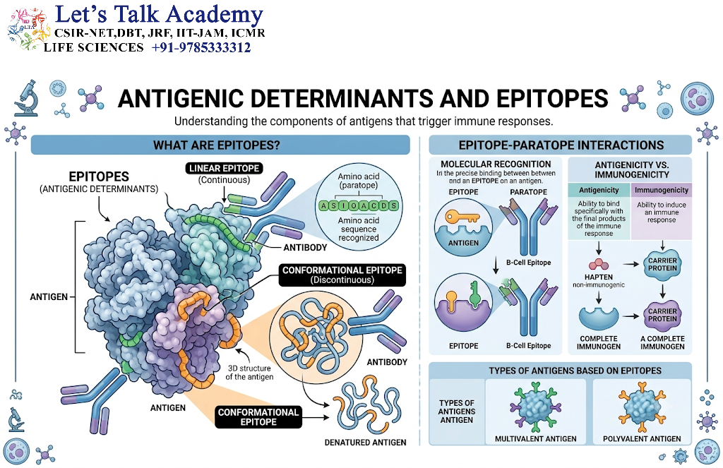
An epitope, also called an antigenic determinant, is the specific region of an antigen that is recognized by an antibody, B-cell receptor, or T-cell receptor.
An antigen may contain several distinct epitopes. Different B-cell clones can recognize different regions of the same antigen, producing antibodies with different specificities.
For example, a viral surface protein may contain multiple antibody-binding regions. One antibody may recognize a site near the receptor-binding region, whereas another may recognize a separate surface region.
The presence of multiple epitopes allows a single antigen to stimulate a diverse population of lymphocytes.
4.2 B-Cell Epitopes
B-cell epitopes are recognized directly by membrane-bound immunoglobulin on B cells or by antibodies released into body fluids.
These epitopes may be:
-
Linear epitopes: Formed by a continuous sequence of amino acids or other chemical units.
-
Conformational epitopes: Formed by amino acids or chemical groups that are separated in the primary sequence but brought together by protein folding.
Many antibody epitopes depend on the three-dimensional structure of a protein. Consequently, denaturation may destroy some conformational epitopes while exposing previously inaccessible linear sequences.
The ability of antibodies to recognize native molecular structures is important in neutralization, diagnostic testing, and antibody-based therapies.
4.3 T-Cell Epitopes
T-cell epitopes are usually short peptide sequences derived from proteins. These peptides are presented on cell surfaces by MHC molecules and recognized by T-cell receptors.
T cells generally do not recognize intact soluble proteins in the same way that antibodies do. Instead, they recognize a combination of:
-
A peptide derived from an antigen.
-
An MHC molecule presenting that peptide.
-
A suitable molecular and cellular activation context.
MHC class I molecules commonly present peptides to CD8-positive T cells, whereas MHC class II molecules commonly present peptides to CD4-positive T cells.
The same protein can therefore generate different T-cell epitopes depending on how it is processed and which MHC molecules are expressed by the host.
4.4 Epitope Density and Antigenic Complexity
A large antigen may contain many epitopes, but not every epitope will generate an equally strong immune response.
The number and accessibility of epitopes can influence:
-
The diversity of antibodies produced.
-
The number of lymphocyte clones activated.
-
The strength of receptor binding.
-
The possibility of cross-reactivity.
-
The development of immune escape variants.
Some epitopes are immunodominant, meaning they generate relatively prominent immune responses compared with other epitopes present in the same antigen.
4.5 Epitope Mapping
Epitope mapping is the process of identifying the precise regions of an antigen recognized by antibodies or T-cell receptors.
Common approaches include:
-
Peptide libraries.
-
Overlapping synthetic peptides.
-
Mutational analysis.
-
X-ray crystallography.
-
Cryo-electron microscopy.
-
Nuclear magnetic resonance studies.
-
Computational structural analysis.
-
Binding assays using monoclonal antibodies.
Epitope mapping is useful in vaccine development, antibody engineering, diagnostic assay design, and the investigation of immune escape.
4.6 Cross-Reactive Epitopes
Cross-reactive epitopes are molecular regions shared by two or more different antigens or sufficiently similar to be recognized by the same immune receptor.
Cross-reactivity can occur between related microbial species or between microbial and host molecules.
The outcome depends on the strength and specificity of the interaction, the abundance of the target, and the biological context. Cross-reactivity may provide partial protection against related pathogens, but it can also complicate diagnostic tests and contribute to certain immune-mediated disorders.
5. Antigenicity and Immunogenicity
5.1 Relationship Between Antigenicity and Immunogenicity
Antigenicity and immunogenicity are closely related concepts, but they describe different biological properties.
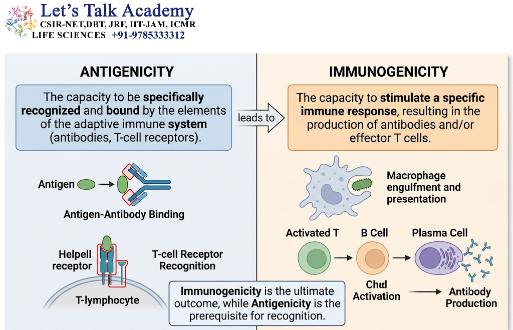
Antigenicity refers to the ability of a molecule to interact specifically with an immune receptor. Immunogenicity refers to the ability of that molecule to induce an immune response.
For example, a hapten may bind to a specific antibody because it possesses a complementary chemical structure. However, the hapten may be unable to activate sufficient numbers of lymphocytes to produce an immune response when administered alone. When attached to a suitable carrier protein, the same hapten may become capable of inducing antibody production.
This example demonstrates that receptor binding and immune activation are not identical processes.
5.2 Factors Determining Immunogenicity
Immunogenicity is influenced by the chemical properties of the antigen, the characteristics of the host, and the circumstances of exposure.
5.2.1 Molecular Size
In general, larger molecules are more likely to be immunogenic than very small molecules. Larger molecules may possess a greater variety of epitopes and exhibit more complex chemical structures.
However, molecular size alone does not determine immunogenicity. A large molecule may be poorly immunogenic if it is chemically simple, rapidly eliminated, or unable to activate the appropriate immune pathways.
5.2.2 Chemical Complexity
Molecules containing a wide variety of amino acids, sugar units, or other chemical groups are often more immunogenic than structurally simple molecules.
Proteins with complex tertiary and quaternary structures can provide numerous potential epitopes. Chemical complexity increases the likelihood that a molecule will contain structures capable of being recognized by diverse lymphocyte populations.
5.2.3 Foreignness
The immune system is more likely to respond to molecules that differ substantially from the host’s own molecular components.
A protein from a distantly related organism may be strongly immunogenic in a particular host. However, molecules derived from closely related species may be less immunogenic because of their structural similarity to host proteins.
Foreignness is relative to the individual organism. A molecule recognized as foreign in one species may be poorly immunogenic in another.
5.2.4 Physical Form and Solubility
The physical state of an antigen can influence its uptake and processing.
Particulate antigens may be efficiently engulfed by phagocytic cells. Soluble antigens may diffuse through tissues and interact with B-cell receptors or be taken up through endocytic mechanisms.
Aggregated antigens can also produce stronger receptor clustering than individual soluble molecules. The effects depend on the antigen and the type of immune response being considered.
5.2.5 Route of Exposure
The route through which an antigen enters the body can influence the type and magnitude of the immune response.
Different routes include:
-
Intravenous exposure.
-
Subcutaneous exposure.
-
Intramuscular exposure.
-
Intradermal exposure.
-
Mucosal exposure.
-
Oral exposure.
The route affects which antigen-presenting cells encounter the antigen, which lymphoid tissues are involved, and whether the resulting response is systemic or localized.
5.2.6 Dose and Frequency of Exposure
The quantity and frequency of antigen exposure can influence immune activation.
An appropriate dose may stimulate a strong immune response, whereas extremely low or extremely high doses may sometimes produce weaker responses or promote immune tolerance. Repeated exposure may enhance immunological memory or, under certain conditions, contribute to tolerance.
The relationship between dose and immune response is not always linear.
5.2.7 Host Genetic Factors
The genetic background of an individual influences antigen recognition and immune responsiveness.
MHC genes are particularly important because they determine which peptide fragments can be presented to T cells. Different individuals may process and present the same protein differently because of variations in their MHC molecules.
Other genetic factors affecting cytokine production, innate immune receptors, lymphocyte development, and antigen-processing proteins may also influence immunogenicity.
5.2.8 Adjuvants and Innate Immune Signals
An adjuvant is a substance that enhances an immune response to an antigen when administered together with it.
Adjuvants may improve antigen uptake, stimulate innate immune receptors, promote the activation of antigen-presenting cells, or prolong the availability of antigen within tissues.
An antigen-specific response is generally more effective when antigen recognition occurs alongside appropriate activation signals. This helps the immune system distinguish potentially harmful stimuli from harmless substances.
6. Haptens and Carrier Molecules
6.1 Definition of a Hapten
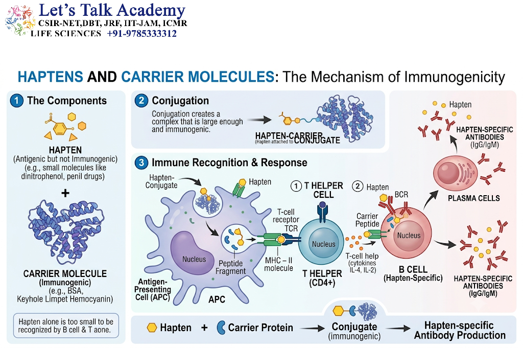
A hapten is a small molecule that can bind specifically to an antibody or antigen receptor but generally cannot induce an effective adaptive immune response by itself.
Haptens are sometimes described as incomplete antigens because they possess antigenicity but may lack sufficient immunogenicity when administered alone.
Examples of molecules that can behave as haptens include certain chemical compounds, drug-derived molecules, and small synthetic chemical groups.
6.2 Hapten–Carrier Conjugates
A hapten can become immunogenic when chemically linked to a larger carrier molecule, usually a protein.
The carrier provides peptide sequences and structural complexity that can support T-cell activation. B cells specific for the hapten may bind the hapten–carrier conjugate through their B-cell receptors, internalize the conjugate, and present carrier-derived peptides to helper T cells.
This mechanism allows the hapten-specific B cell to receive T-cell help and produce antibodies against the hapten.
6.3 Mechanism of Hapten-Induced Antibody Production
The general process can be summarized as follows:
-
A hapten is attached to a carrier protein.
-
A B cell recognizes the hapten portion of the conjugate.
-
The B cell internalizes the hapten–carrier complex.
-
The carrier protein is processed into peptide fragments.
-
Carrier-derived peptides are presented on MHC class II molecules.
-
A suitable helper T cell recognizes the peptide–MHC complex.
-
The B cell receives activation signals and proliferates.
-
Activated B cells differentiate into antibody-secreting plasma cells and memory B cells.
The antibodies produced can bind specifically to the hapten.
6.4 Biological and Clinical Significance of Haptens
Haptens are important in the study of drug allergies and immune-mediated reactions.
Certain drugs or drug metabolites may bind covalently to host proteins and create new antigenic structures. The immune system may recognize these modified proteins as foreign, potentially resulting in an immune response.
Haptens are also used in experimental immunology to investigate antibody specificity, carrier effects, and the molecular basis of immune recognition.
7. Antigen Recognition by B Cells and T Cells
7.1 Overview of Antigen Receptors
B cells and T cells express specialized antigen receptors that provide the basis for adaptive immune specificity.
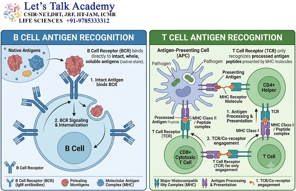
B cells express B-cell receptors, which are membrane-bound immunoglobulins. T cells express T-cell receptors, which recognize antigen-derived structures displayed by MHC molecules.
Both receptor systems are generated through genetic rearrangement during lymphocyte development. This process produces a large repertoire of antigen receptors with different specificities.
7.2 Recognition by B-Cell Receptors
B-cell receptors can recognize intact antigens in their native form.
Depending on the receptor, B cells may recognize:
-
Protein surfaces.
-
Polysaccharides.
-
Lipids.
-
Small chemical structures.
-
Repetitive molecular patterns.
The binding of an antigen to a B-cell receptor can initiate signaling within the B cell. However, B-cell receptor binding alone is often insufficient for the development of a complete, long-lasting antibody response.
Additional signals from helper T cells, innate immune pathways, or other activating mechanisms may be required.
7.3 Recognition by T-Cell Receptors
T-cell receptors generally recognize peptide fragments bound to MHC molecules.
The T-cell receptor interacts with both the presented peptide and the MHC molecule. This is known as MHC-restricted antigen recognition.
The specificity of a T-cell response therefore depends on:
-
The amino acid sequence of the peptide.
-
The ability of the peptide to bind a particular MHC molecule.
-
The structure of the T-cell receptor.
-
The presence of appropriate accessory and costimulatory signals.
T cells usually do not recognize free, intact proteins in the same manner as antibodies.
7.4 Role of Coreceptors
CD4 and CD8 are important T-cell coreceptors.
-
CD4 is commonly associated with helper T cells and interacts with MHC class II molecules.
-
CD8 is commonly associated with cytotoxic T cells and interacts with MHC class I molecules.
Coreceptors strengthen interactions between T cells and antigen-presenting cells and help recruit signaling molecules to the T-cell receptor complex.
7.5 Clonal Selection and Antigen Recognition
The clonal selection theory explains how antigen-specific lymphocytes are activated.
According to this concept, the body contains many lymphocyte clones, each expressing receptors with a particular specificity. When an antigen enters the body, it binds to receptors on lymphocytes capable of recognizing it.
Appropriately activated lymphocytes then undergo clonal expansion, producing a population of cells with the same antigen specificity.
Some of these cells become effector cells, while others become memory cells. Memory cells enable a faster and often stronger response when the same antigen is encountered again.
7.6 Affinity and Avidity
Affinity describes the strength of a single molecular interaction between one antigen-binding site and one epitope.
Avidity describes the combined strength of multiple interactions between a multivalent antibody or receptor system and an antigen containing multiple binding sites.
An antibody may have moderate affinity for an individual epitope but exhibit strong overall avidity when several binding interactions occur simultaneously.
These concepts are important in antibody binding assays, immune-complex formation, and the interpretation of serological tests.
8. Antigen Processing and Presentation
8.1 Meaning of Antigen Processing
Antigen processing refers to the intracellular breakdown of proteins into peptide fragments that can associate with MHC molecules.
Many proteins must be enzymatically degraded before their antigen-derived peptides can be presented to T cells.
Antigen processing is a critical link between the presence of a protein antigen and its recognition by the adaptive immune system.
8.2 Meaning of Antigen Presentation
Antigen presentation is the display of antigen-derived peptide fragments on the surface of cells in association with MHC molecules.
The peptide–MHC complex can then be recognized by a specific T-cell receptor.
Antigen presentation allows T cells to detect proteins originating from pathogens, damaged cells, or other sources that have been processed within cells.
The principal professional antigen-presenting cells are:
-
Dendritic cells.
-
Macrophages.
-
B lymphocytes.
Other cells may present antigen-derived peptides under particular physiological or inflammatory conditions.
8.3 Major Histocompatibility Complex
The major histocompatibility complex, or MHC, is a group of genes encoding molecules that present antigen-derived peptides to T cells.
In humans, MHC molecules are called human leukocyte antigens, or HLA molecules.
MHC molecules are essential for immune surveillance and T-cell activation. Their genetic diversity influences which peptides can be presented and contributes to variation in immune responses among individuals.
MHC molecules are divided into two major classes involved in conventional peptide presentation:
-
MHC class I.
-
MHC class II.
8.4 MHC Class I Antigen Presentation
MHC class I molecules are expressed on nearly all nucleated cells under normal conditions.
They generally present peptides derived from proteins located in the cytosol. These proteins may originate from:
-
Viruses replicating inside cells.
-
Intracellular microorganisms.
-
Normal cellular proteins.
-
Mutated proteins.
-
Abnormally expressed proteins.
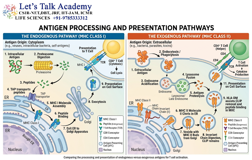
The conventional MHC class I pathway proceeds through the following stages.
8.4.1 Protein Degradation
Proteins in the cytosol are degraded into smaller peptide fragments, often by a multiprotein complex called the proteasome.
During certain inflammatory conditions, specialized proteasomes known as immunoproteasomes can alter the types of peptides generated.
8.4.2 Transport of Peptides
Suitable peptides are transported from the cytosol into the endoplasmic reticulum through transporter associated with antigen processing, commonly abbreviated as TAP.
8.4.3 Peptide Loading
Peptides bind to newly synthesized MHC class I molecules within the endoplasmic reticulum. Several accessory proteins assist in the assembly and quality control of peptide–MHC class I complexes.
8.4.4 Transport to the Cell Surface
Stable peptide–MHC class I complexes move through the secretory pathway to the plasma membrane.
8.4.5 Recognition by CD8-Positive T Cells
CD8-positive cytotoxic T cells recognize suitable peptide–MHC class I complexes through their T-cell receptors.
When appropriately activated, these cells can destroy infected or abnormal target cells through mechanisms involving cytotoxic granules and death-inducing signaling pathways.
8.5 MHC Class II Antigen Presentation
MHC class II molecules are primarily expressed by professional antigen-presenting cells, although their expression may be induced in certain other cells during inflammation.
MHC class II molecules generally present peptides derived from proteins taken up from the extracellular environment.
These proteins may originate from:
-
Extracellular bacteria.
-
Soluble microbial proteins.
-
Endocytosed toxins.
-
Environmental proteins.
-
Extracellular debris.
-
Proteins internalized by B cells.
8.5.1 Antigen Uptake
Antigen-presenting cells take up extracellular material through processes such as phagocytosis, receptor-mediated endocytosis, or macropinocytosis.
B cells can also internalize specific antigens through their B-cell receptors.
8.5.2 Protein Breakdown in Vesicles
Internalized proteins enter endosomal or lysosomal compartments, where proteolytic enzymes break them down into peptide fragments.
The resulting peptides are generated within an acidic intracellular environment containing proteases.
8.5.3 MHC Class II Assembly
MHC class II molecules are synthesized in the endoplasmic reticulum. An associated invariant chain helps protect the peptide-binding groove during transport and directs the MHC class II molecules toward the appropriate intracellular compartments.
8.5.4 Peptide Loading and Surface Expression
The invariant-chain-derived fragment is removed or exchanged, allowing suitable antigen-derived peptides to bind MHC class II molecules.
The resulting peptide–MHC class II complexes are transported to the cell surface.
8.5.5 Recognition by CD4-Positive T Cells
CD4-positive helper T cells recognize peptide–MHC class II complexes.
Activated helper T cells can release cytokines and provide essential signals to other immune cells, including B cells and macrophages.
8.6 Cross-Presentation
Cross-presentation is a specialized process in which certain antigen-presenting cells, particularly dendritic cells, present extracellularly acquired antigens through MHC class I molecules.
This process allows CD8-positive T cells to respond to antigens that may not have been produced directly inside the presenting dendritic cell.
Cross-presentation is particularly important in the initiation of immune responses against some viruses, tumors, and other sources of extracellular antigen.
8.7 Antigen Presentation and T-Cell Activation
The recognition of a peptide–MHC complex is essential but may not be sufficient to activate a naïve T cell.
Effective naïve T-cell activation usually requires several signals.
Signal 1: Antigen recognition
The T-cell receptor binds the appropriate peptide–MHC complex.
Signal 2: Costimulation
Costimulatory molecules on the antigen-presenting cell interact with receptors on the T cell. A well-known example is the interaction between CD80 or CD86 and CD28.
Signal 3: Cytokine-mediated instruction
Cytokines produced by antigen-presenting cells and other immune cells influence T-cell proliferation, differentiation, and functional specialization.
The integration of these signals helps regulate whether a T cell becomes activated, remains unresponsive, or develops another functional state.
8.8 Antigen Presentation and Immune Tolerance
Antigen presentation is involved not only in immune activation but also in immune tolerance.
When antigens are presented in the absence of appropriate costimulatory signals, or under specific tolerogenic conditions, antigen-specific lymphocytes may become functionally unresponsive.
Tolerance mechanisms help prevent excessive immune responses against harmless environmental molecules and self-antigens.
The outcome of antigen presentation therefore depends on the presenting cell, the tissue environment, the type of antigen, and the signals accompanying recognition.
9. Antigen–Antibody Interactions
9.1 Basic Principle
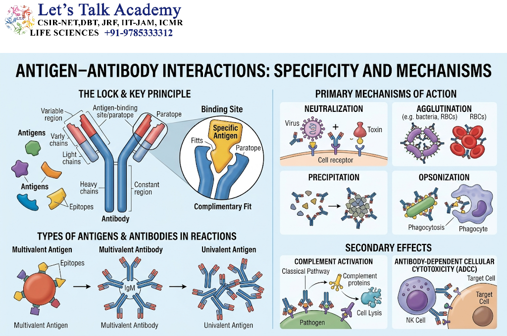
Antibodies are immunoglobulin molecules produced by plasma cells. They bind specifically to epitopes on antigens through complementary molecular interactions.
The antigen-binding region of an antibody is located in the variable regions of its heavy and light chains. These regions form a binding surface called the paratope.
The antigenic region that interacts with the paratope is called the epitope.
Antigen–antibody binding is generally noncovalent and reversible.
9.2 Forces Involved in Binding
Several types of weak molecular forces contribute to antigen–antibody interactions.
9.2.1 Hydrogen Bonds
Hydrogen bonds form between suitable hydrogen-donor and hydrogen-acceptor groups in the antigen and antibody.
9.2.2 Electrostatic Interactions
Oppositely charged groups can attract each other and contribute to binding specificity.
9.2.3 Hydrophobic Interactions
Nonpolar surfaces may interact in ways that reduce their exposure to water and stabilize the antigen–antibody complex.
9.2.4 Van der Waals Forces
Short-range attractions between atoms can contribute to the overall stability of the binding interface.
The strength of the interaction depends on the combined effect of these forces and the degree of structural complementarity.
9.3 Precipitation Reactions
Precipitation occurs when soluble antigens interact with antibodies to form sufficiently large immune complexes that become insoluble under appropriate conditions.
The formation of a visible precipitate depends on factors such as:
-
Antigen concentration.
-
Antibody concentration.
-
Valency of the interacting molecules.
-
Relative proportions of antigen and antibody.
-
Environmental conditions.
The zone of equivalence refers to a range in which antigen and antibody proportions favor the formation of large immune-complex networks.
An excess of antibody or antigen may reduce the formation of large lattices. These situations are referred to as the prozone and postzone phenomena, respectively.
9.4 Agglutination Reactions
Agglutination is the visible clumping of particulate antigens, such as cells or particles, caused by antibody-mediated cross-linking.
Examples include:
-
Red blood cell agglutination.
-
Bacterial agglutination.
-
Latex particle agglutination.
Agglutination is widely used in laboratory diagnostics and blood group testing.
9.5 Neutralization
Neutralization occurs when antibodies bind to a pathogen or toxin and interfere with its biological activity.
Neutralizing antibodies may:
-
Block viral attachment to host-cell receptors.
-
Prevent viral entry into cells.
-
Inhibit bacterial toxin binding.
-
Interfere with the activity of soluble microbial factors.
Neutralization depends on the location of the epitope and whether antibody binding interferes with an essential biological function.
9.6 Opsonization
Opsonization is the process by which an antigen-bearing particle is coated with molecules that promote its recognition and uptake by phagocytic cells.
Antibodies can function as opsonins when their antigen-binding regions attach to a target and their Fc regions interact with Fc receptors on phagocytes.
Complement proteins can also act as opsonins. Opsonization improves the efficiency of phagocytosis and contributes to the clearance of certain microorganisms.
9.7 Complement Activation
Antigen–antibody complexes can activate the classical complement pathway when appropriate antibody classes and molecular arrangements are present.
Complement activation can lead to:
-
Enhanced phagocytosis.
-
Inflammatory mediator release.
-
Recruitment of immune cells.
-
Formation of membrane attack complexes in susceptible targets.
-
Removal of immune complexes.
Not every antigen–antibody interaction activates complement. The outcome depends on antibody class, antibody arrangement, and the specific molecular context.
9.8 Immune Complexes
An immune complex is a molecular assembly formed by the interaction of antigen and antibody.
Small immune complexes may remain soluble, whereas larger complexes may be removed by phagocytic cells or deposited in tissues under certain conditions.
Persistent or improperly cleared immune complexes can contribute to inflammation and tissue damage in some immune-mediated diseases.
10. Classification of Antigens According to Their Source
10.1 Exogenous Antigens
Exogenous antigens originate outside the cell or organism in which they are encountered.
Examples include proteins from extracellular bacteria, environmental allergens, and soluble microbial toxins.
Exogenous antigens are commonly taken up by antigen-presenting cells and processed through the MHC class II pathway.
However, some exogenous antigens may also enter MHC class I presentation pathways through cross-presentation.
10.2 Endogenous Antigens
Endogenous antigens originate within a cell.
Examples include:
-
Viral proteins produced during intracellular infection.
-
Mutated cellular proteins.
-
Abnormally expressed proteins.
-
Some proteins derived from intracellular microorganisms.
These proteins can be processed through the MHC class I pathway and recognized by CD8-positive T cells.
10.3 Autoantigens
Autoantigens are molecules derived from the host’s own cells or tissues that can be recognized by the immune system.
Under normal circumstances, immune tolerance limits harmful responses against self-antigens.
When tolerance mechanisms fail, immune responses against autoantigens may contribute to autoimmune disease.
Examples of self-molecules that can become targets in autoimmune disorders include nuclear proteins, cell-surface receptors, and tissue-specific proteins.
The presence of an autoantigen does not itself imply disease. Autoimmunity involves additional factors, including immune activation, genetic susceptibility, and tissue-specific mechanisms.
10.4 Alloantigens
Alloantigens are antigens that differ between genetically distinct individuals of the same species.
They are important in:
-
Blood transfusion.
-
Organ transplantation.
-
Tissue transplantation.
-
Maternal–fetal immune interactions.
For example, differences in red blood cell antigens or HLA molecules may lead to immune recognition of cells or tissues from another individual.
10.5 Xenoantigens
Xenoantigens are antigens originating from a different species.
When tissues or biological materials from one species are introduced into another species, xenoantigens may stimulate immune responses.
Xenoantigens are particularly important in xenotransplantation research and the study of interspecies immune recognition.
10.6 Tumor-Associated and Tumor-Specific Antigens
Tumor-associated antigens are molecules expressed by tumor cells or expressed at unusually high levels in tumors compared with normal tissues.
Tumor-specific antigens are restricted to tumor cells or particular tumor types in the relevant biological context.
These antigens may arise from:
-
Mutated genes.
-
Abnormal gene expression.
-
Viral proteins in virus-associated cancers.
-
Overexpressed normal proteins.
-
Altered protein-processing mechanisms.
Tumor antigens are studied extensively in cancer immunology, immunodiagnostics, and the development of antigen-directed therapies.
11. Classification of Antigens According to Their Ability to Activate T Cells
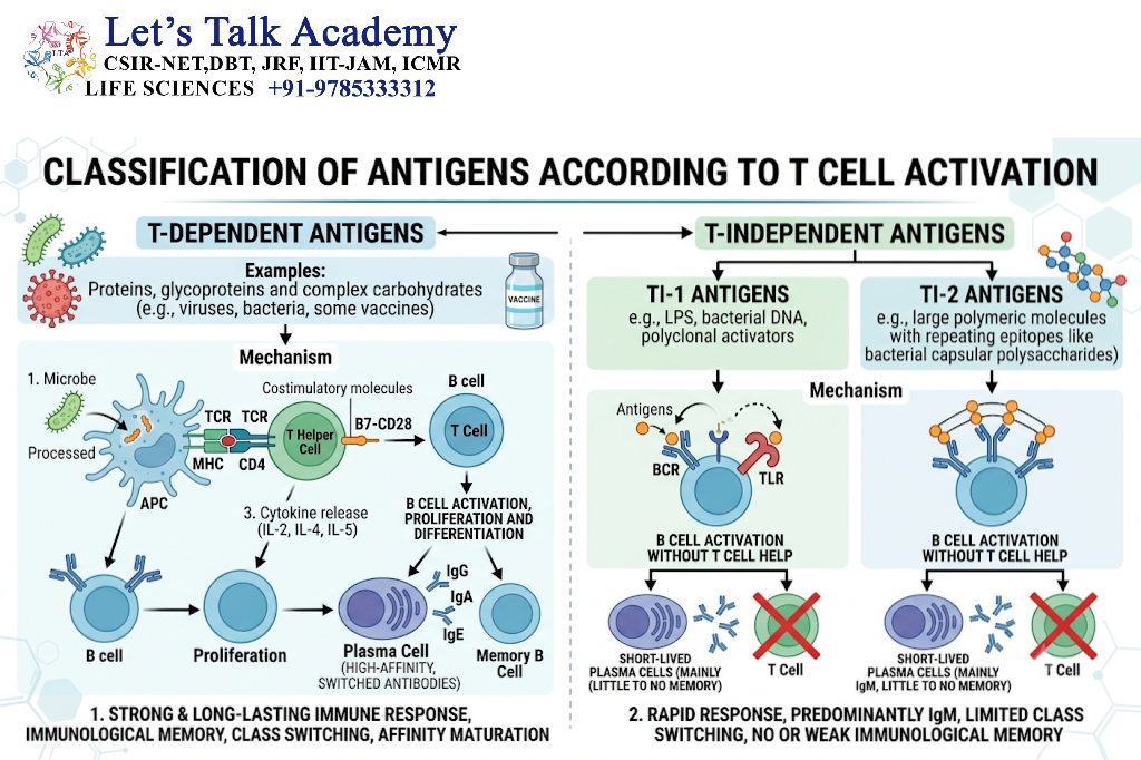
11.1 T-Dependent Antigens
T-dependent antigens generally require assistance from helper T cells to produce strong, long-lasting antibody responses.
Most protein antigens fall into this category.
The response commonly involves:
-
Recognition of antigen by a B-cell receptor.
-
Internalization and processing of the antigen by the B cell.
-
Presentation of carrier-derived peptides through MHC class II.
-
Recognition by a helper T cell.
-
Delivery of costimulatory signals and cytokines.
-
B-cell proliferation and differentiation.
-
Antibody class switching, affinity maturation, and memory formation.
T-dependent responses are important for the generation of high-affinity antibodies and long-term immunological memory.
11.2 T-Independent Antigens
T-independent antigens can stimulate B cells without the conventional requirement for peptide-specific helper T-cell assistance.
These antigens often contain repetitive molecular structures capable of extensively cross-linking B-cell receptors.
Some bacterial polysaccharides are examples of antigens that can stimulate T-independent antibody responses.
T-independent responses may produce substantial IgM production. Depending on the antigen and the type of B-cell activation, some additional antibody class switching or memory responses may occur, but these responses often differ from the classical T-dependent response.
11.3 Type 1 T-Independent Antigens
Certain T-independent antigens can activate B cells through a combination of B-cell receptor engagement and innate immune receptor signaling.
Some microbial components may stimulate Toll-like receptors or other innate immune receptors while also interacting with B-cell receptors.
The combined signaling can promote B-cell activation without conventional antigen-specific T-cell help.
11.4 Type 2 T-Independent Antigens
Type 2 T-independent antigens often possess highly repetitive molecular structures.
These structures can cross-link many B-cell receptors simultaneously, generating a strong activation signal.
Examples include certain repetitive polysaccharide antigens found on bacterial surfaces.
The resulting response is often associated with rapid antibody production, especially IgM, but may produce less extensive affinity maturation and memory than typical T-dependent responses.
12. Antigens and the Immune Response
12.1 Innate Recognition of Microbial Structures
Before the adaptive immune system produces antigen-specific responses, the innate immune system detects molecular patterns associated with microorganisms or tissue damage.
These structures are recognized by pattern-recognition receptors.
Examples of innate immune sensors include:
-
Toll-like receptors.
-
NOD-like receptors.
-
RIG-I-like receptors.
-
C-type lectin receptors.
-
Cytosolic DNA sensors.
These receptors do not generally possess the same rearranged antigen-specific recognition system as B-cell and T-cell receptors. Instead, they detect conserved molecular patterns or signals associated with infection and tissue damage.
Innate recognition promotes inflammation and helps activate antigen-presenting cells.
12.2 Activation of Dendritic Cells
Dendritic cells are specialized antigen-presenting cells that connect innate and adaptive immunity.
When dendritic cells encounter appropriate microbial or inflammatory signals, they may undergo maturation.
Mature dendritic cells commonly exhibit:
-
Increased expression of antigen-presenting molecules.
-
Enhanced expression of costimulatory molecules.
-
Changes in chemokine receptor expression.
-
Improved migration toward lymphoid tissues.
-
Increased ability to activate naïve T cells.
The antigen-processing and presentation functions of dendritic cells are essential for initiating many primary T-cell responses.
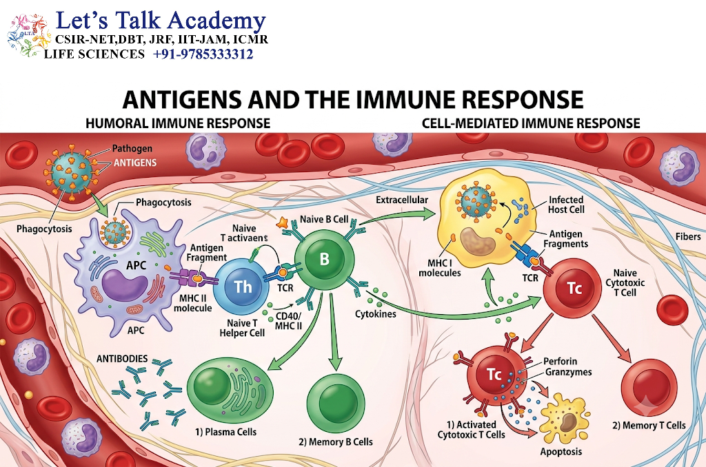
12.3 B-Cell Activation
B cells recognize specific antigenic structures through their B-cell receptors.
Activation may require:
-
Antigen binding.
-
Coreceptor signaling.
-
Innate immune signals.
-
Helper T-cell interactions.
-
Cytokine stimulation.
Activated B cells may differentiate into plasma cells that secrete antibodies or memory B cells that persist for future encounters.
The nature of the response depends on the antigen and the signals received during activation.
12.4 T-Cell Activation
T-cell activation occurs when a suitable T-cell receptor recognizes a peptide–MHC complex in the appropriate cellular context.
The response may include:
-
Clonal expansion.
-
Cytokine production.
-
Differentiation into specialized T-cell subsets.
-
Cytotoxic activity.
-
Formation of memory T cells.
CD4-positive T cells can support other immune cells, whereas CD8-positive T cells can destroy infected or abnormal cells.
12.5 Immunological Memory
Immunological memory is the ability of the adaptive immune system to respond more rapidly or effectively following a subsequent exposure to the same or a related antigen.
Memory cells may persist for extended periods after the initial response.
Memory B cells can generate antibodies with improved affinity in many T-dependent responses. Memory T cells can respond rapidly when they encounter their specific peptide–MHC complexes again.
The strength and duration of memory depend on the antigen, the type of immune response, and the conditions under which the initial response developed.
12.6 Immune Tolerance
Immune tolerance is a state in which the immune system does not mount a damaging response against a particular antigen.
Tolerance may develop through mechanisms acting during lymphocyte development or after lymphocytes have entered peripheral tissues.
Tolerance is essential for preventing excessive responses against self-antigens and many harmless substances.
Antigen presentation without the appropriate activating environment can contribute to tolerance, deletion of reactive lymphocytes, or functional unresponsiveness.
13. Antigens in Vaccination and Immunological Applications
13.1 Role of Antigens in Vaccines
Vaccines expose the immune system to an antigen or antigen-encoding material in a manner designed to stimulate protective immunity.
The antigen may be provided as:
-
An inactivated microorganism.
-
An attenuated microorganism.
-
A purified protein.
-
A polysaccharide.
-
A protein–polysaccharide conjugate.
-
A recombinant antigen.
-
A viral-vector-encoded antigen.
-
A nucleic acid that directs antigen production.
The aim is to stimulate protective immune responses without causing the disease associated with the natural infectious agent.
13.2 Antigen Selection in Vaccine Development
Selecting suitable antigens is a major step in vaccine design.
Researchers consider:
-
Whether the antigen is present in the target pathogen.
-
Whether the antigen is sufficiently conserved.
-
Whether antibodies against the antigen can block infection.
-
Whether the antigen can stimulate useful T-cell responses.
-
Whether the antigen is safely produced.
-
Whether the antigen is accessible to the immune system.
-
Whether the antigen may undergo substantial variation.
A vaccine may contain one antigen or multiple antigens to broaden the immune response.
13.3 Recombinant Antigens
Recombinant antigens are produced using molecular cloning and gene-expression technologies.
A gene encoding an antigen is inserted into a suitable expression system, such as a bacterial, yeast, insect, or mammalian cell system.
The resulting protein can be purified and used in research, diagnostic assays, or vaccine formulations.
Recombinant antigen technology allows researchers to produce selected proteins without requiring the cultivation of the complete infectious organism.
13.4 Antigenic Variation and Immune Evasion
Some microorganisms modify their antigenic structures to avoid recognition by pre-existing antibodies or T cells.
Antigenic variation can occur through:
-
Genetic mutation.
-
Recombination.
-
Gene conversion.
-
Altered gene expression.
-
Genome segment reassortment in certain viruses.
Antigenic drift refers to gradual changes in antigenic structures caused by the accumulation of mutations.
Antigenic shift refers to a major change in antigenic properties associated with genetic reassortment in certain viruses with segmented genomes.
These processes can affect immunity, vaccine effectiveness, and the epidemiology of infectious diseases.
13.5 Antigens in Diagnostic Tests
Antigens are widely used in laboratory diagnosis.
Diagnostic assays may detect:
-
Microbial proteins.
-
Viral structural proteins.
-
Bacterial toxins.
-
Parasite-specific molecules.
-
Tumor-associated molecules.
-
Autoantigens.
-
Antigen–antibody complexes.
Antigen detection tests may use antibodies to identify specific molecular targets in patient samples.
Examples of techniques include enzyme-linked immunosorbent assays, immunochromatographic tests, immunofluorescence assays, and immunohistochemical methods.
The sensitivity and specificity of an antigen-based test depend on the quality of the antibodies, the abundance of the target antigen, sample quality, and the design of the assay.
13.6 Antigens in Blood Group Immunology
Blood group antigens are molecular structures expressed on the surface of red blood cells.
Some blood group antigens are proteins, whereas others are carbohydrate structures.
The ABO blood group system is primarily determined by carbohydrate differences on red blood cell-associated molecules. The Rh blood group system includes important protein antigens.
If an individual lacks a particular blood group antigen but possesses antibodies against it, transfusion of incompatible red blood cells may cause an immune-mediated transfusion reaction.
Blood group antigen analysis is therefore essential for safe transfusion practices.
13.7 Antigens in Transplantation
Transplantation introduces cells or tissues that may express antigens different from those of the recipient.
HLA molecules are particularly important in transplant immunology because they can be recognized as foreign by the recipient’s immune system.
Immune responses against donor antigens may contribute to graft rejection.
Transplant compatibility testing and immunosuppressive strategies aim to reduce harmful immune responses while preserving essential immune defense.
13.8 Antigens in Cancer Immunotherapy
Cancer immunotherapy uses immune mechanisms to recognize or eliminate malignant cells.
Some approaches target tumor-associated or tumor-specific antigens through:
-
Monoclonal antibodies.
-
Antibody–drug conjugates.
-
T-cell receptor-based therapies.
-
Chimeric antigen receptor T-cell therapies.
-
Therapeutic cancer vaccines.
The effectiveness of an antigen-targeted therapy depends on the antigen’s expression pattern, accessibility, degree of tumor specificity, and the ability of the immune system to reach the target.
14. Advanced Concepts in Antigen Biology
14.1 Immunodominance
Immunodominance refers to the phenomenon in which certain epitopes generate stronger or more prominent immune responses than other epitopes within the same antigen.
An antigen may contain many possible epitopes, but only a subset may dominate the response in a particular individual.
Immunodominance is influenced by:
-
Peptide generation during antigen processing.
-
MHC binding affinity.
-
T-cell receptor repertoire.
-
Epitope accessibility.
-
Competition among lymphocyte clones.
-
Prior immune exposure.
Immunodominance is important in vaccine design and the study of pathogen immune evasion.
14.2 Antigenic Competition
Antigenic competition occurs when different antigens or antigenic determinants influence one another’s immune responses.
When multiple antigens are presented simultaneously, they may compete for:
-
Antigen uptake.
-
Processing machinery.
-
MHC-binding sites.
-
Antigen-presenting cell resources.
-
Available lymphocyte populations.
The outcome depends on antigen concentration, structure, timing, and the immune environment.
14.3 Epitope Spreading
Epitope spreading refers to the development of immune responses against additional epitopes during the progression of an immune response.
It may occur when tissue damage releases new antigens or exposes previously inaccessible epitopes.
Epitope spreading can be observed in certain chronic infections, autoimmune disorders, and experimental models of tissue inflammation.
The process may broaden the antigenic targets recognized by the immune system over time.
14.4 Cryptic Epitopes
Cryptic epitopes are antigenic determinants that are poorly recognized under normal conditions but become accessible or immunologically relevant after changes in antigen processing, protein structure, or tissue conditions.
These epitopes may become exposed after:
-
Protein unfolding.
-
Proteolytic cleavage.
-
Chemical modification.
-
Changes in antigen processing.
-
Tissue injury.
Cryptic epitopes are relevant to the study of immune tolerance and autoimmune responses.
14.5 Altered Peptide Ligands
Altered peptide ligands are modified peptide sequences that interact with T-cell receptors but produce signaling outcomes that differ from those induced by the original antigenic peptide.
Depending on the molecular interaction and cellular context, altered peptide ligands may cause:
-
Full T-cell activation.
-
Partial activation.
-
Antagonism of a T-cell response.
-
Functional changes in cytokine production.
These molecules are useful in investigating T-cell receptor signaling and the relationship between peptide structure and T-cell function.
14.6 Superantigens
Superantigens are molecules that can activate unusually large numbers of T cells by binding to particular regions of T-cell receptors and MHC class II molecules outside the conventional peptide-binding interaction.
Unlike conventional antigens, superantigens do not require recognition of a specific peptide epitope by each activated T-cell receptor.
Their activity can lead to extensive T-cell activation and cytokine release.
Certain bacterial toxins exhibit superantigenic properties. Excessive immune activation by such molecules may contribute to severe inflammatory responses.
14.7 Neoantigens
Neoantigens are newly generated antigenic structures that arise from changes in proteins, often through mutations or abnormal gene expression.
In cancer, mutations may produce altered peptide sequences that are presented by MHC molecules and recognized by T cells as different from normal self-peptides.
Neoantigens are being investigated as potential targets for personalized cancer immunotherapies.
Their usefulness depends on factors such as tumor-specific expression, peptide presentation, and recognition by the patient’s T-cell repertoire.
14.8 Antigenic Modifications
Antigens can undergo chemical or structural modifications that alter their immune recognition.
Examples include:
-
Glycosylation.
-
Phosphorylation.
-
Oxidation.
-
Citrullination.
-
Proteolytic cleavage.
-
Denaturation.
-
Covalent binding to other molecules.
Such modifications may create new epitopes, eliminate existing epitopes, or change the accessibility of recognized regions.
Post-translational modifications are particularly important in autoimmune research because modified self-proteins may be recognized differently from their unmodified counterparts.
14.9 Antigen Processing Beyond Conventional Pathways
Although MHC class I and MHC class II pathways account for many conventional peptide-presentation events, additional pathways also exist.
Examples include:
-
Presentation of peptides generated by alternative proteolytic processes.
-
Presentation of peptides produced through autophagy-related mechanisms.
-
Cross-presentation of extracellular antigens.
-
Presentation of lipid antigens by CD1 molecules.
-
Presentation of certain metabolites by specialized antigen-presenting molecules.
These pathways expand the range of molecular structures that can be surveyed by immune cells.
15. Experimental Techniques for Studying Antigens
15.1 Enzyme-Linked Immunosorbent Assay
The enzyme-linked immunosorbent assay, commonly called ELISA, is a laboratory technique used to detect or quantify antigens or antibodies.
In a sandwich ELISA, a capture antibody binds the target antigen. A second antibody recognizes another epitope on the same antigen and allows the target to be detected through an enzyme-mediated signal.
ELISA is widely used in research, diagnostic testing, and the measurement of immune molecules.
15.2 Western Blotting
Western blotting is a technique used to detect specific proteins in a complex mixture.
The general procedure involves:
-
Separation of proteins by gel electrophoresis.
-
Transfer of proteins to a membrane.
-
Blocking of nonspecific binding sites.
-
Incubation with a primary antibody.
-
Incubation with a labeled secondary antibody or detection reagent.
-
Visualization of the target protein.
Western blotting can provide information about protein size and relative abundance.
15.3 Immunofluorescence
Immunofluorescence uses fluorescently labeled antibodies to identify antigens in cells or tissue sections.
The technique may be direct, using a labeled primary antibody, or indirect, using a labeled secondary antibody that binds the primary antibody.
Immunofluorescence helps determine the cellular or tissue distribution of specific antigens.
15.4 Flow Cytometry
Flow cytometry is used to analyze individual cells based on physical and molecular characteristics.
Fluorescently labeled antibodies can identify cell-surface or intracellular antigens.
The technique is useful for:
-
Identifying immune cell populations.
-
Measuring expression of cell-surface molecules.
-
Studying antigen-specific immune responses.
-
Characterizing tumor-associated markers.
-
Evaluating cellular activation states.
15.5 Immunohistochemistry
Immunohistochemistry detects antigens in tissue sections using specific antibodies.
The antibodies may be linked to enzymes or fluorescent labels. The technique helps determine where a particular antigen is located within a tissue and may reveal changes in antigen expression associated with disease.
15.6 Immunoprecipitation
Immunoprecipitation is a method for isolating a target antigen from a complex biological mixture using a specific antibody.
The antibody–antigen complex is separated from the mixture, often with the help of protein-binding beads.
Immunoprecipitation can be used to study protein expression, protein–protein interactions, and molecular complexes.
15.7 Surface Plasmon Resonance
Surface plasmon resonance is a biophysical technique used to study molecular binding interactions in real time.
It can provide information about:
-
Association rates.
-
Dissociation rates.
-
Binding affinity.
-
Interaction kinetics.
The technique is useful in the characterization of antigen–antibody interactions and the development of therapeutic antibodies.
15.8 Peptide–MHC Tetramers
Peptide–MHC tetramers are laboratory reagents used to identify T cells that recognize a particular peptide–MHC complex.
They contain multiple copies of a peptide–MHC complex linked together and labeled with a detectable marker.
These reagents allow researchers to estimate the frequency of antigen-specific T cells in a sample and investigate antigen-specific cellular immunity.
16. Factors Influencing Antigen Recognition and Immune Response
16.1 Antigen Concentration
The concentration of an antigen can influence receptor occupancy, antigen uptake, and the degree of immune activation.
At low concentrations, an antigen may fail to provide sufficient stimulation. At higher concentrations, it may promote stronger activation or, in some circumstances, tolerance.
The outcome depends on the antigen, the immune cell type, and the surrounding biological conditions.
16.2 Antigen Accessibility
An antigen must be accessible to the immune receptor or to the cellular machinery responsible for antigen processing.
An epitope buried within a folded protein may not be accessible to an antibody. Similarly, a peptide may fail to be presented if the relevant antigen-processing pathway does not generate it efficiently.
Changes in protein conformation can therefore affect antigen recognition.
16.3 Molecular Stability
The stability of an antigen influences its persistence, distribution, and processing.
Highly unstable proteins may be rapidly degraded, whereas stable proteins may persist longer in tissues or body fluids.
The relationship between stability and immune response is complex because antigen degradation can both eliminate antigenic structures and generate peptides required for T-cell presentation.
16.4 Tissue Distribution
The location of an antigen influences which immune cells encounter it.
An antigen restricted to a particular tissue may stimulate a localized immune response, whereas a widely distributed antigen may interact with immune cells in multiple locations.
Tissue distribution is particularly important in autoimmunity, transplantation, and tumor immunology.
16.5 Antigen Persistence
The duration of antigen presence can influence the development of immune responses.
Short-lived antigen exposure may stimulate a transient response, whereas persistent antigen exposure can produce prolonged immune stimulation.
Chronic antigen persistence may also contribute to immune exhaustion, altered immune regulation, or tissue inflammation in particular settings.
16.6 Host Age and Physiological State
Age, nutritional status, hormonal conditions, and general physiological state can influence immune responsiveness.
The immune system changes throughout life, and these changes can affect antigen presentation, lymphocyte activation, antibody production, and the development of immunological memory.
16.7 Inflammatory Environment
The cytokines and signaling molecules present during antigen exposure can influence the nature of the resulting immune response.
Inflammatory signals may promote antigen-presenting cell activation and T-cell differentiation. In contrast, anti-inflammatory or tolerogenic environments may reduce immune activation or encourage immune tolerance.
17. Important Differences in Antigen-Related Concepts
17.1 Antigen and Immunogen
An antigen is recognized by a specific immune receptor. An immunogen is capable of inducing an immune response under suitable conditions.
17.2 Epitope and Paratope
An epitope is the antigenic region recognized by an antibody or antigen receptor.
A paratope is the antigen-binding region of an antibody or other receptor that physically interacts with the epitope.
17.3 Antigenicity and Immunogenicity
Antigenicity refers to receptor-binding capacity.
Immunogenicity refers to the ability to induce an immune response.
17.4 Antigen and Antibody
An antigen is the molecular target recognized by an antibody.
An antibody is an immunoglobulin produced by plasma cells that binds specifically to an antigen.
17.5 Hapten and Carrier
A hapten is a small molecule that may bind an antibody but generally requires a carrier to induce a strong immune response.
A carrier is a larger molecule, commonly a protein, that provides the immunogenic context required for a response to the hapten.
17.6 MHC Class I and MHC Class II
MHC class I molecules generally present peptides derived from intracellular proteins to CD8-positive T cells.
MHC class II molecules generally present peptides derived from internalized extracellular proteins to CD4-positive T cells.
These are general pathways, and specialized mechanisms can create exceptions.


