
1. Introduction to the Immune System
1.1 Definition of Immunity
Immunity is the ability of an organism to recognize, resist, and respond to potentially harmful agents, including viruses, bacteria, fungi, parasites, and abnormal cells.
The immune system is a complex biological network consisting of specialized cells, tissues, organs, receptors, signaling molecules, and effector mechanisms. Together, these components protect the body against infection, remove damaged cells, and contribute to tissue homeostasis.
The immune system must perform two seemingly opposing functions. It must respond effectively to harmful agents while avoiding unnecessary damage to the body’s own tissues.
Immune responses are broadly divided into two interconnected categories:
-
Innate immunity: The immediate and evolutionarily ancient arm of host defense, which recognizes common molecular features of pathogens and tissue damage.
-
Adaptive immunity: An antigen-specific system that develops highly diverse receptors and generates immunological memory.
Although innate and adaptive immunity are discussed separately, they function as an integrated system. Innate immune cells help activate and direct adaptive responses, while adaptive immune cells and antibodies can enhance innate effector mechanisms.
1.2 Major Functions of the Immune System
The immune system performs several essential functions.
1.2.1 Recognition of foreign substances
Immune cells identify pathogens and other potentially harmful substances through specialized receptors.
These receptors recognize either conserved molecular patterns or specific antigenic structures.
1.2.2 Elimination of pathogens
The immune system destroys invading microorganisms through phagocytosis, cytotoxicity, complement activation, antibody-mediated mechanisms, and other processes.
1.2.3 Removal of damaged cells
Immune cells help eliminate dead, damaged, or abnormal cells and contribute to the clearance of cellular debris.
1.2.4 Immunological memory
Adaptive immunity can retain information about previously encountered antigens. This allows a subsequent response to be faster or more effective in many circumstances.
1.2.5 Maintenance of self-tolerance
The immune system develops mechanisms that limit harmful responses against the body’s own tissues.
Failure of self-tolerance may contribute to autoimmune disease.
1.2.6 Regulation of inflammation
Inflammation helps recruit immune cells and other protective resources to sites of infection or injury.
The immune system also possesses mechanisms that resolve inflammation and prevent excessive tissue damage.
1.3 Primary and Secondary Lymphoid Organs
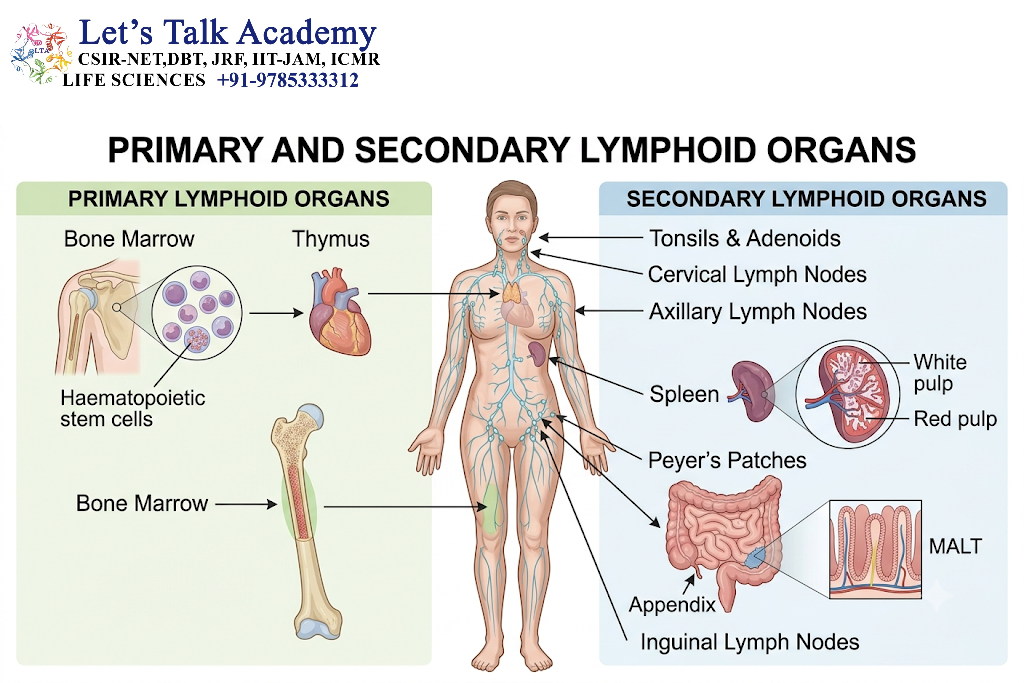
Immune cells develop, mature, or become activated within specialized tissues known as lymphoid organs.
1.3.1 Primary lymphoid organs
Primary lymphoid organs are sites where important stages of immune cell development occur.
-
Bone marrow: Produces blood cells and supports the development of B lymphocytes and many innate immune cells.
-
Thymus: Supports the maturation and selection of T lymphocytes.
1.3.2 Secondary lymphoid organs
Secondary lymphoid organs are sites where mature lymphocytes encounter antigens and initiate many adaptive immune responses.
Examples include:
-
Lymph nodes.
-
Spleen.
-
Mucosa-associated lymphoid tissues.
-
Other organized lymphoid structures.
The lymphatic system transports antigens, fluid, and immune cells between tissues and lymphoid organs, facilitating immune surveillance.
2. Overview of Innate and Adaptive Immunity
2.1 Innate Immunity
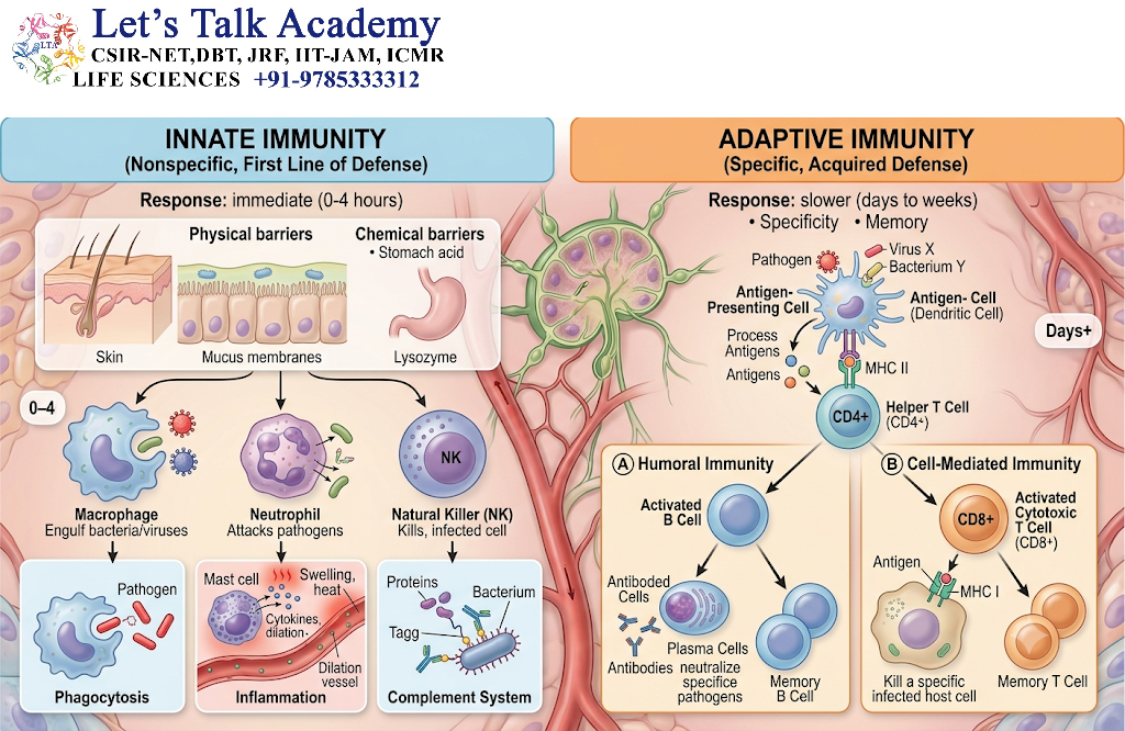
Innate immunity is the first-line defense system that responds rapidly to invading microorganisms and tissue injury.
It is present from birth and relies on germline-encoded receptors that recognize conserved molecular patterns.
Important characteristics include:
-
Rapid activation.
-
Recognition of pathogen-associated and damage-associated molecular patterns.
-
Use of phagocytes, innate lymphoid cells, complement, and inflammatory mediators.
-
Broad recognition rather than the highly diverse antigen-specific recognition characteristic of adaptive immunity.
-
Limited conventional antigen-specific memory, although trained immunity and other forms of innate immune memory can occur.
Innate immunity does not simply provide a nonspecific response. It uses highly regulated receptors and signaling pathways to distinguish different types of threats.
2.2 Adaptive Immunity
Adaptive immunity is mediated primarily by B and T lymphocytes.
These cells express antigen receptors generated through somatic gene rearrangement, producing a highly diverse repertoire.
The main characteristics of adaptive immunity include:
-
Antigen-specific recognition.
-
Receptor diversity.
-
Clonal expansion.
-
Differentiation into specialized effector cells.
-
Immunological memory.
-
Mechanisms of self-tolerance.
Adaptive responses generally require more time to develop during a first exposure to a new antigen than many innate responses. However, memory responses can develop more rapidly and effectively following subsequent exposure to the same or a related antigen.
2.3 Comparison of Innate and Adaptive Immunity
|
Feature |
Innate immunity |
Adaptive immunity |
|---|---|---|
|
Recognition |
Conserved molecular patterns and other danger signals |
Specific antigens recognized by diverse receptors |
|
Main receptors |
Germline-encoded pattern-recognition receptors |
Rearranged B-cell and T-cell antigen receptors |
|
Initial response |
Usually rapid |
Usually slower during primary activation |
|
Main cells |
Neutrophils, macrophages, dendritic cells, NK cells, and others |
B cells, T cells, and antibody-secreting plasma cells |
|
Memory |
Trained immunity and other forms can occur |
Classical antigen-specific memory |
|
Major soluble components |
Complement, cytokines, chemokines, acute-phase proteins |
Antibodies, cytokines, and other regulatory molecules |
|
Principal role |
Early defense and initiation of inflammation |
Specific elimination and long-term immune protection |
2.4 Integration of Innate and Adaptive Immunity
Innate immunity and adaptive immunity are interconnected at almost every stage of an immune response.
For example, dendritic cells recognize microbial molecules through innate receptors and then present antigen-derived peptides to T lymphocytes. Cytokines produced during innate activation influence the type of adaptive response that develops.
Similarly, antibodies produced by adaptive immune cells can activate complement, enhance phagocytosis, and support the destruction of infected cells.
The immune response is therefore best understood as a coordinated network rather than two completely independent systems.
3. Cells Involved in Innate Immunity
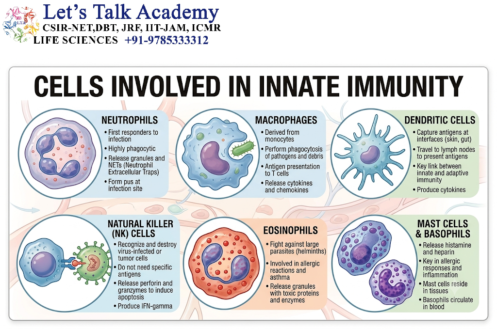
Innate immune cells provide the initial cellular defense against pathogens and tissue damage. Most of these cells arise from haematopoietic stem and progenitor cells in the bone marrow, although some tissue-resident populations are established during development and maintained locally.
Innate immune cells recognize danger signals, release inflammatory mediators, engulf microorganisms, destroy infected cells, and regulate the activation of adaptive immunity.
The major cells involved in innate immunity include neutrophils, monocytes, macrophages, dendritic cells, natural killer cells, eosinophils, basophils, mast cells, and innate lymphoid cells.
3.1 Neutrophils
Neutrophils are abundant circulating granulocytes and important effector cells of the innate immune system. They are among the earliest leukocytes recruited to many sites of acute infection and tissue injury.
Neutrophils are produced in the bone marrow through granulopoiesis. Their production is regulated by growth factors such as granulocyte colony-stimulating factor.
3.1.1 Structural characteristics
Neutrophils typically possess a multilobed nucleus and cytoplasmic granules containing enzymes and antimicrobial proteins.
Their granules contain substances such as:
-
Myeloperoxidase.
-
Proteases.
-
Defensins.
-
Lysozyme.
-
Lactoferrin.
-
Antimicrobial proteins and enzymes.
The contents of these granules contribute to the destruction of microorganisms.
3.1.2 Functions of neutrophils
Phagocytosis: Neutrophils engulf microorganisms and cellular debris into intracellular compartments called phagosomes.
Oxidative killing: Neutrophils generate reactive oxygen species through the activity of the NADPH oxidase complex. These molecules contribute to microbial destruction.
Degranulation: Granules release antimicrobial substances into phagosomes and, in some circumstances, into the extracellular environment.
Formation of neutrophil extracellular traps: Neutrophils can release structures composed of DNA and associated antimicrobial proteins called neutrophil extracellular traps, or NETs. NETs may help immobilize microorganisms, although excessive NET formation can contribute to tissue injury and thrombosis.
Recruitment of additional immune cells: Neutrophils release inflammatory mediators that influence the activity of other immune cells.
3.1.3 Regulation of neutrophil activity
Neutrophil recruitment is influenced by chemokines such as CXCL8, also called interleukin-8, in humans, as well as by complement fragments and other inflammatory mediators.
Neutrophils must be tightly regulated because their antimicrobial mechanisms can also damage healthy tissues.
3.2 Monocytes
Monocytes are circulating myeloid cells that can migrate into tissues and differentiate into macrophages or certain dendritic cell populations.
They are produced in the bone marrow and circulate in the blood before entering tissues in response to specific signals.
3.2.1 Types of monocytes
In humans, monocytes are commonly classified into three major subsets:
-
Classical monocytes: Generally characterized by high CD14 expression and relatively low CD16 expression.
-
Intermediate monocytes: Express both CD14 and CD16 at intermediate or variable levels.
-
Non-classical monocytes: Generally express lower CD14 levels and higher CD16 levels.
These subsets differ in their migratory behavior, inflammatory responses, and interactions with blood vessels and tissues.
3.2.2 Functions of monocytes
-
Phagocytosis of microorganisms and cellular debris.
-
Production of inflammatory cytokines.
-
Migration into inflamed tissues.
-
Differentiation into macrophages or selected dendritic cell populations.
-
Contribution to antigen presentation.
-
Participation in tissue repair and inflammatory regulation.
3.3 Macrophages
Macrophages are phagocytic cells found in almost all tissues. They recognize and engulf microorganisms, damaged cells, and foreign particles.
Some macrophages develop from circulating monocytes, whereas many tissue-resident macrophages originate from embryonic precursors and can maintain themselves through local proliferation.
3.3.1 Structural and functional features
Macrophages are generally large cells with abundant cytoplasm and specialized intracellular organelles.
They contain lysosomes, phagosomes, and other structures that enable them to digest engulfed material.
Macrophages express multiple receptors that recognize microorganisms, antibodies, complement fragments, apoptotic cells, and other molecules.
3.3.2 Major functions
Phagocytosis: Macrophages engulf and digest pathogens and cellular debris.
Antigen presentation: Macrophages can process antigens and present peptide fragments through major histocompatibility complex class II molecules to appropriate CD4-positive T cells.
Cytokine secretion: They produce inflammatory mediators such as tumour necrosis factor, interleukin-1, and interleukin-6 under appropriate stimulatory conditions.
Tissue repair: Macrophages participate in the removal of dead cells, tissue remodeling, and repair processes.
Clearance of apoptotic cells: Macrophages recognize and remove apoptotic cells through a process called efferocytosis.
Homeostatic functions: Tissue-resident macrophages perform specialized tasks. For example, liver macrophages participate in the clearance of blood-borne material, while lung macrophages help maintain the pulmonary environment.
3.3.3 Macrophage activation states
Macrophage activation is highly dependent on the local tissue environment.
The terms M1 and M2 are commonly used in simplified experimental descriptions:
-
M1-like activation: Associated with certain inflammatory and antimicrobial responses.
-
M2-like activation: Associated with selected tissue repair, remodeling, and immunoregulatory functions.
However, macrophages in living tissues exhibit a broad spectrum of activation states. The M1/M2 classification does not fully represent their biological diversity.
3.4 Dendritic Cells
Dendritic cells are specialized antigen-presenting cells that connect innate immune recognition with adaptive immune activation.
They are particularly important for initiating primary T-cell responses.
Dendritic cells detect pathogens through pattern-recognition receptors, capture antigens, and migrate to lymphoid organs, where they present antigen-derived peptides to naïve T lymphocytes.
3.4.1 Major dendritic cell populations
Conventional dendritic cells: These cells are highly efficient at antigen presentation and are important for the activation of naïve T cells.
-
cDC1 populations are particularly specialized for cross-presentation of certain extracellular antigens on MHC class I molecules.
-
cDC2 populations are involved in the presentation of antigens to CD4-positive T cells and can contribute to different types of helper T-cell responses.
Plasmacytoid dendritic cells: These cells are important producers of type I interferons, especially during certain viral infections.
Monocyte-derived dendritic cells: These cells can arise under inflammatory conditions and may acquire dendritic cell-like functions.
3.4.2 Functions of dendritic cells
-
Recognition of pathogens and danger signals.
-
Capture and processing of antigens.
-
Migration to draining lymph nodes.
-
Presentation of peptides through MHC molecules.
-
Activation of naïve T cells.
-
Production of cytokines that influence adaptive immune differentiation.
-
Contribution to immune tolerance under appropriate conditions.
3.4.3 Dendritic cell maturation
When dendritic cells encounter microbial components or inflammatory signals, they undergo functional maturation.
Maturation is associated with:
-
Increased expression of MHC molecules.
-
Increased expression of co-stimulatory molecules such as CD80 and CD86.
-
Enhanced migration toward lymphoid tissues.
-
Changes in cytokine production.
-
Improved ability to activate naïve T cells.
3.5 Natural Killer Cells
Natural killer cells, commonly called NK cells, are innate lymphocytes that recognize and destroy certain infected or transformed cells.
Unlike conventional T lymphocytes, NK cells do not require prior antigen-specific sensitization to carry out many of their cytotoxic functions.
3.5.1 Recognition of target cells
NK cells integrate signals from activating and inhibitory receptors.
A healthy cell may express sufficient ligands for inhibitory receptors, including normal levels of MHC class I molecules, which can help prevent inappropriate NK-cell activation.
Some infected or transformed cells show altered expression of MHC class I molecules or increased expression of stress-associated ligands. These changes may shift the balance toward NK-cell activation.
This principle is commonly described as missing-self recognition, although NK-cell responses also depend on many other activating and inhibitory signals.
3.5.2 Major functions
Cytotoxicity: NK cells kill susceptible target cells by releasing perforin and granzymes, which initiate cell death pathways.
Cytokine production: NK cells produce interferon-gamma, which can activate macrophages and influence immune responses.
Antibody-dependent cellular cytotoxicity: NK cells express CD16, a receptor for the Fc region of certain IgG antibodies. Through CD16, NK cells can recognize antibody-coated target cells and induce cytotoxic responses.
3.5.3 Regulation of NK-cell activity
NK-cell function is determined by the balance between activating and inhibitory receptor signals.
Important receptor families include:
-
Killer-cell immunoglobulin-like receptors.
-
NKG2 family receptors.
-
Natural cytotoxicity receptors.
-
CD16.
The cytokine IL-15 is particularly important for NK-cell development and survival.
3.6 Eosinophils
Eosinophils are granulocytes involved in immune responses against certain parasites and in allergic and inflammatory conditions.
They contain cytoplasmic granules rich in proteins that can damage parasites and other targets.
3.6.1 Major functions
-
Participation in host defense against certain helminths.
-
Contribution to allergic inflammation.
-
Release of granule proteins.
-
Interaction with T-helper type 2-associated cytokine networks.
-
Participation in tissue immune regulation.
3.6.2 Important regulatory molecules
Interleukin-5 is a major cytokine involved in eosinophil development, survival, and activation.
Eosinophil activity is also influenced by chemokines, including eotaxin family members.
Excessive eosinophil activation can contribute to tissue damage in certain allergic and inflammatory disorders.
3.7 Basophils
Basophils are rare circulating granulocytes involved in allergic and type 2 immune responses.
They possess high-affinity IgE receptors called FcεRI.
When appropriately activated, basophils can release histamine, lipid mediators, and cytokines.
3.7.1 Functions
-
Participation in immediate hypersensitivity reactions.
-
Release of histamine and other inflammatory mediators.
-
Contribution to type 2 immune responses.
-
Modulation of immune cell recruitment.
-
Participation in certain responses to parasites.
Basophils are distinct from mast cells, even though both express FcεRI and can participate in IgE-associated reactions.
3.8 Mast Cells
Mast cells are tissue-resident immune cells that are particularly abundant near blood vessels, nerves, and epithelial surfaces.
They play important roles in immediate allergic reactions, host defense, and tissue regulation.
3.8.1 Structural characteristics
Mast cells contain numerous cytoplasmic granules filled with biologically active mediators.
Their granules may contain:
-
Histamine.
-
Proteases such as tryptase.
-
Heparin-associated molecules.
-
Other inflammatory mediators.
3.8.2 Major functions
Immediate mediator release: Mast cells can rapidly release preformed mediators following appropriate activation.
Lipid mediator synthesis: They produce leukotrienes and prostaglandins, which influence vascular permeability, smooth muscle activity, and inflammation.
Cytokine production: They release cytokines that can influence immune cell recruitment and tissue responses.
Allergic reactions: Cross-linking of FcεRI-bound IgE by an appropriate allergen can trigger mast cell activation.
Barrier defense: Mast cells contribute to immune surveillance at tissue interfaces.
3.8.3 Mast cell activation and regulation
Mast cells can be activated by several mechanisms, including IgE-dependent and IgE-independent pathways.
Because mast cell mediators can produce powerful effects on blood vessels and smooth muscle, their activation is tightly regulated.
3.9 Innate Lymphoid Cells
Innate lymphoid cells, or ILCs, are lymphocyte-like cells that lack conventional rearranged antigen receptors but produce cytokines that influence tissue immunity.
They are found in tissues such as the intestine, lungs, and skin.
3.9.1 Major groups
-
ILC1: Often associated with interferon-gamma production and type 1 immune responses.
-
ILC2: Associated with type 2 cytokines such as IL-5 and IL-13.
-
ILC3: Associated with IL-17- and IL-22-related responses in appropriate contexts.
Natural killer cells are generally classified separately from helper-like ILC groups, although both belong to the broader innate lymphoid cell family.
3.9.2 Functions
-
Maintenance of epithelial barrier integrity.
-
Rapid cytokine production.
-
Regulation of tissue inflammation.
-
Contribution to defense against pathogens.
-
Communication with epithelial cells, stromal cells, and adaptive immune cells.
4. Molecules Involved in Innate Immunity
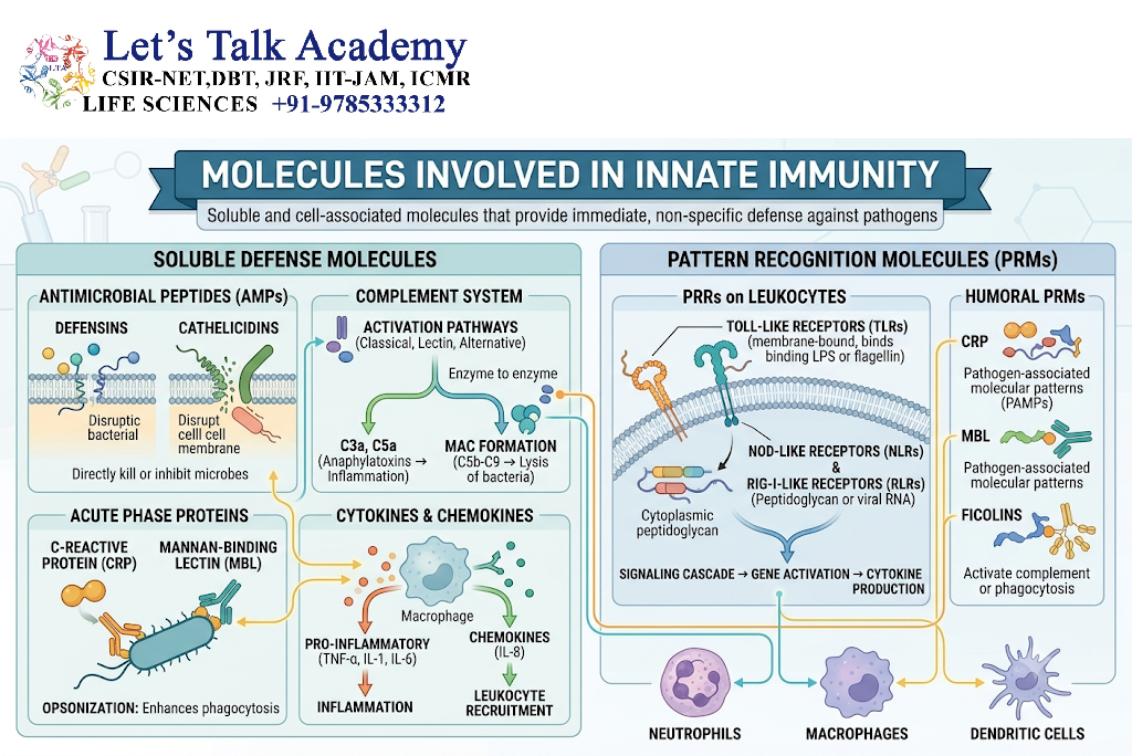
Innate immunity relies on a wide range of molecules that recognize pathogens, activate inflammatory pathways, recruit immune cells, and directly destroy microorganisms.
The major molecular components include pattern-recognition receptors, complement proteins, cytokines, chemokines, acute-phase proteins, antimicrobial peptides, and intracellular signaling proteins.
4.1 Pattern-Recognition Receptors
Pattern-recognition receptors, commonly called PRRs, are receptors of the innate immune system that detect conserved molecular structures associated with pathogens or cellular damage.
The recognized structures are called:
-
Pathogen-associated molecular patterns (PAMPs).
-
Damage-associated molecular patterns (DAMPs).
PRRs are found on the cell surface, within endosomal compartments, or in the cytoplasm.
4.1.1 Functions of PRRs
PRRs can activate:
-
Inflammatory cytokine production.
-
Type I interferon responses.
-
Phagocytosis.
-
Antimicrobial mechanisms.
-
Inflammasome assembly.
-
Dendritic cell maturation.
-
Expression of co-stimulatory molecules.
4.2 Toll-Like Receptors
Toll-like receptors, or TLRs, are a major family of pattern-recognition receptors.
They recognize microbial components and activate intracellular signaling pathways that regulate inflammation and antimicrobial defense.
4.2.1 Major Toll-like receptors and their ligands
|
Receptor |
Major recognized structure or ligand |
|---|---|
|
TLR2 |
Lipoproteins and selected microbial lipids, in cooperation with other receptors |
|
TLR3 |
Double-stranded RNA |
|
TLR4 |
Lipopolysaccharide from many Gram-negative bacteria, with accessory molecules |
|
TLR5 |
Bacterial flagellin |
|
TLR7 |
Single-stranded RNA |
|
TLR8 |
Single-stranded RNA and related nucleic acid signals |
|
TLR9 |
Unmethylated CpG-rich DNA in appropriate contexts |
The precise ligand recognition of some TLRs depends on accessory proteins and the cellular environment.
4.2.2 Signaling mechanism
Many TLRs signal through the adaptor protein MyD88, while TLR3 primarily uses TRIF. TLR4 can signal through both MyD88-associated and TRIF-associated pathways.
The resulting signals activate transcription factors such as:
-
NF-κB.
-
AP-1.
-
Interferon regulatory factors.
These transcription factors regulate the expression of inflammatory cytokines, chemokines, and interferons.
4.3 NOD-Like Receptors
NOD-like receptors, or NLRs, are intracellular pattern-recognition receptors.
They detect microbial components, changes in cellular homeostasis, or other danger-associated signals.
4.3.1 NOD1 and NOD2
NOD1 and NOD2 recognize specific fragments of bacterial peptidoglycan.
Their activation can stimulate NF-κB and MAPK signaling, leading to the production of inflammatory mediators.
NOD2 is particularly important in intestinal immune regulation.
4.3.2 Inflammasome-associated NLRs
Some NLRs participate in the formation of inflammasomes.
Inflammasomes are multiprotein signaling complexes that activate inflammatory caspases and promote the maturation of certain cytokines.
The NLRP3 inflammasome is one of the most extensively studied examples.
4.4 RIG-I-Like Receptors
RIG-I-like receptors, or RLRs, are cytoplasmic sensors of viral RNA.
Important members include:
-
RIG-I.
-
MDA5.
-
LGP2.
RIG-I and MDA5 detect different types of RNA-associated molecular patterns and activate signaling through the mitochondrial antiviral-signaling protein, also called MAVS.
This signaling promotes the production of type I and, in some settings, type III interferons and other antiviral mediators.
4.5 cGAS–STING Pathway
The cGAS–STING pathway is an important intracellular DNA-sensing system.
Cyclic GMP–AMP synthase, or cGAS, detects certain forms of cytosolic DNA and generates the second messenger cyclic GMP–AMP.
Cyclic GMP–AMP activates STING, an adaptor protein associated with intracellular membranes.
STING signaling can activate TBK1 and interferon regulatory factor 3, resulting in type I interferon production.
This pathway participates in antiviral defense and responses to abnormal cytosolic DNA.
4.6 C-Type Lectin Receptors
C-type lectin receptors are a diverse group of receptors that recognize carbohydrate structures and other molecular patterns.
They are expressed on several myeloid cell populations, including macrophages and dendritic cells.
Examples include:
-
Dectin-1.
-
Dectin-2.
-
The mannose receptor.
-
DC-SIGN.
Some C-type lectin receptors recognize fungal cell wall components and contribute to antifungal immunity.
Their signaling can influence phagocytosis, cytokine production, and the development of adaptive immune responses.
4.7 Complement System
The complement system is a network of plasma proteins and cell-associated regulators that contributes to innate and antibody-associated immune defense.
Complement proteins circulate in inactive forms and become activated through a series of tightly regulated proteolytic reactions.
4.7.1 Major functions
-
Opsonization of microorganisms.
-
Recruitment of inflammatory cells.
-
Enhancement of phagocytosis.
-
Direct membrane damage to susceptible targets.
-
Clearance of immune complexes and cellular debris.
-
Amplification of immune responses.
4.7.2 Complement activation pathways
There are three major complement activation pathways.
Classical pathway: Usually activated when C1q recognizes appropriate antibody–antigen complexes. It can also be activated through certain antibody-independent interactions.
Lectin pathway: Initiated when pattern-recognition molecules such as mannose-binding lectin recognize suitable carbohydrate structures on microbial surfaces.
Alternative pathway: Involves spontaneous low-level complement activation and amplification on surfaces that permit complement activation.
All three pathways converge on the activation of complement component C3.
4.7.3 Major complement components
|
Component |
Major function |
|---|---|
|
C3 |
Central complement component; cleavage generates C3a and C3b |
|
C3b |
Opsonization and participation in convertase formation |
|
C3a |
Anaphylatoxin with inflammatory effects |
|
C5a |
Potent inflammatory mediator and leukocyte chemoattractant |
|
C5b–C9 |
Formation of the membrane attack complex |
|
C1q |
Recognition component of the classical pathway |
|
Factor B |
Component of the alternative pathway convertase |
|
Factor D |
Protease involved in alternative pathway activation |
|
Properdin |
Stabilizes selected alternative pathway convertases |
|
Factor H and factor I |
Important regulators of complement activity |
4.7.4 Membrane attack complex
The membrane attack complex is formed by complement components C5b, C6, C7, C8, and multiple copies of C9.
It creates a pore-like structure in susceptible target membranes.
The membrane attack complex is particularly effective against certain susceptible Gram-negative bacteria, although its effectiveness varies among organisms.
4.7.5 Regulation of complement
Complement activation must be controlled to prevent damage to healthy host tissues.
Regulatory proteins include:
-
CD55, also known as decay-accelerating factor.
-
CD59.
-
Factor H.
-
Factor I.
-
C1 inhibitor.
Defects in complement regulation can contribute to inflammatory disease and tissue injury.
4.8 Cytokines of Innate Immunity
Cytokines are signaling proteins that coordinate immune responses and communication between cells.
They influence inflammation, cell survival, proliferation, differentiation, and the development of adaptive immune responses.
4.8.1 Tumour necrosis factor
Tumour necrosis factor, or TNF, is an important inflammatory cytokine.
It is produced by macrophages, T cells, and other cells under appropriate conditions.
Functions include:
-
Activation of vascular endothelial cells.
-
Promotion of leukocyte recruitment.
-
Induction of inflammatory gene expression.
-
Regulation of cell survival and death pathways.
-
Contribution to systemic inflammatory responses.
Excessive TNF activity can contribute to tissue damage and systemic inflammation.
4.8.2 Interleukin-1
Interleukin-1 includes cytokines such as IL-1α and IL-1β.
IL-1β is commonly produced as an inactive precursor that requires processing, often involving inflammatory caspases.
Functions include:
-
Induction of fever.
-
Promotion of inflammation.
-
Activation of endothelial cells.
-
Stimulation of acute-phase responses.
-
Influence on haematopoietic and immune cell activity.
4.8.3 Interleukin-6
IL-6 is a multifunctional cytokine that participates in acute inflammation, immune regulation, and the acute-phase response.
It stimulates hepatocytes to produce acute-phase proteins such as C-reactive protein.
IL-6 can also influence B-cell differentiation, T-cell responses, and haematopoiesis.
4.8.4 Type I interferons
Type I interferons primarily include interferon-alpha and interferon-beta.
They are produced in response to viral infection and other stimuli.
Major functions include:
-
Induction of antiviral gene expression.
-
Enhancement of antigen presentation under appropriate conditions.
-
Regulation of natural killer cell activity.
-
Modulation of dendritic cell and adaptive immune responses.
Type I interferons act through the IFNAR receptor and activate the JAK–STAT pathway.
4.8.5 Interleukin-12
IL-12 is produced mainly by activated antigen-presenting cells.
It promotes interferon-gamma production by NK cells and T cells and supports the development of type 1 immune responses.
IL-12 is an important link between innate immune activation and cell-mediated adaptive immunity.
4.9 Chemokines
Chemokines are signaling proteins that regulate the migration of immune cells.
They bind to chemokine receptors, many of which are G-protein-coupled receptors.
4.9.1 Major chemokines
|
Chemokine |
Major role |
|---|---|
|
CXCL8 (IL-8 in humans) |
Recruitment of neutrophils |
|
CCL2 |
Recruitment of monocytes through CCR2-associated mechanisms |
|
CXCL10 |
Recruitment of selected activated T cells and other responsive cells |
|
CXCL12 |
Regulation of cell migration and retention, including HSC positioning |
|
CCL5 |
Recruitment of selected T cells, NK cells, and other immune populations |
|
CCL11 |
Recruitment of eosinophils in appropriate inflammatory settings |
Chemokines establish concentration gradients that guide immune cells toward sites of infection or tissue injury.
4.10 Acute-Phase Proteins
Acute-phase proteins are plasma proteins whose concentrations change during inflammation.
Many are produced by the liver in response to cytokines such as IL-6.
4.10.1 C-reactive protein
C-reactive protein, or CRP, binds to certain molecular structures on damaged cells and microorganisms.
It can contribute to opsonization and complement activation through the classical pathway.
CRP is also widely used as a laboratory marker of inflammation, although it does not identify a specific cause.
4.10.2 Mannose-binding lectin
Mannose-binding lectin is a soluble pattern-recognition molecule that binds certain carbohydrate structures on microorganisms.
It can activate the lectin pathway of complement through associated serine proteases.
4.10.3 Serum amyloid A
Serum amyloid A proteins are acute-phase molecules that participate in inflammatory responses and interactions with immune cells.
Persistently elevated serum amyloid A can be associated with chronic inflammatory conditions.
4.11 Antimicrobial Peptides
Antimicrobial peptides are small molecules that contribute to defense against microorganisms.
They are produced by epithelial cells, neutrophils, and other immune and non-immune cells.
4.11.1 Defensins
Defensins are cationic antimicrobial peptides that can disrupt microbial membranes and contribute to host defense.
They include alpha-defensins and beta-defensins.
4.11.2 Cathelicidins
Cathelicidins are antimicrobial peptides produced in several tissues and immune cell populations.
The human cathelicidin precursor is encoded by the CAMP gene and gives rise to LL-37 after processing.
Cathelicidins can directly affect microorganisms and influence inflammation, cell migration, and tissue repair.
4.11.3 Lysozyme
Lysozyme is an antimicrobial enzyme that hydrolyses specific bonds in bacterial peptidoglycan.
It is found in secretions such as tears and saliva and in the granules of certain immune cells.
5. Cells Involved in Adaptive Immunity
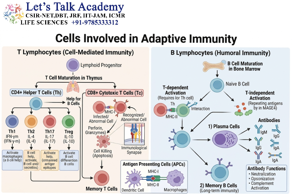
Adaptive immunity is primarily mediated by B lymphocytes and T lymphocytes.
These cells possess highly diverse antigen receptors generated through somatic gene rearrangement. Following antigen recognition and appropriate activation, lymphocytes proliferate and differentiate into effector and memory populations.
5.1 B Lymphocytes
B lymphocytes, or B cells, are specialized immune cells responsible for antibody-mediated immunity.
They develop through several stages in the bone marrow and subsequently migrate to peripheral lymphoid tissues.
5.1.1 B-cell receptor
The B-cell receptor, or BCR, is a membrane-bound immunoglobulin associated with signaling proteins.
The BCR recognizes a specific molecular structure on an antigen.
Unlike T-cell receptors, B-cell receptors can recognize intact antigens, including proteins, polysaccharides, lipids, and other molecular structures.
5.1.2 Functions of B cells
-
Recognition of antigens.
-
Antibody production following differentiation into plasma cells.
-
Antigen presentation to T cells.
-
Cytokine production in selected contexts.
-
Formation of memory B cells.
-
Contribution to immune regulation.
5.2 B-Cell Subpopulations
5.2.1 Naïve B cells
Naïve B cells are mature B cells that have not yet undergone activation by their cognate antigen.
They circulate between blood, lymph, and secondary lymphoid organs.
5.2.2 Plasma cells
Plasma cells are specialized antibody-secreting cells derived from activated B lymphocytes.
They possess abundant rough endoplasmic reticulum and a cellular structure adapted for the production of large quantities of immunoglobulins.
Some plasma cells are short-lived, whereas others survive for long periods in supportive tissue environments.
5.2.3 Memory B cells
Memory B cells are long-lived or persistent antigen-experienced B cells that can respond to subsequent exposure to the same or related antigen.
They contribute to the enhanced antibody response associated with many secondary immune responses.
5.2.4 Regulatory B cells
Some B-cell populations can produce immunoregulatory molecules, including IL-10, and influence immune responses.
The term regulatory B cell describes functional properties rather than one universally defined cell population.
5.3 T Lymphocytes
T lymphocytes, or T cells, mature in the thymus and are essential for cell-mediated immunity and the regulation of adaptive immune responses.
T cells express T-cell receptors that recognize antigen-derived peptides presented by major histocompatibility complex molecules.
5.3.1 T-cell receptor
The T-cell receptor, or TCR, is a membrane-bound antigen receptor.
Most conventional T cells express an αβ T-cell receptor composed of alpha and beta chains.
Some T cells express γδ T-cell receptors and have distinct recognition properties.
Conventional αβ T cells generally recognize peptide antigens presented by MHC molecules.
5.3.2 Functions of T cells
-
Activation of macrophages.
-
Regulation of B-cell responses.
-
Killing of infected or abnormal cells.
-
Production of cytokines.
-
Development of immunological memory.
-
Maintenance of immune tolerance through regulatory populations.
5.4 CD4-Positive T Helper Cells
CD4-positive T cells recognize antigen-derived peptides presented by MHC class II molecules.
Following activation, they can differentiate into distinct functional subsets according to the cytokine environment and transcriptional programs.
5.4.1 Th1 cells
Th1 cells are associated with cellular immune responses against certain intracellular pathogens.
They produce interferon-gamma and other mediators that support macrophage activation and cell-mediated immunity.
The transcription factor T-bet is an important regulator of Th1-associated differentiation.
5.4.2 Th2 cells
Th2 cells are associated with immune responses involving helminths, allergic inflammation, and type 2 immunity.
They produce cytokines such as IL-4, IL-5, and IL-13.
The transcription factor GATA-3 is an important regulator of Th2-associated differentiation.
5.4.3 Th17 cells
Th17 cells produce cytokines such as IL-17A, IL-17F, and, in appropriate contexts, IL-22.
They contribute to defense against certain extracellular bacteria and fungi, particularly at epithelial barriers.
The transcription factor RORγt is a major regulator of Th17-associated differentiation.
5.4.4 T follicular helper cells
T follicular helper cells, or Tfh cells, support B-cell responses in lymphoid follicles and germinal centres.
They produce signals such as IL-21 and express molecules such as ICOS and CD40 ligand.
Tfh cells help B cells undergo affinity maturation, class-switch recombination, and differentiation into memory B cells or antibody-secreting cells.
The transcription factor BCL-6 is an important regulator of Tfh differentiation.
5.4.5 Regulatory T cells
Regulatory T cells, or Tregs, suppress excessive immune responses and contribute to self-tolerance.
Many conventional Tregs express CD4, CD25, and the transcription factor FOXP3.
They can suppress immune responses through multiple mechanisms, including inhibitory cytokines, modulation of antigen-presenting cells, and consumption of growth-promoting signals.
5.5 CD8-Positive Cytotoxic T Lymphocytes
CD8-positive cytotoxic T lymphocytes, commonly called CTLs, recognize peptide antigens presented by MHC class I molecules.
They are important for the elimination of virus-infected cells and certain abnormal cells.
5.5.1 Major cytotoxic mechanisms
Perforin–granzyme pathway: CTLs release perforin and granzymes. Granzymes enter target cells and activate intracellular cell death mechanisms.
Death receptor pathway: CTLs can express Fas ligand, which interacts with Fas on susceptible target cells and triggers apoptosis.
5.5.2 Activation of CTLs
Naïve CD8-positive T cells typically require antigen recognition, co-stimulatory signals, and appropriate cytokine support for effective activation.
Activated CTLs undergo clonal expansion and differentiate into effector cells.
Some activated cells form memory populations that persist after the initial immune response.
5.6 Other Lymphocyte Populations
5.6.1 Natural killer T cells
Natural killer T cells are a heterogeneous group of T lymphocytes with properties that overlap with conventional T cells and NK cells.
Some NKT populations express semi-invariant T-cell receptors and recognize lipid antigens presented by CD1d.
They can rapidly produce cytokines and participate in immune regulation.
5.6.2 Mucosal-associated invariant T cells
Mucosal-associated invariant T cells, or MAIT cells, recognize certain microbial metabolite-derived antigens presented by MR1.
They are abundant in some mucosal tissues and can respond rapidly to microbial infection.
5.6.3 Gamma-delta T cells
Gamma-delta T cells express γδ T-cell receptors and recognize a range of antigens and stress-associated signals.
They can participate in epithelial surveillance, antimicrobial responses, and tissue repair.
6. Molecules Involved in Adaptive Immunity
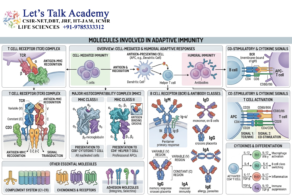
Adaptive immunity depends on molecular recognition systems that provide specificity, diversity, and immunological memory.
The major molecules include antigen receptors, antibodies, MHC proteins, co-stimulatory molecules, cytokines, and signaling proteins.
6.1 Immunoglobulins and Antibodies
Immunoglobulins are proteins produced by B cells and plasma cells.
When secreted, they are commonly called antibodies.
Antibodies bind specific antigens through antigen-binding regions and recruit other mechanisms to help eliminate the target.
6.1.1 Structure of an antibody
A typical antibody molecule consists of four polypeptide chains:
-
Two identical heavy chains.
-
Two identical light chains.
The chains are connected by disulfide bonds.
Each immunoglobulin chain contains variable and constant regions.
Variable regions form the antigen-binding site and determine antigen specificity.
Constant regions determine many of the biological functions of the antibody, including interactions with Fc receptors and complement proteins.
6.1.2 Fab and Fc regions
The antigen-binding portion of an antibody is located in the Fab regions.
The Fc region is responsible for many interactions with immune effector mechanisms.
For example, Fc regions can bind to:
-
Fc receptors on immune cells.
-
Complement-associated proteins.
-
Transport and regulatory receptors.
6.1.3 Major antibody functions
-
Neutralization of toxins and viruses.
-
Opsonization of microorganisms.
-
Activation of the classical complement pathway.
-
Antibody-dependent cellular cytotoxicity.
-
Agglutination of certain antigens.
-
Facilitation of antigen clearance.
-
Regulation of immune responses.
6.2 Immunoglobulin Classes
There are five major immunoglobulin isotypes in humans: IgG, IgA, IgM, IgE, and IgD.
|
Immunoglobulin |
Major features and functions |
|---|---|
|
IgG |
Important in systemic immunity; neutralization, opsonization, and Fc-mediated effector functions |
|
IgA |
Important at mucosal surfaces and in secretions |
|
IgM |
Usually the first major isotype produced during a primary response; efficient complement activation |
|
IgE |
Associated with mast cells, basophils, and type 2 immune responses |
|
IgD |
Primarily expressed on certain mature naïve B cells and involved in B-cell receptor functions |
6.2.1 IgG
IgG is the predominant antibody class in human serum.
Its functions include:
-
Neutralization of pathogens and toxins.
-
Opsonization.
-
Activation of complement by appropriate IgG subclasses.
-
Interaction with Fcγ receptors.
-
Placental transfer of certain IgG antibodies through FcRn-mediated transport.
6.2.2 IgA
IgA is especially important at mucosal surfaces.
It is present in secretions such as saliva, tears, intestinal secretions, and breast milk.
Secretory IgA helps prevent microorganisms from attaching to epithelial surfaces.
6.2.3 IgM
IgM is commonly expressed as a membrane-bound immunoglobulin on naïve B cells.
Secreted IgM often forms a pentameric structure in association with a joining chain.
Pentameric IgM is highly effective at activating the classical complement pathway under suitable conditions.
6.2.4 IgE
IgE binds with high affinity to FcεRI on mast cells and basophils.
Cross-linking of receptor-bound IgE by a relevant antigen can trigger mediator release.
IgE also contributes to immune responses against certain parasites.
6.2.5 IgD
IgD is expressed primarily on the surface of mature naïve B cells, where it contributes to antigen receptor functions.
Its role as a secreted antibody is less prominent than that of IgG, IgA, or IgM.
6.3 Major Histocompatibility Complex Molecules
Major histocompatibility complex molecules, or MHC molecules, present peptide antigens to T lymphocytes.
In humans, MHC proteins are called human leukocyte antigens (HLA).
They are essential for antigen recognition and immune surveillance.
6.4 MHC Class I Molecules
MHC class I molecules are expressed on most nucleated human cells, although expression varies among cell types and conditions.
They present peptides, often derived from intracellular proteins, to CD8-positive T cells.
6.4.1 Structure
A classical MHC class I molecule consists of:
-
An MHC class I heavy chain.
-
β2-microglobulin.
The peptide-binding groove accommodates peptides that are commonly around 8–10 amino acids long, although length variation occurs.
6.4.2 Antigen processing through MHC class I
-
Intracellular proteins are degraded into peptides.
-
Peptides are transported into the endoplasmic reticulum or generated through other relevant processing routes.
-
Peptides bind to MHC class I molecules.
-
Peptide–MHC class I complexes move to the cell surface.
-
CD8-positive T cells survey these complexes through their T-cell receptors.
6.4.3 Importance
MHC class I presentation allows cytotoxic T cells to detect and respond to certain intracellular infections and abnormal cellular changes.
6.5 MHC Class II Molecules
MHC class II molecules are expressed mainly by professional antigen-presenting cells, including dendritic cells, macrophages, and B cells.
They present peptides derived from extracellular proteins to CD4-positive T cells.
6.5.1 Structure
MHC class II molecules consist of two transmembrane chains:
-
An alpha chain.
-
A beta chain.
The peptide-binding groove is open at both ends and can accommodate longer peptides than classical MHC class I molecules.
6.5.2 Antigen processing through MHC class II
-
Extracellular proteins are internalized by an antigen-presenting cell.
-
Proteins are degraded into peptides within endosomal or lysosomal compartments.
-
MHC class II molecules are transported through intracellular compartments.
-
Peptides are loaded onto MHC class II molecules.
-
Peptide–MHC class II complexes are displayed on the cell surface.
-
CD4-positive T cells recognize the complexes through their T-cell receptors.
6.5.3 Importance
MHC class II presentation is essential for the activation of many CD4-positive T-cell responses.
6.6 Antigen Receptors
6.6.1 B-cell receptor
The BCR recognizes intact antigens and transmits signals through associated proteins, including CD79A and CD79B.
The antigen-binding immunoglobulin determines the specificity of the receptor.
6.6.2 T-cell receptor
The TCR recognizes antigen-derived peptides or other antigens presented by specialized molecules.
Conventional αβ TCRs recognize peptide–MHC complexes.
The TCR associates with the CD3 signaling complex, which transmits activation signals into the T cell.
6.7 Co-Stimulatory Molecules
Antigen recognition alone is often insufficient to fully activate a naïve T cell.
Co-stimulatory molecules provide additional signals that influence T-cell activation, differentiation, and survival.
6.7.1 CD28 and B7 molecules
CD28 is a co-stimulatory receptor expressed on T cells.
Its major ligands are CD80 and CD86, which are also known as B7-1 and B7-2.
The interaction between CD28 and CD80 or CD86 supports T-cell activation.
6.7.2 CTLA-4
CTLA-4 is an inhibitory receptor expressed on activated T cells and regulatory T cells.
It binds CD80 and CD86 and can reduce T-cell activation.
CTLA-4 is an important component of immune tolerance.
6.7.3 PD-1 and its ligands
PD-1 is an inhibitory receptor expressed on activated T cells and other immune populations.
Its ligands include PD-L1 and PD-L2.
PD-1 signaling can reduce T-cell activity and contributes to the regulation of immune responses.
These inhibitory pathways are important in normal immune regulation and are also relevant to cancer immunotherapy.
6.8 Cytokines of Adaptive Immunity
Adaptive immune responses are regulated by cytokines produced by T cells, B cells, antigen-presenting cells, and other immune populations.
6.8.1 Interleukin-2
IL-2 is an important T-cell growth and survival factor.
It supports the proliferation of activated T cells and contributes to the development and maintenance of regulatory T cells.
6.8.2 Interferon-gamma
Interferon-gamma is produced by NK cells and several T-cell populations.
It activates macrophages and supports type 1 immune responses.
It also influences antigen presentation and immune cell differentiation.
6.8.3 Interleukin-4
IL-4 is associated with type 2 immune responses.
It contributes to:
-
Th2 differentiation.
-
B-cell class switching toward IgE and certain other isotypes.
-
Regulation of allergic and anti-parasitic immune responses.
6.8.4 Interleukin-5
IL-5 supports eosinophil development and survival.
It is also associated with type 2 immune responses.
6.8.5 Interleukin-17
IL-17A and IL-17F contribute to the recruitment and activation of cells involved in defense against certain extracellular bacteria and fungi.
They stimulate epithelial and stromal cells to produce inflammatory mediators and neutrophil-recruiting chemokines.
6.8.6 Interleukin-21
IL-21 is produced by selected T-cell populations, including Tfh cells.
It supports B-cell responses, plasma cell differentiation, and immune regulation in context-dependent ways.
6.8.7 Transforming growth factor beta
TGF-β contributes to immune regulation, tissue homeostasis, and the differentiation of selected T-cell populations.
It plays an important role in the development and maintenance of regulatory immune responses.
7. Molecular Mechanisms of Antigen Recognition and Lymphocyte Activation
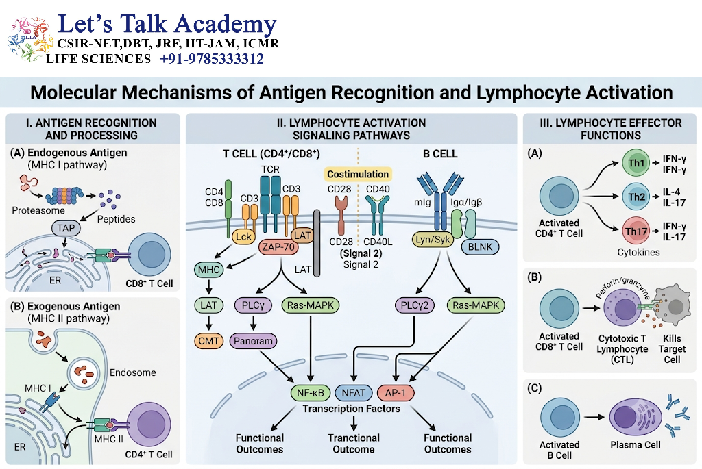
7.1 Antigen Recognition
Antigen recognition is the process through which immune receptors identify molecular structures associated with a target.
B-cell receptors can recognize intact antigens, whereas conventional T-cell receptors recognize antigen fragments presented by MHC molecules.
The specificity of an antigen receptor is determined by its variable region.
7.2 Generation of Antigen Receptor Diversity
Adaptive immune receptor diversity is generated through somatic V(D)J recombination.
During lymphocyte development, selected variable, diversity, and joining gene segments are rearranged to generate distinct antigen receptor genes.
7.2.1 Important enzymes
-
RAG1.
-
RAG2.
-
Terminal deoxynucleotidyl transferase, or TdT, which contributes to junctional diversity in developing lymphocytes.
7.2.2 Significance
V(D)J recombination enables the generation of a very large repertoire of antigen receptors from a limited number of gene segments.
The process is tightly regulated to preserve functional receptor expression and reduce the risk of harmful genomic rearrangements.
7.3 T-Cell Activation
Naïve T-cell activation generally requires three major categories of signals.
7.3.1 Signal 1: Antigen recognition
The T-cell receptor recognizes a specific antigen–MHC complex.
CD4 or CD8 co-receptors contribute to the recognition and signaling process.
7.3.2 Signal 2: Co-stimulation
Co-stimulatory molecules such as CD28 interact with ligands on antigen-presenting cells.
This signal supports full activation of many naïve T cells.
7.3.3 Signal 3: Cytokine-mediated instruction
Cytokines produced in the local environment influence T-cell proliferation, survival, and differentiation into distinct functional subsets.
7.3.4 Intracellular signaling
TCR signaling involves the activation of protein tyrosine kinases and adaptor proteins.
Important signaling components include:
-
Lck.
-
ZAP-70.
-
LAT.
-
PLC-γ1.
-
Calcium-dependent signaling pathways.
-
NFAT.
-
NF-κB.
-
AP-1.
These pathways activate genes involved in proliferation, metabolism, survival, and effector functions.
7.4 B-Cell Activation
B cells can be activated through BCR signaling and additional signals from other receptors and helper cells.
7.4.1 BCR signaling
Antigen binding induces changes in the BCR-associated signaling machinery.
Important signaling proteins include:
-
Lyn.
-
Syk.
-
BLNK.
-
BTK.
-
PLC-γ2.
-
PI3K.
-
NF-κB-associated signaling proteins.
These pathways promote B-cell activation, survival, and proliferation.
7.4.2 T-dependent B-cell activation
Many protein antigens stimulate B cells through a T-dependent pathway.
The process includes:
-
BCR recognition of an antigen.
-
Internalization and processing of the antigen.
-
Presentation of antigen-derived peptides through MHC class II.
-
Recognition by an appropriate helper T cell.
-
CD40–CD40 ligand interaction.
-
Cytokine-mediated regulation of B-cell differentiation.
-
Formation of antibody-secreting and memory B-cell populations.
7.4.3 Germinal centre reaction
Germinal centres form in secondary lymphoid follicles during many T-dependent immune responses.
Within germinal centres, activated B cells undergo:
-
Proliferation.
-
Somatic hypermutation.
-
Selection based on antigen-binding properties.
-
Class-switch recombination.
-
Differentiation into memory B cells and plasma cells.
7.5 Somatic Hypermutation
Somatic hypermutation is a process in which mutations are introduced into immunoglobulin variable-region genes in activated B cells.
The enzyme activation-induced cytidine deaminase, or AID, is essential for this process.
Somatic hypermutation creates antibody variants with different antigen-binding properties.
B cells that produce antibodies with improved binding to the relevant antigen may be preferentially selected during germinal centre reactions.
7.6 Class-Switch Recombination
Class-switch recombination allows activated B cells to change the constant region of the antibody heavy chain while retaining the same variable-region antigen specificity.
The enzyme AID is also essential for class-switch recombination.
Class switching enables an antibody response to acquire different effector properties.
For example, a B cell may switch from producing IgM to producing IgG, IgA, or IgE, depending on the signals it receives.
7.7 Immunological Memory
Immunological memory is the ability of the adaptive immune system to respond differently after previous exposure to an antigen.
Memory B cells, long-lived plasma cells, and memory T cells contribute to this phenomenon.
Memory responses are shaped by the nature of the antigen, the type of initial immune response, the tissue environment, and the persistence of antigenic stimulation.
8. Interaction Between Innate and Adaptive Immunity
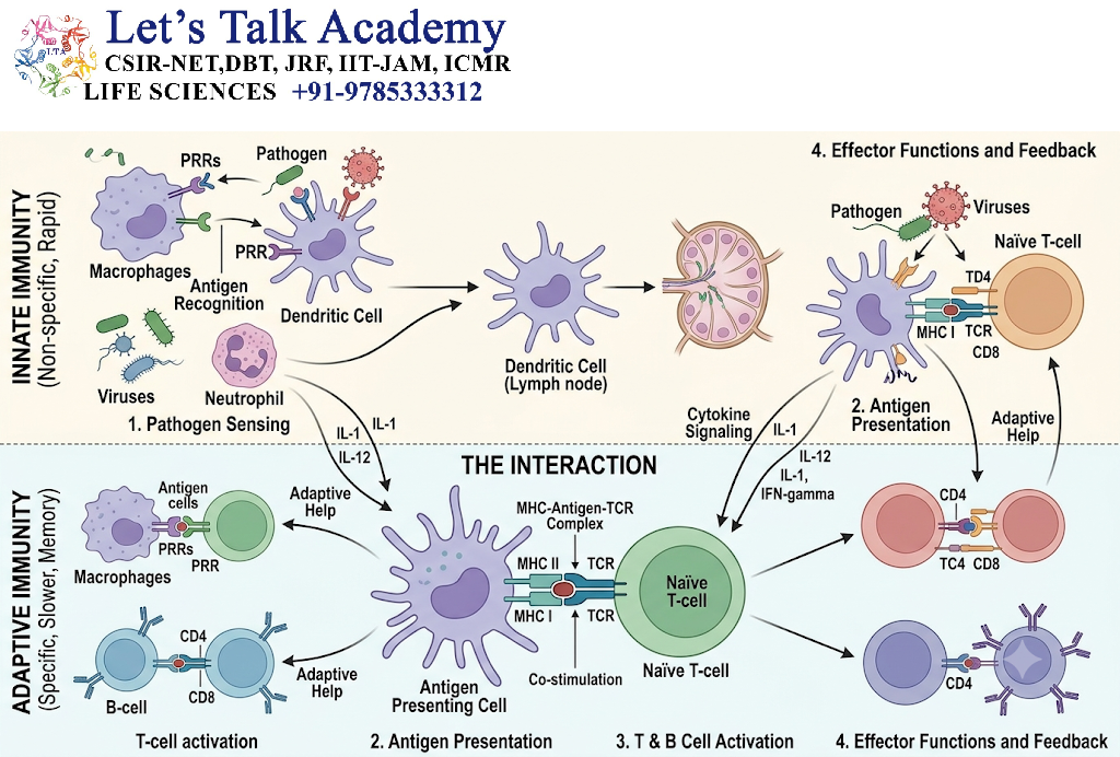
8.1 Role of Dendritic Cells in Bridging Immunity
Dendritic cells recognize pathogens through innate immune receptors and present antigen-derived peptides to T lymphocytes.
They also produce cytokines and express co-stimulatory molecules that influence the type and strength of the adaptive response.
This makes dendritic cells a central link between early pathogen recognition and antigen-specific immunity.
8.2 Role of Complement in Adaptive Immunity
Complement is not restricted to innate defense.
Complement fragments can influence B-cell activation, antigen transport, and the development of antibody responses.
For example, complement component C3d can remain associated with antigenic material and interact with complement receptor 2, or CD21, on B cells.
The interaction between CD21 and complement-tagged antigen can enhance B-cell activation when appropriate BCR signals are also present.
8.3 Antibodies and Innate Effector Cells
Antibodies produced by plasma cells can recruit innate immune mechanisms.
8.3.1 Opsonization
Antibodies coat microorganisms and facilitate their recognition by phagocytes through Fc receptors.
8.3.2 Antibody-dependent cellular cytotoxicity
NK cells can recognize IgG-coated target cells through CD16 and induce cytotoxicity.
Other immune cells can also participate in antibody-dependent effector mechanisms.
8.3.3 Classical complement activation
Appropriate antigen–antibody complexes can activate the classical complement pathway.
This provides an example of adaptive immune recognition triggering innate effector mechanisms.
8.4 T Cells and Macrophage Activation
Th1-associated T cells produce interferon-gamma, which can activate macrophages and enhance their ability to control certain intracellular pathogens.
Macrophages can present antigen-derived peptides to CD4-positive T cells, allowing reciprocal communication between the two cell types.
8.5 Cytokine Networks
Cytokines produced by innate immune cells influence the differentiation and activity of adaptive immune cells.
Conversely, cytokines produced by T cells can alter the function of macrophages, dendritic cells, neutrophils, and other innate populations.
These interconnected cytokine networks help coordinate immune responses at the site of infection and throughout the body.
9. Regulation of Immune Responses
9.1 Importance of Immune Regulation
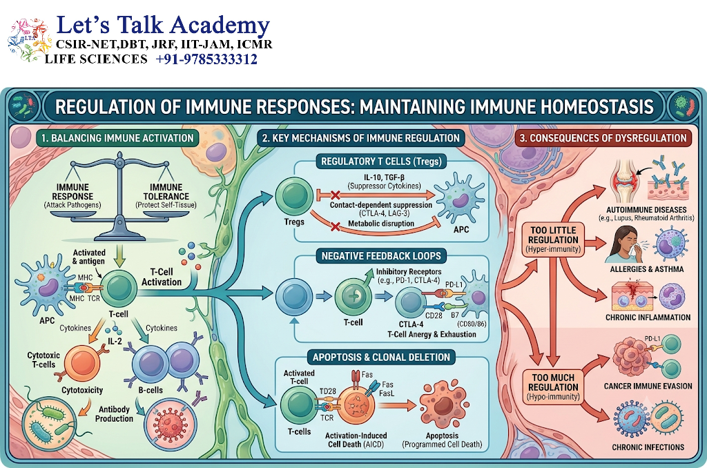
Immune activation must be controlled to ensure effective pathogen elimination without excessive damage to healthy tissues.
Regulatory mechanisms operate at the levels of receptors, intracellular signaling, cytokine production, cell migration, cellular death, and immune suppression.
9.2 Regulatory T Cells
Regulatory T cells are important suppressors of excessive immune responses.
They express FOXP3 in many major conventional regulatory T-cell populations and can suppress immune activity through several mechanisms.
Their functions include:
-
Limiting autoreactive T-cell responses.
-
Reducing excessive inflammation.
-
Contributing to peripheral tolerance.
-
Modulating antigen-presenting cell activity.
-
Supporting tissue immune homeostasis.
9.3 Immune Checkpoints
Immune checkpoints are receptor–ligand systems that regulate the strength and duration of immune responses.
Important examples include:
-
CTLA-4.
-
PD-1.
-
PD-L1.
-
PD-L2.
-
Other inhibitory receptor systems.
These molecules can limit T-cell activation and help prevent excessive immune responses.
9.4 Anti-Inflammatory Cytokines
Certain cytokines contribute to the suppression or resolution of inflammation.
Examples include:
-
IL-10.
-
TGF-β.
Their effects depend on the cell type, tissue, timing, and surrounding signaling environment.
9.5 Resolution of Inflammation
The resolution of inflammation involves active biological processes that restore tissue homeostasis.
These include:
-
Reduction in the production of inflammatory mediators.
-
Clearance of neutrophils.
-
Efferocytosis by macrophages.
-
Production of specialized pro-resolving mediators.
-
Restoration of tissue barrier function.
-
Repair of damaged tissue.
Resolution does not simply mean that inflammation stops. It involves coordinated molecular and cellular mechanisms that promote recovery.
10. Intracellular Signaling Pathways in Immunity
10.1 Introduction to Immune Signaling
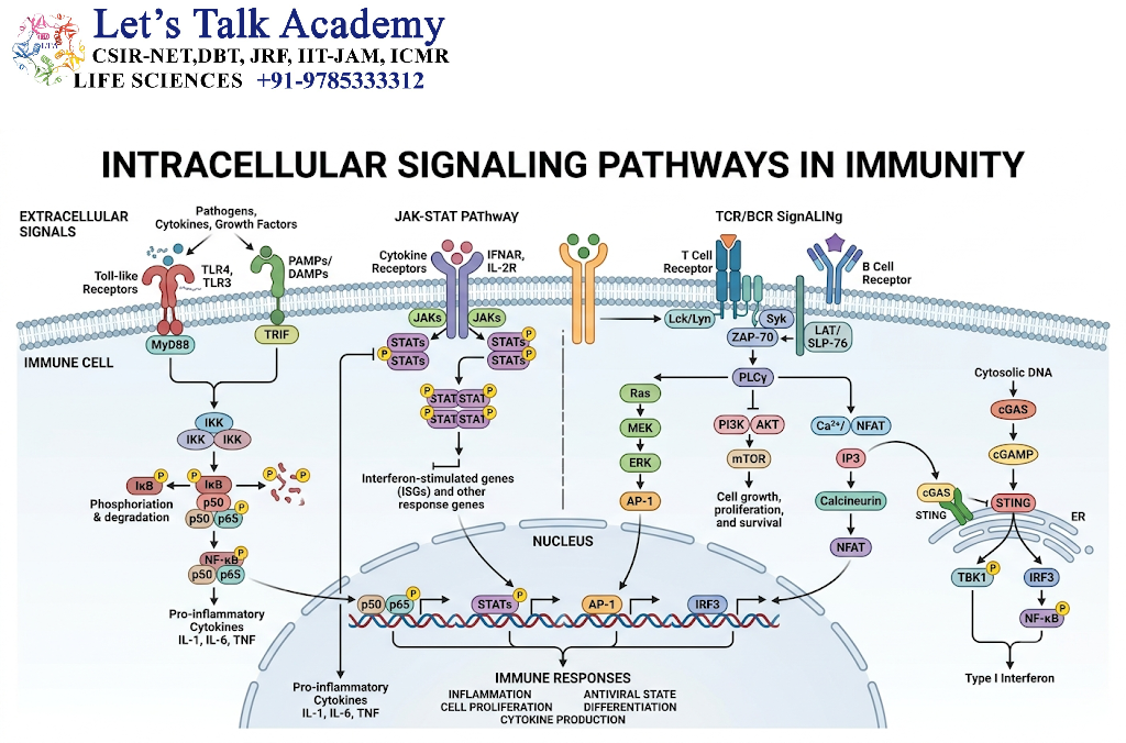
Immune cells communicate with one another through receptors, signaling molecules, and intracellular biochemical pathways. When a receptor recognizes a pathogen-associated molecule, antigen, cytokine, or other signal, it initiates a cascade of intracellular reactions.
These signaling pathways regulate gene expression, cellular metabolism, proliferation, migration, differentiation, and effector functions.
The major signaling pathways involved in immunity include NF-κB, MAPK, JAK–STAT, PI3K–AKT, calcium-dependent signaling, and inflammasome-associated pathways.
10.2 NF-κB Signaling Pathway
NF-κB is a family of transcription factors that regulates inflammatory responses, immune cell survival, and the expression of genes involved in host defense.
10.2.1 Activation mechanism
-
A pathogen-associated molecule binds to an appropriate receptor.
-
The receptor activates intracellular adaptor proteins.
-
A signaling complex activates the IκB kinase complex.
-
IκB proteins are phosphorylated and degraded.
-
NF-κB is released from its inhibitory interaction.
-
NF-κB enters the nucleus.
-
It regulates the transcription of target genes.
10.2.2 Functions
NF-κB signaling contributes to:
-
Production of inflammatory cytokines.
-
Expression of adhesion molecules.
-
Activation of antigen-presenting cells.
-
Cell survival.
-
Innate immune responses.
-
Regulation of lymphocyte activity.
Excessive NF-κB activation can contribute to chronic inflammation and inflammatory disease.
10.3 MAPK Signaling
Mitogen-activated protein kinase pathways transmit signals from cell surface receptors to intracellular targets and the nucleus.
Important MAPK pathways include:
-
ERK.
-
JNK.
-
p38 MAPK.
These pathways influence immune cell activation, cytokine production, proliferation, differentiation, and stress responses.
For example, activation of p38 and JNK can contribute to inflammatory gene expression following innate receptor stimulation.
10.4 JAK–STAT Signaling
The Janus kinase–signal transducer and activator of transcription pathway is important in cytokine-mediated communication.
10.4.1 Mechanism
-
A cytokine binds to its receptor.
-
Receptor-associated JAK proteins become activated.
-
STAT proteins are phosphorylated.
-
STAT proteins form dimers.
-
STAT dimers enter the nucleus.
-
They regulate the expression of target genes.
10.4.2 Examples of immune regulation
|
Signaling molecule |
Important signaling associations |
|---|---|
|
Type I interferons |
STAT1, STAT2, and IRF9-associated signaling |
|
Interferon-gamma |
STAT1 |
|
IL-4 |
STAT6 |
|
IL-6 |
STAT3-associated signaling |
|
IL-12 |
STAT4 |
|
IL-2 |
STAT5-associated signaling |
|
IL-21 |
STAT3 and other context-dependent pathways |
The same STAT protein may participate in different biological responses depending on the receptor, cell type, and signaling environment.
10.5 PI3K–AKT–mTOR Pathway
The PI3K–AKT–mTOR pathway regulates cellular growth, metabolism, survival, and protein synthesis.
In immune cells, it influences:
-
Lymphocyte activation.
-
Cell proliferation.
-
Metabolic reprogramming.
-
Cytokine production.
-
Differentiation.
-
Cellular survival.
The activity of this pathway must be carefully regulated because excessive or inappropriate activation can alter immune homeostasis.
10.6 Inflammasome Signaling
Inflammasomes are intracellular multiprotein complexes that detect specific cellular danger signals and activate inflammatory responses.
The NLRP3 inflammasome is one of the best-studied examples.
10.6.1 Major components
The NLRP3 inflammasome commonly includes:
-
NLRP3 sensor protein.
-
ASC adaptor protein.
-
Inflammatory caspase-1.
10.6.2 Activation and function
Following appropriate activation signals, NLRP3 interacts with ASC, which promotes the recruitment and activation of caspase-1.
Caspase-1 processes pro-IL-1β and pro-IL-18 into mature cytokines.
It can also cleave gasdermin D, whose resulting fragments can form membrane pores and promote a lytic inflammatory form of cell death called pyroptosis.
Inflammasomes help defend against certain infections but can contribute to inflammatory disease when inappropriately activated.
11. Antigen Processing and Presentation
11.1 Definition of Antigen Presentation
Antigen presentation is the process through which antigen-presenting cells display antigen-derived molecular fragments to T lymphocytes.
It is essential for the activation and regulation of adaptive immunity.
Antigens are processed into smaller fragments, commonly peptides, which are then bound to MHC molecules.
The peptide–MHC complex is recognized by the T-cell receptor.
11.2 Professional Antigen-Presenting Cells
The principal professional antigen-presenting cells are:
-
Dendritic cells.
-
Macrophages.
-
B lymphocytes.
Dendritic cells are especially important for the activation of naïve T cells.
Macrophages are particularly important in presenting antigens to activated T cells during tissue immune responses.
B cells can efficiently present antigens captured through their B-cell receptors.
11.3 Exogenous Antigen Processing
Exogenous antigens originate outside the cell.
Examples include bacterial proteins, soluble proteins, and extracellular microbial material.
The general pathway involves:
-
Uptake of antigen through endocytosis or phagocytosis.
-
Breakdown of antigenic proteins within endosomal or lysosomal compartments.
-
Loading of peptides onto MHC class II molecules.
-
Transport of peptide–MHC class II complexes to the cell surface.
-
Recognition by CD4-positive T cells.
11.4 Endogenous Antigen Processing
Endogenous antigens originate within the cell.
Examples include proteins produced by viruses infecting a cell or abnormal proteins produced by transformed cells.
The general pathway involves:
-
Degradation of intracellular proteins, often through the proteasome.
-
Transport of selected peptides into the endoplasmic reticulum or through alternative antigen-processing routes.
-
Loading of peptides onto MHC class I molecules.
-
Transport of peptide–MHC class I complexes to the cell surface.
-
Recognition by CD8-positive T cells.
11.5 Cross-Presentation
Cross-presentation is the process through which certain antigen-presenting cells, particularly specialized dendritic cell populations, present peptides derived from extracellular antigens on MHC class I molecules.
This process allows the activation of CD8-positive T-cell responses against targets that may not directly infect the antigen-presenting cell.
Cross-presentation is important in antiviral immunity, antitumour immunity, and some vaccine responses.
12. Immune Receptors and Accessory Molecules
12.1 Importance of Immune Receptors
Immune receptors enable cells to detect pathogens, antigens, cytokines, antibodies, and signals from other cells.
They are essential for communication between the immune system and its environment.
Receptors can be classified according to their ligands, cellular location, and signaling mechanisms.
12.2 Fc Receptors
Fc receptors bind to the constant Fc regions of antibodies.
They are expressed on various immune cells, including macrophages, neutrophils, NK cells, mast cells, and other leukocytes.
12.2.1 Major Fc receptor groups
|
Receptor |
Major ligand or function |
|---|---|
|
Fcγ receptors |
Bind IgG and regulate phagocytosis, activation, or inhibition |
|
FcεRI |
High-affinity receptor for IgE on mast cells and basophils |
|
Fcα receptors |
Recognize IgA through receptors such as FcαRI on appropriate cells |
|
FcRn |
Regulates IgG transport and protection from degradation |
12.2.2 Functions of Fc receptors
-
Antibody-dependent phagocytosis.
-
Antibody-dependent cellular cytotoxicity.
-
Activation or inhibition of immune cells.
-
Regulation of antibody transport.
-
Clearance of antibody-coated material.
12.3 Adhesion Molecules
Adhesion molecules facilitate interactions between immune cells and other cells or tissues.
They are essential for immune cell migration, tissue entry, and the formation of cellular contacts.
Important adhesion molecule families include:
-
Selectins.
-
Integrins.
-
Immunoglobulin superfamily adhesion molecules.
12.3.1 Selectins
Selectins participate in the initial attachment and rolling of leukocytes along vascular endothelial surfaces.
Examples include:
-
E-selectin.
-
P-selectin.
-
L-selectin.
12.3.2 Integrins
Integrins mediate firm adhesion and cellular interactions.
Examples include:
-
LFA-1.
-
Mac-1.
-
VLA-4.
Integrin activation is important for the migration of immune cells from blood into tissues.
12.4 CD Molecules
Cluster of differentiation, or CD, molecules are cell surface markers and functional proteins used to identify and characterize immune cell populations.
Examples include:
|
CD marker |
Major association |
|---|---|
|
CD3 |
T-cell receptor-associated signaling complex |
|
CD4 |
Co-receptor on helper T cells and other T-cell populations |
|
CD8 |
Co-receptor on cytotoxic T cells and other T-cell populations |
|
CD19 |
B-cell-associated surface molecule |
|
CD20 |
Mature B-cell-associated surface molecule |
|
CD14 |
Commonly associated with classical monocytes |
|
CD16 |
Fcγ receptor III; expressed on NK cells and selected myeloid cells |
|
CD25 |
IL-2 receptor alpha chain; expressed on activated cells and regulatory T cells |
|
CD28 |
T-cell co-stimulatory receptor |
|
CD40 |
Receptor involved in B-cell and antigen-presenting cell regulation |
|
CD80/CD86 |
Co-stimulatory ligands expressed by antigen-presenting cells |
|
CD95 |
Fas death receptor |
CD markers are useful for immunophenotyping, but their expression may vary according to cell activation, tissue location, and developmental state.
13. Immune Responses at Tissue Barriers
13.1 Skin Immunity
The skin is a major physical and immunological barrier.
Its defense mechanisms include:
-
Keratinized epithelial layers.
-
Tight cellular junctions.
-
Antimicrobial peptides.
-
Resident macrophages.
-
Dendritic cells.
-
Tissue-resident lymphocytes.
-
Local cytokine and chemokine networks.
Keratinocytes can produce antimicrobial molecules and inflammatory mediators in response to injury or infection.
13.2 Mucosal Immunity
Mucosal surfaces line organs such as the respiratory tract, digestive tract, and reproductive tract.
These surfaces are continuously exposed to environmental antigens and microorganisms.
Mucosal immunity includes:
-
Epithelial barrier mechanisms.
-
Mucus production.
-
Secretory IgA.
-
Antimicrobial peptides.
-
Macrophages and dendritic cells.
-
Innate lymphoid cells.
-
Tissue-resident lymphocytes.
13.3 Gut-Associated Immune Regulation
The intestine contains a large population of immune cells that interact with the gut microbiota.
The immune system must protect against harmful microorganisms while maintaining tolerance to many harmless dietary and microbial antigens.
Important components include:
-
Intestinal epithelial cells.
-
Paneth cells.
-
Dendritic cells.
-
Macrophages.
-
Regulatory T cells.
-
Th17-associated responses.
-
Secretory IgA.
-
Innate lymphoid cells.
Disruption of intestinal immune regulation may contribute to inflammatory and autoimmune disorders.
13.4 Respiratory Tract Immunity
The respiratory tract uses mucociliary clearance, epithelial barriers, antimicrobial molecules, alveolar macrophages, and adaptive immune responses to defend against inhaled particles and pathogens.
Alveolar macrophages remove particulate material and contribute to immune surveillance within the lungs.
Excessive inflammation in the respiratory tract can damage the alveolar and airway structures.
14. Immune Tolerance and Autoimmunity
14.1 Definition of Immune Tolerance
Immune tolerance is the ability of the immune system to avoid inappropriate responses against specific antigens, particularly self-antigens.
Tolerance is established through central and peripheral mechanisms.
14.2 Central Tolerance
Central tolerance develops during lymphocyte maturation.
14.2.1 T-cell tolerance
T cells undergo selection in the thymus.
-
Positive selection: Helps ensure that developing T cells can recognize self-MHC molecules.
-
Negative selection: Eliminates or functionally alters many T cells that recognize self-antigens too strongly.
Medullary thymic epithelial cells express tissue-restricted antigens under the influence of regulatory proteins such as AIRE.
This helps expose developing T cells to a wider range of self-antigens.
14.2.2 B-cell tolerance
Developing B cells undergo mechanisms that limit the emergence of strongly self-reactive cells.
These mechanisms can include receptor editing, deletion, and functional unresponsiveness.
14.3 Peripheral Tolerance
Peripheral tolerance controls potentially autoreactive lymphocytes that escape central selection.
Important mechanisms include:
-
Anergy.
-
Regulatory T-cell suppression.
-
Inhibitory receptor signaling.
-
Deletion of certain autoreactive cells.
-
Lack of appropriate co-stimulation.
14.4 Autoimmunity
Autoimmunity occurs when immune responses target the body’s own tissues.
Autoimmune disease can involve genetic susceptibility, environmental influences, defects in tolerance, and abnormal inflammatory signaling.
Examples of autoimmune disorders include:
-
Type 1 diabetes mellitus.
-
Rheumatoid arthritis.
-
Systemic lupus erythematosus.
-
Multiple sclerosis.
-
Myasthenia gravis.
The molecular and cellular mechanisms differ among autoimmune diseases.
15. Clinical Significance of Immune Cells and Molecules
15.1 Immunodeficiency
Immunodeficiency occurs when one or more components of the immune system fail to function adequately.
It may be inherited or acquired.
Examples of affected components include:
-
B cells and antibody production.
-
T cells.
-
Phagocytes.
-
Complement proteins.
-
Cytokine signaling pathways.
Immunodeficiency can increase susceptibility to particular types of infection.
15.2 Hypersensitivity Reactions
Hypersensitivity reactions are excessive or inappropriate immune responses that can cause tissue damage.
Common classifications include four broad types:
|
Type |
Main mechanism |
|---|---|
|
Type I |
IgE-associated immediate hypersensitivity |
|
Type II |
Antibody-mediated responses against cellular or extracellular targets |
|
Type III |
Immune complex-associated tissue injury |
|
Type IV |
T-cell-mediated delayed hypersensitivity |
These categories are useful for understanding immune mechanisms, although many clinical disorders involve overlapping pathways.
15.3 Vaccination and Immune Memory
Vaccination stimulates adaptive immune responses against selected antigens.
The objective is to establish protective immune memory without requiring the individual to experience the full disease caused by the pathogen.
Vaccines can promote:
-
Antibody production.
-
Memory B-cell formation.
-
Long-lived plasma cell development.
-
Memory T-cell responses.
-
Innate immune activation that supports adaptive responses.
The strength and duration of vaccine-induced protection depend on the vaccine platform, antigen, host factors, and the infectious agent.
15.4 Monoclonal Antibodies
Monoclonal antibodies are laboratory-produced antibodies derived from a single B-cell clone or engineered to have a defined binding specificity.
They are used in research, diagnosis, and treatment.
Their applications include:
-
Blocking inflammatory cytokines.
-
Targeting cancer-associated molecules.
-
Modulating immune checkpoints.
-
Preventing or treating selected infections.
-
Depleting specific immune cell populations.
-
Delivering therapeutic agents to particular targets.
15.5 Immune Checkpoint Inhibitors
Immune checkpoint inhibitors are therapeutic agents that block selected inhibitory immune pathways.
Examples include agents targeting CTLA-4 or the PD-1/PD-L1 pathway.
These treatments can enhance antitumour T-cell responses in selected cancers.
However, increased immune activity may also produce immune-related adverse effects affecting normal tissues.
15.6 Complement-Related Disorders
Defects in complement components can increase susceptibility to infection or contribute to immune dysregulation.
Examples include:
-
Deficiencies in terminal complement components, which can increase susceptibility to certain Neisseria infections.
-
Defects in early classical pathway components, which can be associated with immune complex-associated disease.
-
Abnormal complement regulation, which can contribute to conditions involving excessive complement activation.
16. Experimental Techniques Used to Study Immunity
16.1 Flow Cytometry
Flow cytometry is widely used to identify and quantify immune cell populations.
It measures cell-associated markers and other characteristics using fluorescently labelled antibodies or other probes.
Applications include:
-
Identification of T-cell and B-cell subsets.
-
Measurement of CD4-positive and CD8-positive T cells.
-
Detection of activation markers.
-
Analysis of cytokine production.
-
Measurement of apoptosis.
-
Assessment of intracellular signaling proteins.
16.2 Enzyme-Linked Immunosorbent Assay
The enzyme-linked immunosorbent assay, or ELISA, is used to detect and quantify antigens, antibodies, and soluble proteins.
It is commonly used to measure:
-
Cytokine concentrations.
-
Serum antibodies.
-
Hormones.
-
Microbial antigens.
-
Inflammatory markers.
The assay relies on antigen–antibody binding and an enzyme-linked detection system.
16.3 Western Blotting
Western blotting is used to detect specific proteins in biological samples.
It can be used to study:
-
Expression of immune receptors.
-
Signaling proteins.
-
Transcription factors.
-
Cytokines.
-
Protein phosphorylation.
16.4 Immunohistochemistry
Immunohistochemistry uses antibodies to detect specific proteins in tissue sections.
It helps identify the distribution of immune cells and molecules within tissues.
It is useful in the study of lymphoid organs, inflammatory diseases, and tumour immune microenvironments.
16.5 Enzyme-Linked Immunospot Assay
The enzyme-linked immunospot assay, or ELISpot, detects individual cells that secrete a particular protein.
It can be used to measure:
-
Antibody-secreting cells.
-
Cytokine-producing T cells.
-
Antigen-specific immune responses.
16.6 Polymerase Chain Reaction and Gene Expression Analysis
Polymerase chain reaction and related molecular techniques are used to analyze genes and gene expression.
Applications include:
-
Detection of immune-related gene expression.
-
Analysis of cytokine transcripts.
-
Identification of pathogen genetic material.
-
Study of antigen receptor gene rearrangements.
-
Measurement of changes in signaling pathways.
16.7 Single-Cell Sequencing
Single-cell RNA sequencing allows researchers to study gene expression at the level of individual immune cells.
It helps reveal:
-
Immune cell heterogeneity.
-
Rare cell populations.
-
Cellular activation states.
-
Differentiation trajectories.
-
Tumour immune microenvironments.
-
Responses to infection or therapy.
16.8 Immunofluorescence Microscopy
Immunofluorescence uses fluorescently labelled antibodies to visualize specific proteins within cells or tissues.
It can be used to investigate:
-
Protein localization.
-
Cellular interactions.
-
Immune cell distribution.
-
Antigen presentation.
-
Tissue inflammation.
17. Integrated Summary of Immune Cells and Molecules
17.1 Major Cellular and Molecular Components
The immune system depends on a coordinated interaction between cells and molecular mediators.
|
Category |
Important examples |
Main functions |
|---|---|---|
|
Phagocytes |
Neutrophils, macrophages, monocytes |
Engulfment and destruction of pathogens |
|
Antigen-presenting cells |
Dendritic cells, macrophages, B cells |
Antigen processing and T-cell activation |
|
Innate lymphocytes |
NK cells, ILCs |
Cytotoxicity and rapid cytokine production |
|
Granulocytes |
Neutrophils, eosinophils, basophils |
Antimicrobial and inflammatory responses |
|
Tissue-resident immune cells |
Mast cells, macrophages |
Barrier defense and tissue surveillance |
|
B lymphocytes |
Naïve B cells, memory B cells, plasma cells |
Antibody-mediated immunity |
|
T lymphocytes |
Helper, cytotoxic, regulatory, memory T cells |
Cellular immunity and immune regulation |
|
Pattern-recognition receptors |
TLRs, NLRs, RLRs, cGAS |
Detection of microbial and danger signals |
|
Complement proteins |
C3, C5, C5a, C3b, C5b–C9 |
Opsonization, inflammation, and membrane attack |
|
Antibodies |
IgG, IgA, IgM, IgE, IgD |
Specific antigen recognition and effector functions |
|
MHC molecules |
MHC class I and class II |
Presentation of antigens to T cells |
|
Cytokines |
TNF, IL-1, IL-6, IL-2, IL-12, IFN-γ |
Cellular communication and immune regulation |
|
Chemokines |
CXCL8, CCL2, CXCL10 |
Immune cell migration |
|
Co-stimulatory molecules |
CD28, CD80, CD86, CD40 |
Activation and regulation of lymphocytes |
|
Inhibitory molecules |
CTLA-4, PD-1, PD-L1 |
Regulation of immune activation |
|
Antimicrobial peptides |
Defensins, cathelicidins, lysozyme |
Direct antimicrobial defense |
17.2 Integrated Mechanism of an Immune Response
From Pathogen Recognition to Immune Memory
1. Pathogen entry or tissue injury
Microbial molecules and damage signals appear.
2. Innate immune recognition
PRRs, complement, epithelial cells and phagocytes respond.
3. Inflammation and antigen presentation
Cytokines recruit cells; dendritic cells process antigens.
4. Adaptive immune activation
B and T lymphocytes recognize antigens and undergo clonal expansion.
5. Effector responses
Antibodies, cytotoxic T cells and activated phagocytes eliminate targets.
6. Resolution and memory
Inflammation decreases, and memory cells or long-lived plasma cells may persist.


