
1. Introduction
Neurotransmission is the process by which neurons communicate with one another or with target cells such as muscle cells and glandular cells. It allows the nervous system to rapidly transmit information, coordinate body functions, generate behavior, control movement, and maintain physiological homeostasis.
Neurons communicate primarily through electrical signals within the cell and chemical or electrical signals between cells. When an electrical impulse called an action potential reaches the terminal of a neuron, it can trigger the release of neurotransmitters into a specialized junction called a synapse. These neurotransmitters bind to receptors on the target cell and produce a cellular response.
Neurotransmission is highly regulated. The nervous system must control when neurotransmitters are released, how strongly receptors respond, how long the signal persists, and how the signal is terminated. This regulation allows neural circuits to process information precisely and adapt to changing conditions.
2. Definition of Neurotransmission
Neurotransmission is the process through which a neuron transfers information to another neuron or to an effector cell through a synapse.
The basic sequence is:
Electrical signal → Presynaptic terminal → Neurotransmitter release → Synaptic cleft → Receptor activation → Postsynaptic response
Neurotransmission involves several coordinated events:
- Generation of an action potential
- Propagation of the action potential along the axon
- Arrival of the action potential at the presynaptic terminal
- Opening of voltage-gated calcium channels
- Calcium entry into the presynaptic terminal
- Fusion of synaptic vesicles with the plasma membrane
- Release of neurotransmitter
- Diffusion across the synaptic cleft
- Binding to postsynaptic receptors
- Generation of a postsynaptic response
- Termination or modulation of the signal
3. Organization of a Neuron
A typical neuron consists of three major structural regions:
3.1 Cell Body
The cell body, or soma, contains the nucleus and most of the cellular organelles. It maintains the metabolic activities of the neuron.
3.2 Dendrites
Dendrites receive signals from other neurons or sensory receptors. They contain numerous receptors and are major sites of synaptic input.
3.3 Axon
The axon carries electrical signals away from the cell body. Its terminal branches form axon terminals, where neurotransmitters are commonly released.
3.4 Presynaptic and Postsynaptic Regions
The neuron releasing the neurotransmitter is called the presynaptic neuron.
The cell receiving the signal is called the postsynaptic cell.
The small gap between them is called the synaptic cleft.
4. Synapse
A synapse is a specialized junction through which information is transmitted from one cell to another.
Synapses can broadly be divided into:
| Type | Main Feature | Signal Transmission |
|---|---|---|
| Chemical synapse | Uses neurotransmitters | Chemical |
| Electrical synapse | Uses gap junctions | Direct electrical current |
Chemical synapses are particularly important because they allow extensive regulation and modification of neuronal signaling.
5. Structure of a Chemical Synapse
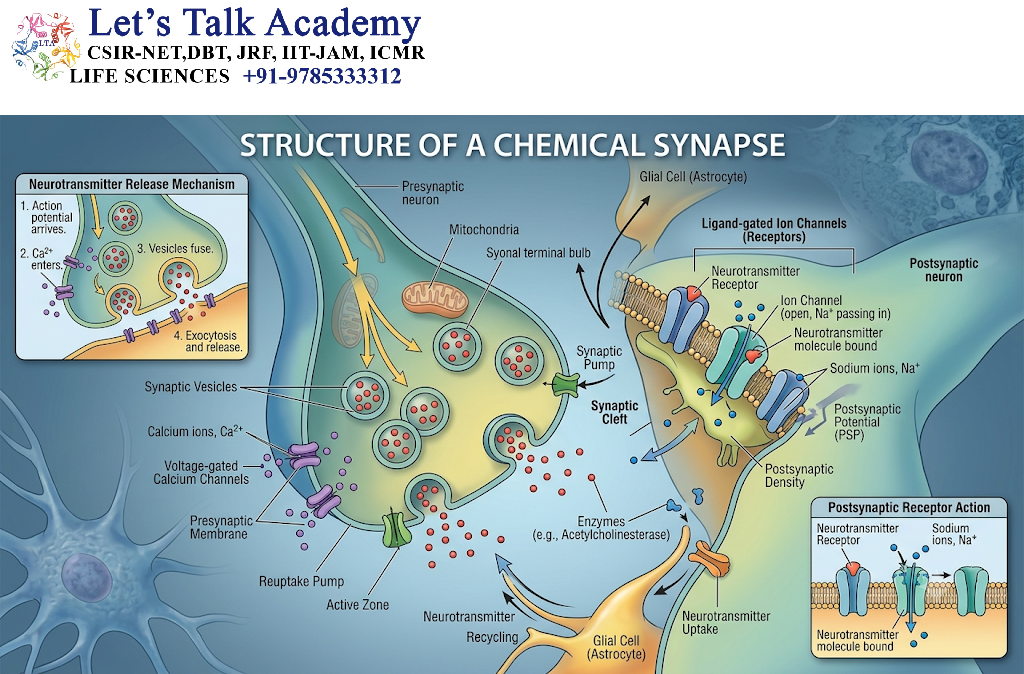
A typical chemical synapse contains three major components:
5.1 Presynaptic Terminal
The presynaptic terminal contains:
- Synaptic vesicles
- Voltage-gated calcium channels
- Mitochondria
- Neurotransmitter-synthesizing machinery
- Proteins involved in vesicle docking and fusion
5.2 Synaptic Cleft
The synaptic cleft is the narrow extracellular space between the presynaptic and postsynaptic membranes.
Neurotransmitters diffuse through this region after their release.
5.3 Postsynaptic Membrane
The postsynaptic membrane contains receptors and signaling proteins that detect the neurotransmitter.
6. Generation and Propagation of the Action Potential
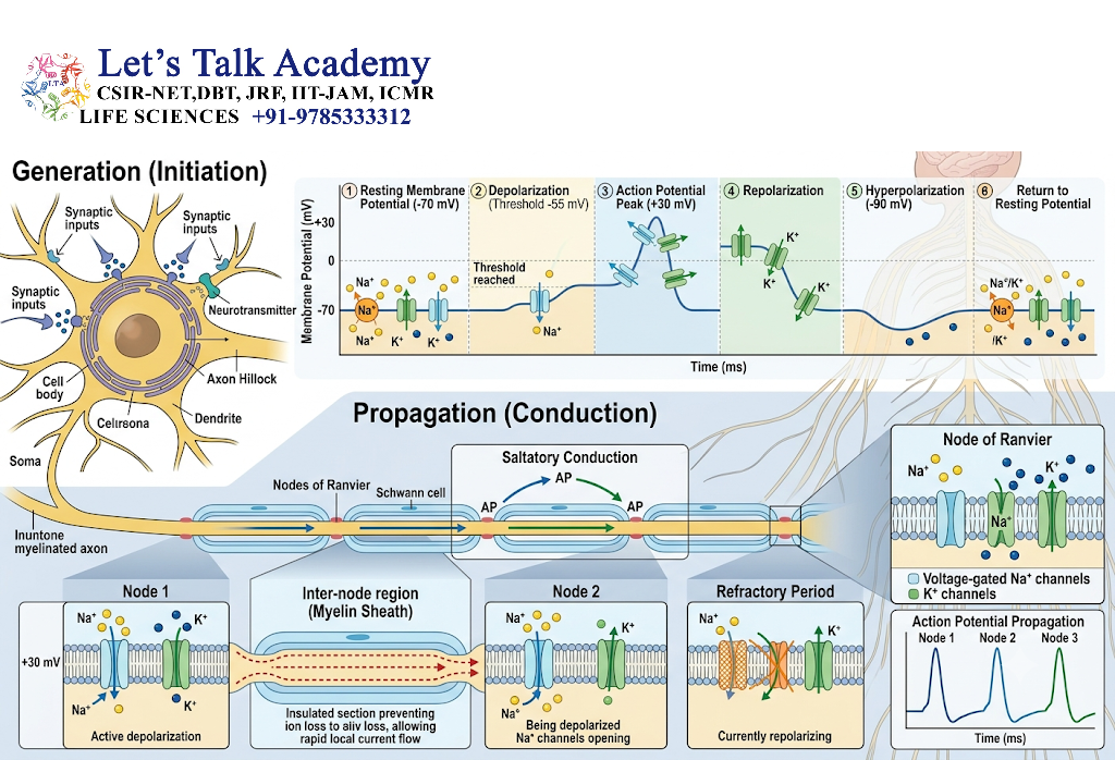
Neurotransmission generally begins with an action potential.
At rest, a neuron maintains a membrane potential, commonly around −70 mV, although the exact value varies among neurons.
When the membrane reaches threshold, voltage-gated sodium channels open.
6.1 Depolarization
Sodium ions enter the neuron, making the membrane potential more positive.
6.2 Repolarization
Sodium channels become inactive and voltage-gated potassium channels open. Potassium leaves the cell, causing the membrane potential to become negative again.
6.3 Hyperpolarization
Potassium channels may remain open briefly, causing the membrane potential to become more negative than the resting potential.
The action potential then propagates along the axon.
7. Arrival of the Action Potential at the Presynaptic Terminal
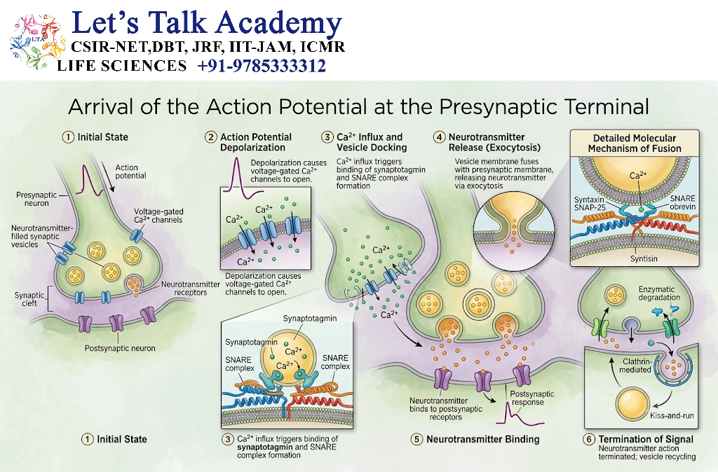
When the action potential reaches the axon terminal, it causes depolarization of the presynaptic membrane.
This activates voltage-gated calcium channels.
Calcium ions then enter the presynaptic terminal.
The increase in intracellular Ca²⁺ concentration is the key trigger for neurotransmitter release.
8. Calcium-Dependent Neurotransmitter Release
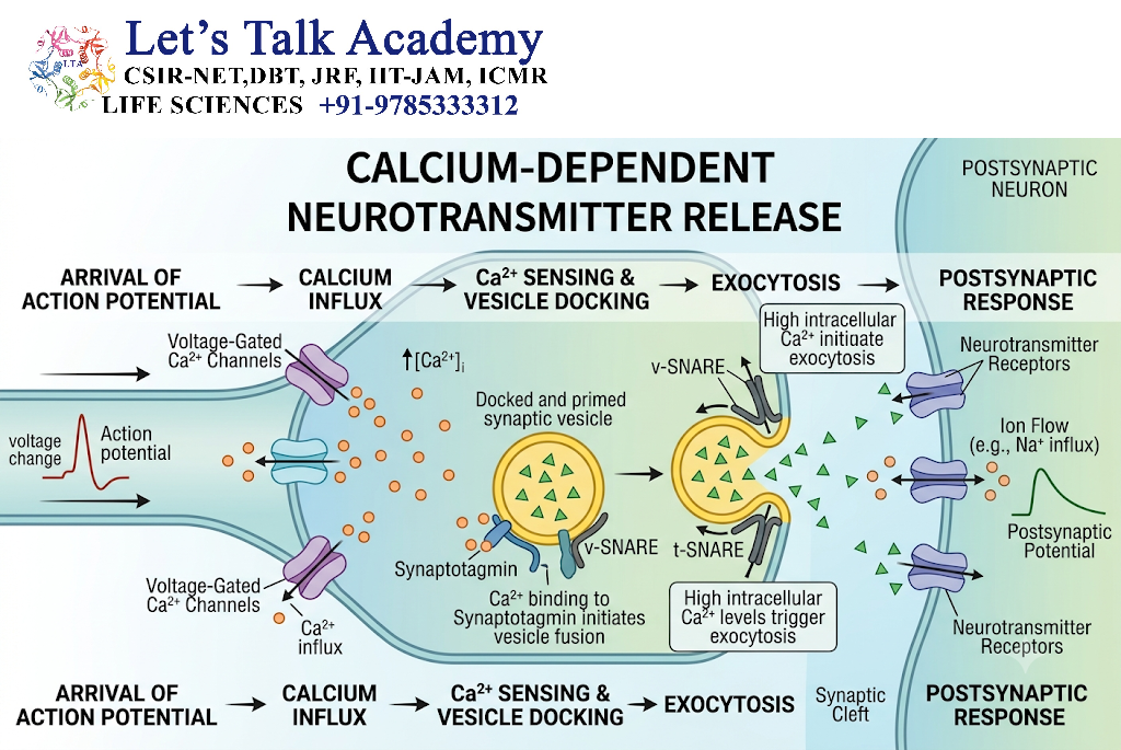
Neurotransmitters are commonly stored inside membrane-bound structures called synaptic vesicles.
When calcium enters the presynaptic terminal, it interacts with specialized proteins involved in vesicle fusion.
One important calcium sensor is synaptotagmin.
The vesicle-fusion machinery includes proteins such as:
- Synaptobrevin/VAMP
- Syntaxin
- SNAP-25
- Synaptotagmin
Together, these proteins facilitate fusion of the synaptic vesicle with the presynaptic membrane.
Mechanism
Action potential → Ca²⁺ channel opening → Ca²⁺ influx → Synaptotagmin activation → SNARE-mediated vesicle fusion → Neurotransmitter release
The neurotransmitter is released into the synaptic cleft by exocytosis.
9. Neurotransmitters
Neurotransmitters are chemical signaling molecules released by neurons to communicate with target cells.
Major neurotransmitters include:
| Neurotransmitter | Major Roles |
|---|---|
| Acetylcholine | Muscle activation, autonomic signaling, cognitive functions |
| Glutamate | Major excitatory neurotransmission |
| GABA | Major inhibitory neurotransmission in the brain |
| Glycine | Important inhibitory neurotransmission, especially in spinal cord |
| Dopamine | Movement, motivation, reward and endocrine regulation |
| Serotonin | Mood, sleep, appetite and sensory processing |
| Norepinephrine | Attention, arousal and autonomic regulation |
| Histamine | Wakefulness and physiological regulation |
The effect of a neurotransmitter depends strongly on the receptor it activates.
10. Classification of Neurotransmitters
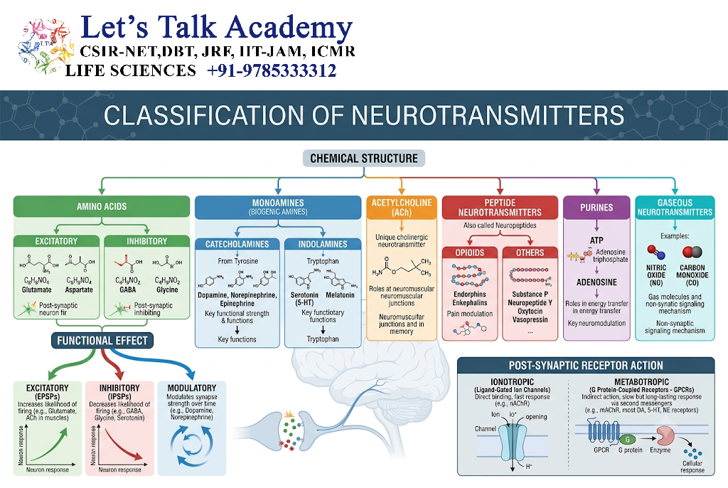
Neurotransmitters can be classified into several groups.
10.1 Amino Acid Neurotransmitters
Examples include:
- Glutamate
- GABA
- Glycine
- Aspartate
10.2 Acetylcholine
Acetylcholine is synthesized from choline and acetyl-CoA by the enzyme choline acetyltransferase.
10.3 Monoamines
Important monoamines include:
- Dopamine
- Norepinephrine
- Epinephrine
- Serotonin
- Histamine
10.4 Neuropeptides
Examples include:
- Substance P
- Endorphins
- Enkephalins
- Neuropeptide Y
10.5 Other Signaling Molecules
Neural signaling can also involve:
- Nitric oxide
- Endocannabinoids
- ATP
- Adenosine
11. Neurotransmitter Receptors
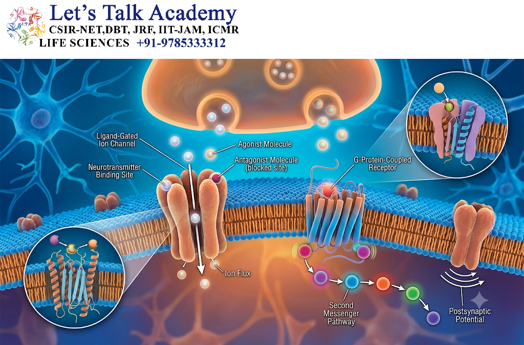
After release, neurotransmitters bind to receptors on the target cell.
Receptors can be broadly divided into:
- Ionotropic receptors
- Metabotropic receptors
12. Ionotropic Receptors
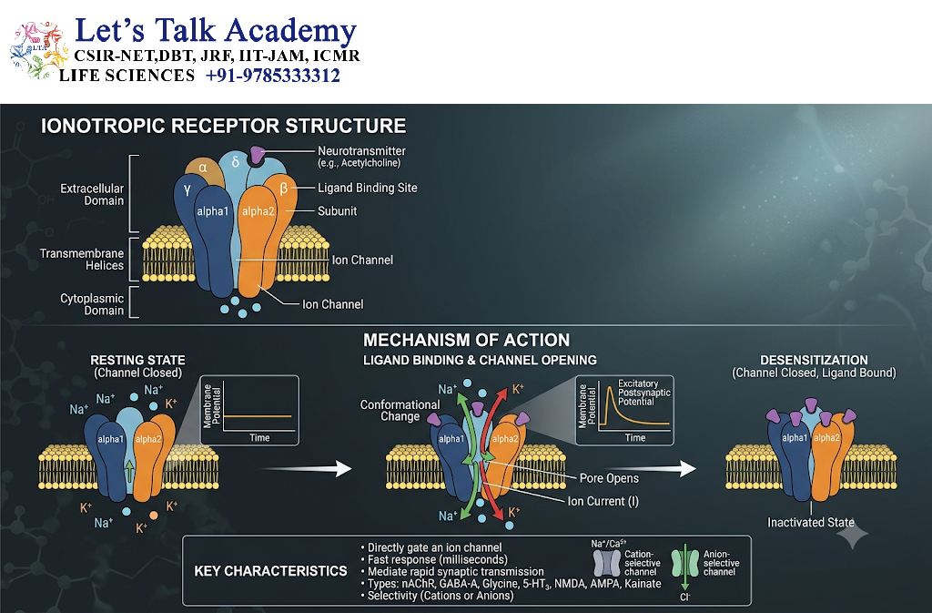
Ionotropic receptors are ligand-gated ion channels.
When a neurotransmitter binds to the receptor, the channel opens and allows specific ions to cross the membrane.
Depending on the receptor and ion involved, this can rapidly alter the membrane potential.
Example
Glutamate can activate AMPA receptors, allowing cations such as Na⁺ to enter the postsynaptic neuron.
This can produce rapid depolarization.
Ionotropic signaling is usually fast and is particularly important for rapid neuronal communication.
13. Metabotropic Receptors
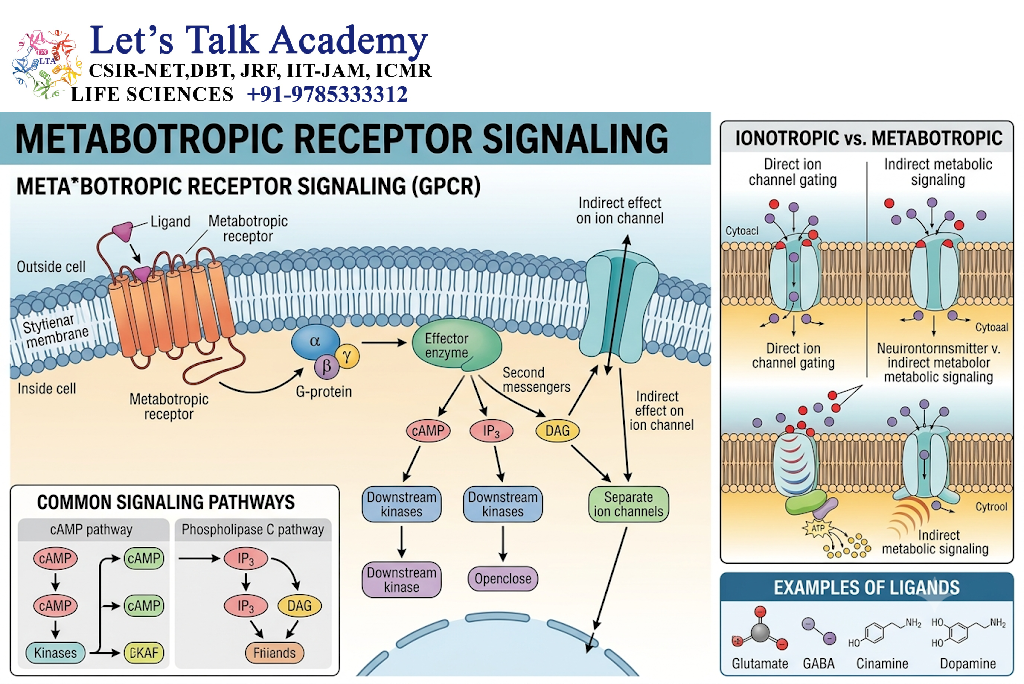
Metabotropic receptors do not directly form ion channels.
Many metabotropic neurotransmitter receptors are G-protein coupled receptors (GPCRs).
When a neurotransmitter binds:
Neurotransmitter → GPCR → G protein → Effector enzyme/channel → Second messenger → Cellular response
Examples include:
- Dopamine receptors
- Muscarinic acetylcholine receptors
- Many serotonin receptors
- Adrenergic receptors
Metabotropic signaling is generally slower than ionotropic signaling but can produce longer-lasting and more complex effects.
14. Excitatory and Inhibitory Neurotransmission
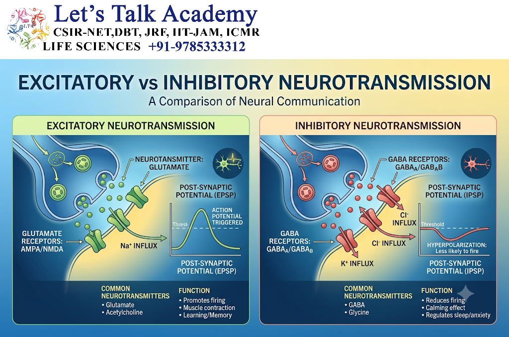
Neurotransmitters can produce excitatory or inhibitory effects depending on the receptor and ionic mechanisms involved.
14.1 Excitatory Postsynaptic Potential
An excitatory postsynaptic potential (EPSP) makes the postsynaptic membrane more likely to generate an action potential.
For example, opening channels that allow inward positive current can produce depolarization.
14.2 Inhibitory Postsynaptic Potential
An inhibitory postsynaptic potential (IPSP) makes the postsynaptic neuron less likely to generate an action potential.
This can occur through:
- Cl⁻ influx
- K⁺ efflux
- Reduction of excitatory currents
Important Principle
A neurotransmitter is not inherently “excitatory” or “inhibitory” in every situation.
Its effect depends on:
- Receptor type
- Ion permeability
- Membrane potential
- Intracellular signaling pathways
- Location of the receptor
15. Synaptic Integration
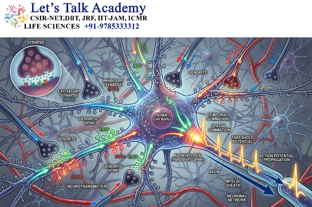
A neuron usually receives signals from many other neurons simultaneously.
The neuron integrates these inputs before determining whether to generate an action potential.
Two major forms of integration are:
15.1 Temporal Summation
Repeated signals arriving rapidly from the same synapse can add together.
15.2 Spatial Summation
Signals arriving simultaneously from different synapses can combine.
The balance between excitatory and inhibitory inputs determines the net effect on the postsynaptic neuron.
16. Termination of Neurotransmitter Signaling
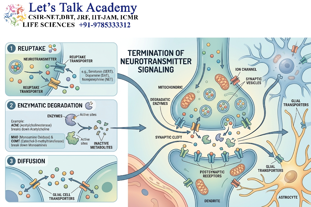
Neurotransmitter signaling must be terminated efficiently to prevent continuous stimulation.
Major mechanisms include:
- Reuptake
- Enzymatic degradation
- Diffusion
- Uptake by glial cells
17. Reuptake
Transport proteins can remove neurotransmitters from the synaptic cleft.
Examples include transporters for:
- Dopamine
- Serotonin
- Norepinephrine
- GABA
- Glutamate
Reuptake is an important mechanism for controlling the duration and intensity of neurotransmitter action.
18. Enzymatic Degradation
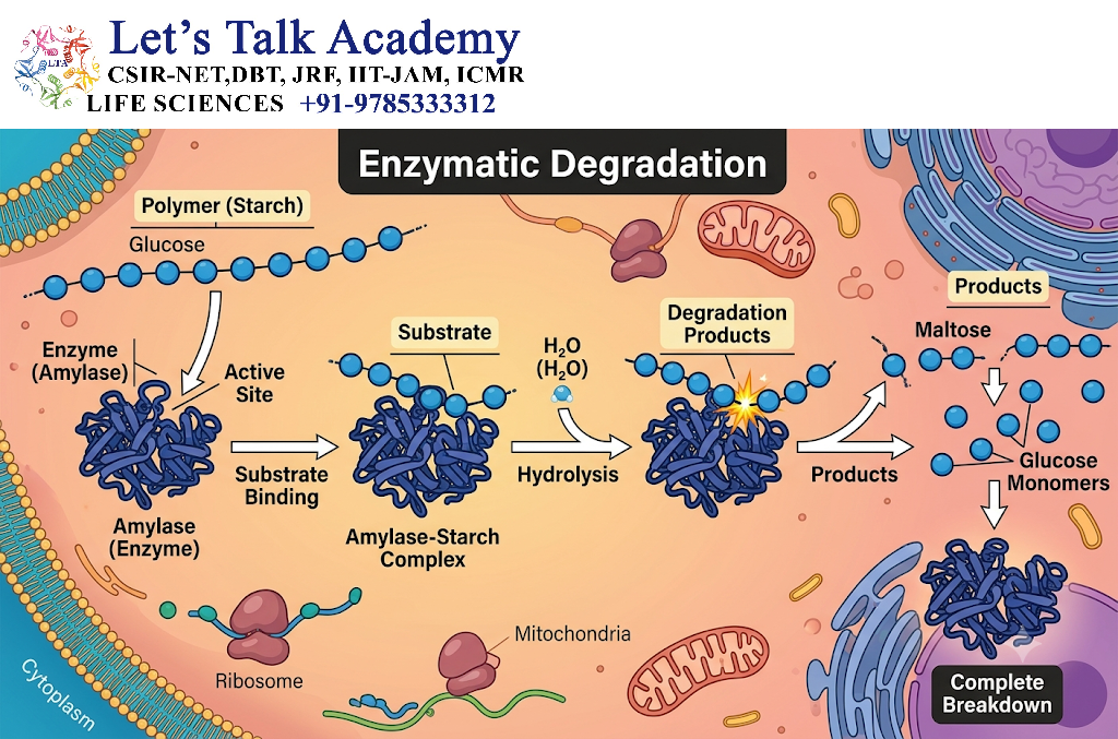
Some neurotransmitters are broken down by enzymes.
A major example is acetylcholine.
The enzyme acetylcholinesterase rapidly hydrolyzes acetylcholine in many cholinergic synapses.
This allows the signal to terminate efficiently.
19. Diffusion
Some neurotransmitter molecules can diffuse away from the synaptic cleft into surrounding extracellular space.
This reduces their concentration near the receptors.
20. Glial Uptake
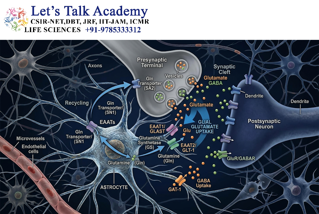
Glial cells, particularly astrocytes, participate in neurotransmitter clearance.
Glutamate is an important example.
Astrocytes remove extracellular glutamate and convert it into glutamine, which can subsequently participate in the neuronal glutamate-glutamine cycle.
21. Regulation of Neurotransmitter Synthesis
Neurotransmission can be regulated before neurotransmitter release by controlling neurotransmitter synthesis.
Factors include:
- Availability of precursors
- Activity of biosynthetic enzymes
- Enzyme phosphorylation
- Feedback regulation
- Cellular metabolic state
For example, the synthesis of catecholamines is influenced by the activity of enzymes involved in dopamine and norepinephrine production.
22. Regulation of Neurotransmitter Storage
Neurotransmitters must be transported into synaptic vesicles.
Specific vesicular transporters control this process.
Examples include:
- Vesicular monoamine transporter
- Vesicular GABA transporter
- Vesicular glutamate transporters
- Vesicular acetylcholine transporter
Changes in vesicular storage can alter the amount of neurotransmitter available for release.
23. Regulation of Neurotransmitter Release
Neurotransmitter release is tightly controlled by calcium-dependent mechanisms.
Important regulatory factors include:
- Number of available vesicles
- Calcium concentration
- Calcium-channel activity
- Vesicle docking
- SNARE proteins
- Presynaptic receptors
Even small changes in presynaptic calcium concentration can strongly influence neurotransmitter release.
24. Presynaptic Regulation
Presynaptic receptors can regulate neurotransmitter release.
These receptors may function as:
- Autoreceptors
- Heteroreceptors
24.1 Autoreceptors
Autoreceptors detect neurotransmitters released by the same neuron.
They commonly provide negative feedback.
For example:
Neurotransmitter release → Autoreceptor activation → Reduced further release
This prevents excessive neurotransmitter secretion.
24.2 Heteroreceptors
Heteroreceptors respond to neurotransmitters released by other neurons and can modify the release of the first neurotransmitter.
25. Postsynaptic Regulation
Postsynaptic regulation occurs through changes in receptor:
- Number
- Distribution
- Sensitivity
- Phosphorylation
- Internalization
- Recycling
Increasing receptor number can enhance responsiveness, whereas receptor removal can reduce responsiveness.
26. Receptor Desensitization
Continuous exposure to a neurotransmitter can reduce receptor responsiveness.
This process is called desensitization.
Mechanisms include:
- Receptor phosphorylation
- Interaction with regulatory proteins
- Receptor internalization
- Reduced receptor availability
Desensitization protects cells from excessive or prolonged stimulation.
27. Receptor Internalization and Recycling
Activated receptors may be removed from the plasma membrane through endocytosis.
The internalized receptors may then:
- Return to the plasma membrane
- Remain in intracellular compartments
- Undergo degradation
This provides an important mechanism for controlling synaptic sensitivity.
28. Second-Messenger Regulation
Metabotropic neurotransmitter receptors can regulate intracellular second messengers.
Important second messengers include:
- cAMP
- IP₃
- DAG
- Ca²⁺
- cGMP
For example:
Neurotransmitter → GPCR → G protein → Adenylyl cyclase → cAMP → PKA → Cellular response
These pathways can modify ion channels, enzymes, transcription factors and other proteins.
29. Regulation by Protein Phosphorylation
Protein kinases and phosphatases regulate many components of neurotransmission.
Kinases
Kinases add phosphate groups to proteins.
Phosphatases
Phosphatases remove phosphate groups.
This reversible modification can regulate:
- Receptor activity
- Ion channels
- Vesicle proteins
- Transcription factors
- Synaptic structural proteins
30. Short-Term Synaptic Plasticity
Synaptic strength can change over short periods.
Important forms include:
Facilitation
Repeated stimulation can increase neurotransmitter release because residual Ca²⁺ remains in the presynaptic terminal.
Depression
Repeated stimulation can reduce neurotransmitter release when readily releasable vesicles become temporarily depleted or through other presynaptic mechanisms.
Post-Tetanic Potentiation
Strong repetitive stimulation can produce a temporary increase in synaptic strength due to persistent presynaptic calcium-related changes.
31. Long-Term Synaptic Plasticity
Long-term changes in synaptic strength are important for learning, memory and neural adaptation.
Two major forms are:
- Long-term potentiation (LTP)
- Long-term depression (LTD)
32. Long-Term Potentiation
LTP is a persistent increase in synaptic strength following appropriate patterns of activity.
A well-studied mechanism involves NMDA receptors.
Glutamate activates NMDA receptors, but their channel is voltage-dependent because Mg²⁺ can block the channel at resting membrane potentials.
Strong postsynaptic depolarization removes this Mg²⁺ block.
Ca²⁺ then enters the postsynaptic neuron.
This activates signaling pathways that can increase AMPA receptor function or number.
Glutamate → NMDA receptor → Ca²⁺ influx → Kinase activation → Increased AMPA receptor function → Increased synaptic strength
33. Long-Term Depression
LTD is a long-lasting reduction in synaptic strength.
It can involve different patterns of calcium signaling and activation of phosphatases and other signaling pathways.
The balance between LTP and LTD allows neuronal circuits to modify their strength according to patterns of activity.
34. Regulation by Neurotrophic Factors
Neurotrophic factors regulate neuronal survival, development and synaptic function.
Important examples include:
- Nerve growth factor (NGF)
- Brain-derived neurotrophic factor (BDNF)
- Neurotrophin-3 (NT-3)
These molecules activate receptor signaling pathways that influence:
- Neuronal survival
- Axonal growth
- Dendritic development
- Synaptic plasticity
- Gene expression
35. Role of Glial Cells in Neurotransmission
Neurotransmission is not controlled only by neurons.
Glial cells, particularly astrocytes, contribute to the regulation of synaptic activity.
Astrocytes can:
- Remove neurotransmitters
- Regulate extracellular ions
- Supply metabolic substrates
- Influence synaptic signaling
- Participate in neurotransmitter recycling
This concept is sometimes described as part of the tripartite synapse, involving:
Presynaptic neuron + Postsynaptic neuron + Astrocyte
36. Neuromodulation
Neuromodulation refers to changes in neuronal activity produced by signaling systems that modify the responsiveness of neurons or synapses.
Neuromodulators can influence large neural networks rather than producing only a rapid point-to-point response.
Examples include:
- Dopamine
- Serotonin
- Norepinephrine
- Acetylcholine
- Neuropeptides
Neuromodulation can affect:
- Attention
- Motivation
- Sleep-wake states
- Learning
- Emotional processing
- Motor activity
37. Electrical Synapses
Electrical synapses use gap junctions to directly connect neighboring cells.
Small ions and electrical currents can pass through intercellular channels formed by connexin proteins.
Advantages include:
- Very rapid transmission
- Low synaptic delay
- Synchronization of neuronal activity
Chemical synapses generally provide greater flexibility and regulatory control.
38. Chemical vs Electrical Synapses
| Feature | Chemical Synapse | Electrical Synapse |
|---|---|---|
| Signal | Neurotransmitter-mediated | Direct ionic current |
| Junction | Synaptic cleft | Gap junction |
| Speed | Relatively slower | Very rapid |
| Directionality | Usually one-way | Often bidirectional |
| Modulation | Extensive | More limited |
| Plasticity | High | More limited |
| Main advantage | Flexible information processing | Rapid synchronization |
39. Regulation of Neurotransmission at Multiple Levels
Neurotransmission can be regulated at several stages.
| Level | Regulatory Mechanism |
|---|---|
| Neurotransmitter synthesis | Enzyme activity and precursor availability |
| Vesicle loading | Vesicular transporters |
| Vesicle docking | SNARE-associated machinery |
| Ca²⁺ entry | Voltage-gated calcium channels |
| Release | Calcium-dependent exocytosis |
| Receptor activation | Receptor affinity and number |
| Intracellular signaling | Kinases, phosphatases and second messengers |
| Signal termination | Reuptake, degradation and diffusion |
| Synaptic structure | Spine and adhesion molecule remodeling |
| Gene expression | Activity-dependent transcription |
40. Integrated Mechanism of Neurotransmission
Flowchart
Action potential generated
↓
Action potential travels along axon
↓
Depolarization of presynaptic terminal
↓
Voltage-gated Ca²⁺ channels open
↓
Ca²⁺ enters presynaptic terminal
↓
Synaptic vesicles dock and fuse
↓
Neurotransmitter released
↓
Neurotransmitter crosses synaptic cleft
↓
Receptor activation
↓
Ion channel opening or intracellular signaling
↓
EPSP/IPSP or other cellular response
↓
Signal termination by reuptake, degradation or diffusion
↓
Synaptic recovery and regulation
41. Factors Affecting Neurotransmission
Neurotransmission is influenced by many cellular and environmental factors, including:
- Ion concentrations
- Membrane potential
- Calcium availability
- Neurotransmitter synthesis
- Vesicle number
- Receptor density
- Receptor sensitivity
- Reuptake activity
- Enzymatic degradation
- Neuromodulators
- Synaptic plasticity
- Cellular energy availability
42. Neurotransmission and Gene Expression
Neuronal signaling can ultimately influence gene expression.
For example:
Receptor activation → Second messenger → Protein kinase → Transcription factor → Gene expression
One important transcription factor is CREB.
Activation of pathways involving cAMP and Ca²⁺ can activate CREB and influence the expression of genes involved in neuronal adaptation and long-term synaptic changes.
43. Neurotransmission and Learning
Learning involves changes in neural circuits.
Activity-dependent modifications of synaptic strength allow neurons to alter their responses to repeated patterns of stimulation.
Processes involved include:
- LTP
- LTD
- Receptor trafficking
- Dendritic spine remodeling
- Gene expression
- Protein synthesis
- Neurotrophic signaling
Thus, neurotransmission provides the basic signaling framework upon which many forms of neural plasticity are built.
44. Neurotransmission and Disease
Abnormal neurotransmission can contribute to neurological and neuropsychiatric disorders.
Examples include:
| Condition | Associated Neurotransmission Changes |
|---|---|
| Parkinsonian disorders | Altered dopaminergic signaling |
| Epilepsy | Imbalance between excitatory and inhibitory signaling |
| Myasthenia gravis | Autoimmune disruption of neuromuscular acetylcholine receptors |
| Alzheimer’s disease | Alterations in several neurotransmitter systems and synaptic function |
| Some mood disorders | Changes involving monoaminergic and other signaling systems |
| Neuromuscular disorders | Abnormal communication at neuromuscular junctions |
These associations are complex, and individual disorders usually involve multiple molecular and cellular mechanisms.


