
1. Introduction
Cells in multicellular organisms are surrounded by a complex network of proteins, carbohydrates and other macromolecules known as the extracellular matrix (ECM).
The extracellular matrix is present outside cells and provides a structural and biochemical environment in which cells live, communicate and perform their functions.
The ECM is not simply an inert framework that holds cells together. It is a dynamic and biologically active system that regulates:
- Cell adhesion
- Cell shape
- Cell migration
- Cell proliferation
- Cell differentiation
- Cell survival
- Tissue organization
- Mechanical properties
- Signal transduction
- Tissue repair
Different tissues contain different types and proportions of ECM components. For example, bone contains a highly mineralized matrix, cartilage contains abundant proteoglycans, and tendons contain large amounts of organized collagen fibers.
Thus, the ECM is an essential component of tissue structure and cellular regulation.
2. Definition of Extracellular Matrix
The extracellular matrix (ECM) is a complex network of macromolecules secreted by cells and present outside the plasma membrane, where it provides structural support and regulates cellular behavior.
The ECM is composed mainly of:
- Fibrous proteins
- Proteoglycans
- Glycosaminoglycans
- Adhesive glycoproteins
- Specialized matrix-associated molecules
- Water and ions
The composition of the ECM varies according to tissue type and physiological condition.
3. General Organization of the Extracellular Matrix
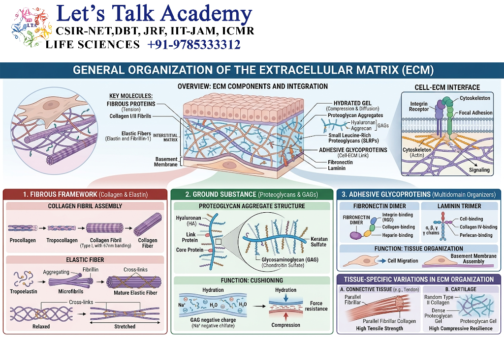
The ECM can be broadly organized into:
3.1 Interstitial Matrix
The interstitial matrix surrounds cells within tissues.
It contains components such as:
- Collagen
- Elastin
- Fibronectin
- Proteoglycans
- Hyaluronan
It provides structural support and creates the extracellular environment surrounding cells.
3.2 Basement Membrane
The basement membrane is a specialized, thin ECM layer located beneath epithelial cells and around certain other cell types.
It contains important components such as:
- Type IV collagen
- Laminins
- Nidogens
- Heparan sulfate proteoglycans
The basement membrane provides structural support and regulates cell attachment, polarity, filtration and signaling.
4. Major Components of the Extracellular Matrix
The major components of ECM can be grouped into:
| Component | Major function |
|---|---|
| Collagen | Tensile strength |
| Elastin | Elasticity |
| Fibronectin | Cell adhesion and organization |
| Laminin | Basement membrane organization |
| Proteoglycans | Hydration and signaling |
| Glycosaminoglycans | Water retention and matrix organization |
| Hyaluronan | Hydration, migration and tissue organization |
These components interact with each other to form tissue-specific extracellular networks.
5. Collagen
Collagen is the major structural protein of the extracellular matrix.
It is particularly important for providing tensile strength.
Collagen is abundant in:
- Skin
- Tendons
- Ligaments
- Bone
- Cartilage
- Blood vessels
- Basement membranes
5.1 Structure of Collagen
Collagen molecules commonly contain three polypeptide chains arranged into a characteristic triple helix.
The chains contain abundant glycine and proline-related residues.
A simplified organization is:
Collagen chains
↓
Triple helix
↓
Collagen molecules
↓
Fibrils
↓
Fibers
↓
Tissue-strengthening network
6. Major Types of Collagen
More than 20 collagen types have been identified.
Some important examples include:
| Collagen type | Major location/function |
|---|---|
| Type I | Skin, tendon, bone, ligaments |
| Type II | Cartilage |
| Type III | Reticular fibers and tissues associated with type I collagen |
| Type IV | Basement membrane |
| Type V | Associated with type I collagen and tissue organization |
6.1 Type I Collagen
Type I collagen is a major fibrillar collagen and provides strong tensile support.
It is abundant in:
- Bone
- Skin
- Tendons
- Ligaments
6.2 Type II Collagen
Type II collagen is particularly important in cartilage.
It contributes to the structural framework of cartilage ECM.
6.3 Type III Collagen
Type III collagen forms reticular fibers and is commonly associated with tissues containing type I collagen.
6.4 Type IV Collagen
Type IV collagen forms a network rather than typical thick fibrils.
It is an important component of the basement membrane.
7. Elastin
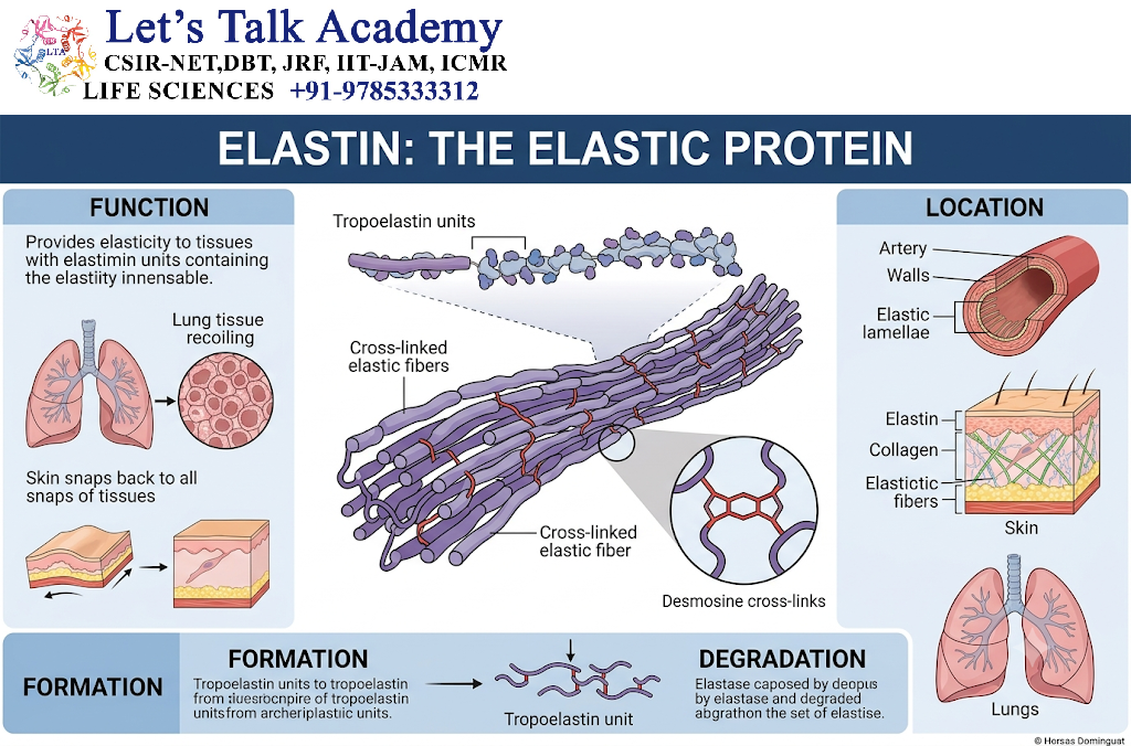
Elastin is an extracellular matrix protein that provides elasticity to tissues.
It allows tissues to stretch and return toward their original shape.
Elastin is especially important in:
- Large arteries
- Lungs
- Skin
- Elastic ligaments
7.1 Structure and Organization
Elastin is deposited as elastic fibers together with associated microfibrillar proteins.
The general organization is:
Elastin + microfibrillar components
↓
Elastic fibers
↓
Stretchable tissue
The elastic properties of ECM are particularly important in organs that undergo repeated deformation.
8. Fibronectin
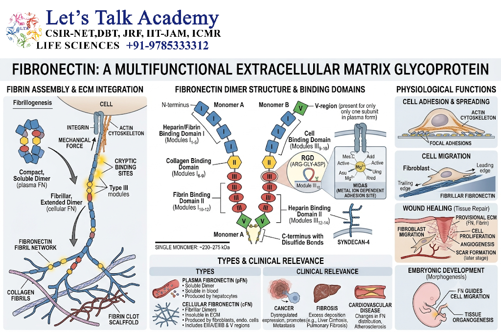
Fibronectin is a large adhesive glycoprotein that participates in cell-ECM interactions and matrix organization.
It can bind to:
- Integrins
- Collagen
- Heparan sulfate
- Other ECM components
Fibronectin is particularly important during:
- Cell adhesion
- Cell migration
- Wound healing
- Embryonic development
- ECM assembly
9. Fibronectin and Integrins
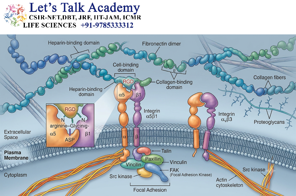
Fibronectin interacts with specific integrins on the cell surface.
A simplified pathway is:
Fibronectin
↓
Integrin
↓
Focal adhesion proteins
↓
Actin cytoskeleton
↓
Cell adhesion + signaling
This interaction connects the extracellular environment to intracellular structures.
10.Laminin
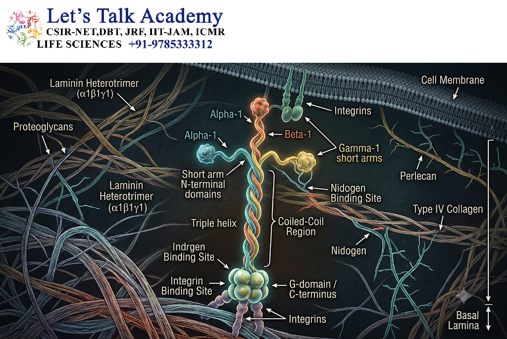
Laminins are large extracellular glycoproteins that are major components of basement membranes.
They interact with:
- Integrins
- Dystroglycan
- Nidogens
- Other basement membrane components
Laminins contribute to:
- Cell attachment
- Basement membrane assembly
- Cell polarity
- Tissue organization
- Development
11. Proteoglycans
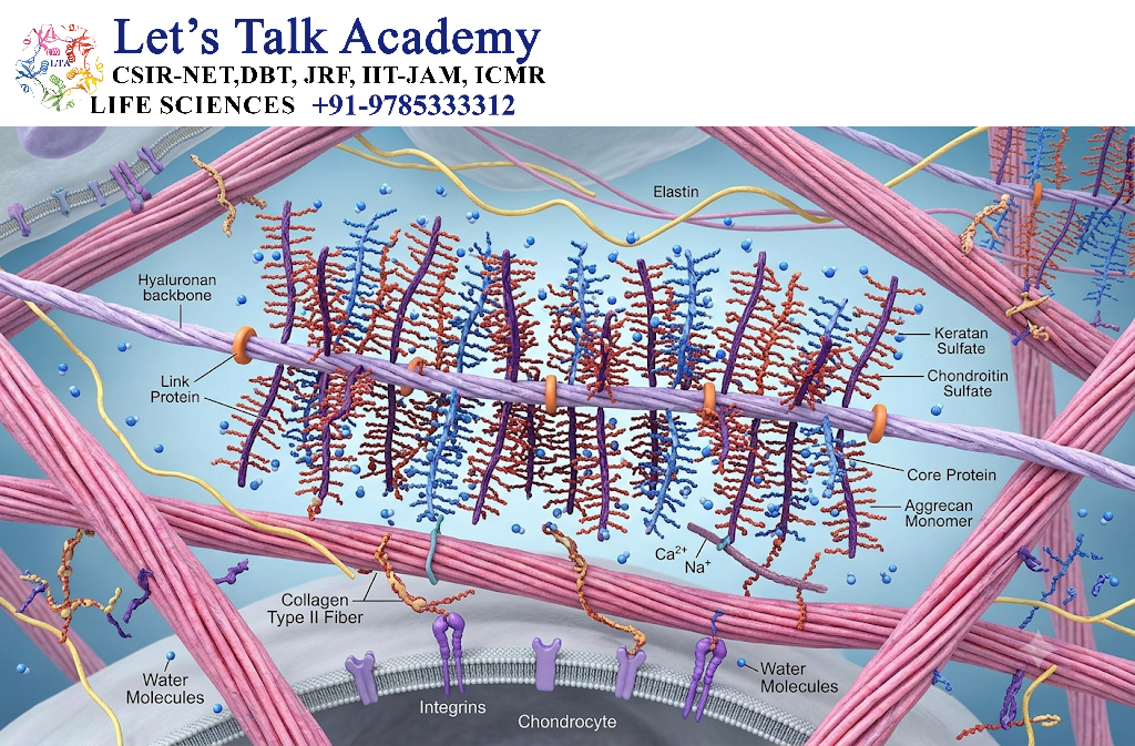
Proteoglycans are extracellular macromolecules consisting of a core protein associated with one or more glycosaminoglycan (GAG) chains.
They are important for:
- Water retention
- Matrix organization
- Resistance to compression
- Growth-factor storage
- Cell signaling
- Regulation of molecular diffusion
A simplified structure is:
Core protein
↓
GAG chains
↓
Proteoglycan
12. Glycosaminoglycans
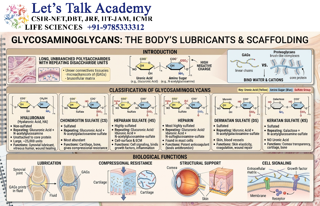
Glycosaminoglycans (GAGs) are long, unbranched polysaccharides composed of repeating disaccharide units.
Important GAGs include:
- Hyaluronan
- Chondroitin sulfate
- Dermatan sulfate
- Keratan sulfate
- Heparan sulfate
Most GAGs are negatively charged, which allows them to attract water and cations.
13. Hyaluronan
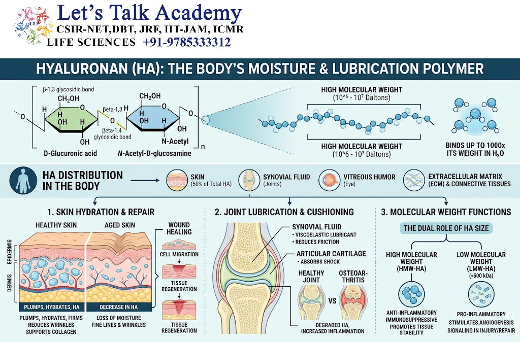
Hyaluronan is a large, non-sulfated glycosaminoglycan that contributes strongly to ECM hydration and organization.
It can form a hydrated extracellular environment that supports:
- Cell migration
- Tissue expansion
- Cell proliferation
- Wound repair
- Morphogenesis
Hyaluronan also interacts with cell-surface receptors such as CD44 and can influence signaling.
14. Heparan Sulfate Proteoglycans
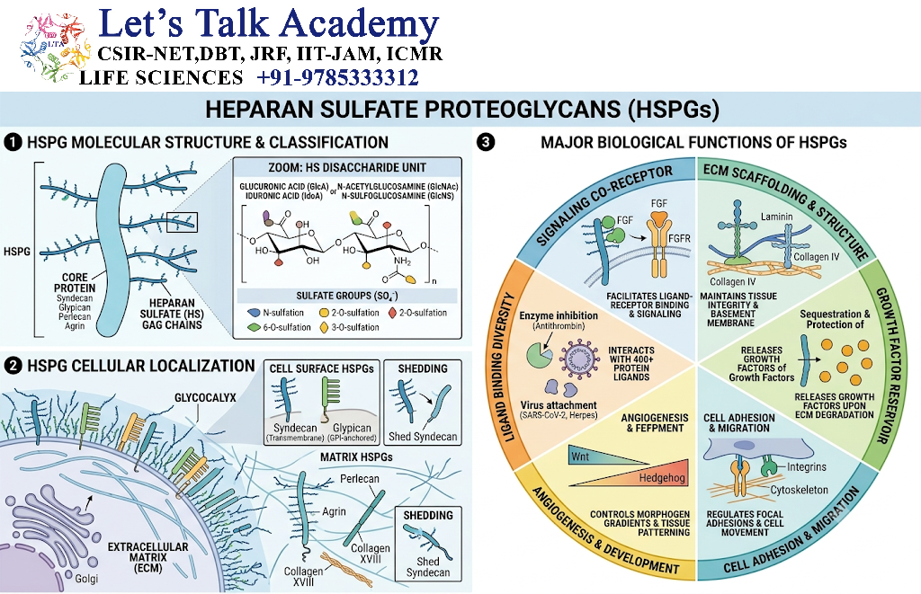
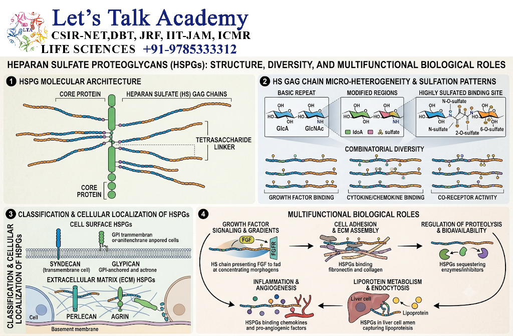
Heparan sulfate proteoglycans contain heparan sulfate chains that can interact with many extracellular signaling molecules.
They can influence:
- Growth-factor availability
- Cell adhesion
- Cell migration
- Development
- Signaling gradients
They are important components of both interstitial and basement-membrane ECM.
15.Heparan Sulfate Proteoglycans
The basement membrane is a specialized ECM layer associated with epithelial and other cells.
Major components include:
- Type IV collagen
- Laminin
- Nidogen
- Perlecan and other proteoglycans
The basement membrane provides:
- Structural support
- Cell anchorage
- Tissue organization
- Filtration
- Signaling
- Polarity cues
16. Structure of Basement Membrane
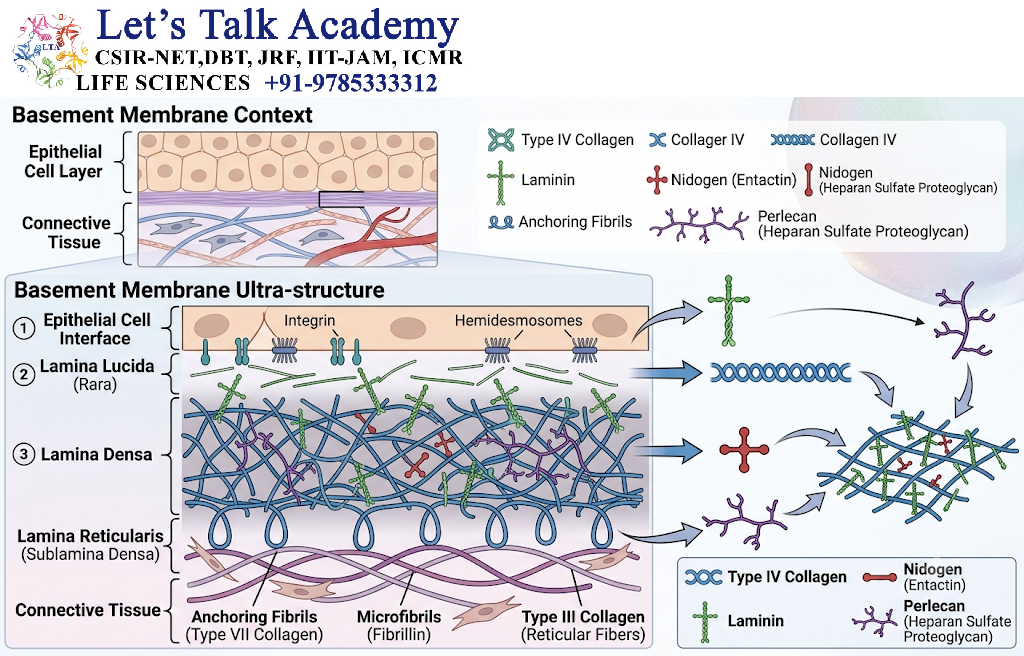
A simplified organization is:
Cell
↓
Cell adhesion receptors
↓
Laminin-rich network
↓
Nidogen and proteoglycan interactions
↓
Type IV collagen network
↓
Underlying connective tissue ECM
The exact organization varies among tissues.
17. Cell-ECM Interaction
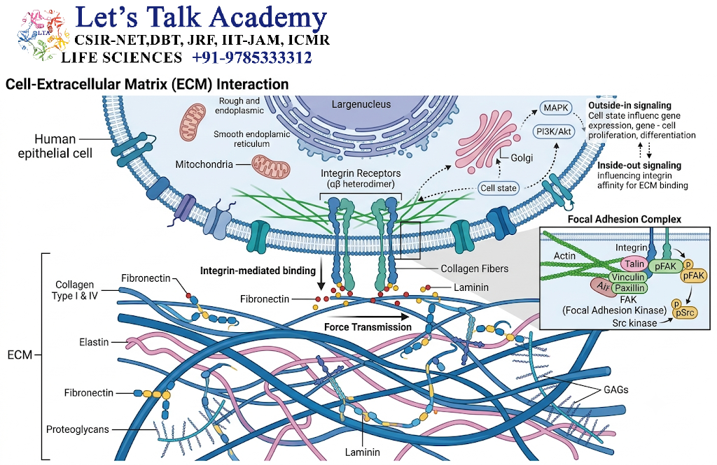
Cells interact with the ECM through specialized receptors.
The major receptors are integrins.
Other cell-surface molecules can also recognize ECM components.
A general interaction is:
ECM ligand
↓
Cell-surface receptor
↓
Adaptor proteins
↓
Cytoskeleton
↓
Signal transduction
↓
Cellular response
Thus, the ECM can influence intracellular behavior.
18. Integrins as ECM Receptors
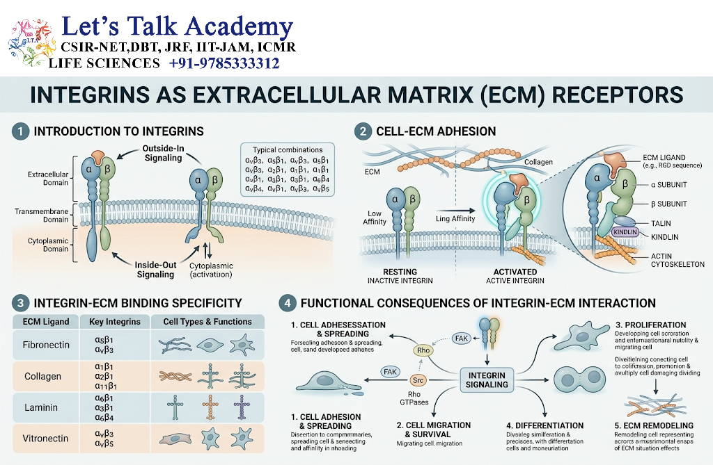
Integrins are heterodimeric receptors consisting of:
- α subunit
- β subunit
Different αβ combinations recognize different ECM ligands.
For example, certain integrins recognize:
- Fibronectin
- Laminin
- Collagen
- Vitronectin
Integrins provide a physical connection between ECM and the cytoskeleton.
19. Focal Adhesions
When cells attach to ECM through integrins, they can form focal adhesions.
Important proteins associated with focal adhesions include:
- Integrins
- Talin
- Kindlin
- Vinculin
- Paxillin
- Focal adhesion kinase (FAK)
- Src-family kinases
A simplified arrangement is:
ECM
↓
Integrin
↓
Talin / kindlin
↓
Vinculin and other adaptor proteins
↓
Actin cytoskeleton
These structures function in both adhesion and signal transduction.
20. ECM and Cytoskeleton
The ECM is mechanically connected to the intracellular cytoskeleton.
This connection can involve:
ECM → Integrins → Focal adhesion proteins → Actin
or, in certain adhesion complexes:
ECM → Integrins → Intermediate filament-associated proteins
This allows cells to sense and respond to mechanical forces.
21. ECM as a Signaling Environment
The ECM can regulate cell signaling in several ways.
It can:
- Bind growth factors
- Present signaling molecules to receptors
- Control ligand availability
- Influence receptor activation
- Regulate cell adhesion
- Affect cytoskeletal organization
Therefore, the ECM acts as an important signaling environment rather than merely a structural scaffold.
22. ECM and Growth Factors
Some growth factors interact with ECM components, particularly proteoglycans.
The ECM can:
- Store growth factors
- Protect them from degradation
- Control their diffusion
- Present them near cell-surface receptors
- Help establish signaling gradients
This mechanism is important during development and tissue repair.
23. ECM and Cell Shape
The composition and mechanical properties of the ECM influence cell shape.
For example:
ECM stiffness
↓
Integrin engagement
↓
Cytoskeletal organization
↓
Cell spreading and shape
↓
Changes in cellular behavior
Therefore, cells continuously respond to their physical environment.
24. ECM and Mechanotransduction
Mechanotransduction is the process by which cells convert mechanical forces into biochemical signals.
ECM stiffness, tension and physical organization can influence cell behavior.
A simplified pathway is:
Mechanical force
↓
ECM deformation
↓
Integrin/focal adhesion response
↓
Cytoskeletal tension
↓
Signaling pathways
↓
Gene expression
↓
Cellular adaptation
Important signaling pathways can include:
- FAK/Src
- Rho-family GTPases
- MAPK
- PI3K-AKT
- YAP/TAZ-associated mechanosensitive signaling
25. ECM and Cell Migration
The ECM provides both physical tracks and chemical signals for cell movement.
During migration:
- Cells form new adhesions.
- Actin cytoskeleton reorganizes.
- Cells generate traction against ECM.
- The cell body moves forward.
- Rear adhesions are released.
- New adhesion sites form.
Fibronectin, laminin, collagen and hyaluronan can all influence migration depending on the tissue and cellular context.
26. ECM and Cell Proliferation
ECM interactions can influence cell-cycle activity.
Integrin signaling can activate pathways that regulate:
- Cyclins
- Kinases
- Growth-factor responses
- Cell survival
Therefore, ECM composition and mechanical properties can influence whether cells proliferate.
27. ECM and Cell Differentiation
The ECM can influence the differentiation state of cells.
Cell differentiation is affected by:
- Matrix composition
- Matrix stiffness
- Cell-ECM adhesion
- Growth-factor availability
- Mechanical signaling
- Cytoskeletal organization
Thus, the same cell type may behave differently when exposed to different ECM environments.
28. ECM and Cell Survival
Appropriate ECM interactions can provide survival signals.
Integrin-mediated signaling can activate pathways such as:
Integrin
↓
FAK/Src
↓
PI3K-AKT
↓
Pro-survival signaling
Loss of proper ECM attachment can lead to anoikis in many normal cells.
29. ECM Remodeling
The ECM is continuously modified.
This process is called ECM remodeling.
It includes:
- Synthesis of new matrix
- Cross-linking
- Degradation
- Reorganization
- Modification of ECM proteins
- Changes in matrix stiffness
ECM remodeling is important in:
- Development
- Growth
- Wound healing
- Tissue repair
- Inflammation
30. Matrix Metalloproteinases
Matrix metalloproteinases (MMPs) are enzymes that degrade various ECM components.
They can act on:
- Collagens
- Gelatin
- Proteoglycans
- Other ECM proteins
MMP activity must be tightly regulated because excessive ECM degradation can damage tissue, whereas controlled degradation is important for normal remodeling.
31. Tissue Inhibitors of Metalloproteinases
TIMPs (tissue inhibitors of metalloproteinases) regulate MMP activity.
Therefore:
MMPs → ECM degradation
TIMPs → Inhibition of MMP activity
The balance between matrix synthesis and degradation helps maintain tissue homeostasis.
32. ECM in Wound Healing
ECM remodeling is essential during wound repair.
A simplified sequence is:
Tissue injury
↓
Inflammatory response
↓
ECM degradation and remodeling
↓
Cell migration
↓
New ECM deposition
↓
Tissue organization
↓
Remodeling and maturation
Fibronectin is particularly important during early stages of tissue repair.
Collagen deposition contributes to strengthening of the repaired tissue.
33. ECM in Development
During embryonic development, ECM controls:
- Cell migration
- Cell differentiation
- Tissue boundaries
- Organ formation
- Morphogenesis
- Growth-factor distribution
Changes in ECM composition can provide positional and mechanical information to developing cells.
34. ECM in Bone
Bone contains a specialized mineralized ECM.
Major organic components include:
- Type I collagen
- Non-collagenous proteins
The organic matrix becomes associated with mineral, primarily calcium-phosphate-containing mineral phases.
This combination provides:
Collagen → Tensile properties
Mineral → Compressive and hardness-related properties
Together, these properties make bone strong yet capable of withstanding mechanical forces.
35. ECM in Cartilage
Cartilage contains abundant:
- Type II collagen
- Proteoglycans
- Hyaluronan
- Water
Proteoglycans attract water and contribute to resistance against compression.
Thus:
Type II collagen → Structural framework
Proteoglycans + water → Resistance to compression
36. ECM in Tendons and Ligaments
Tendons and ligaments contain large amounts of type I collagen arranged in organized bundles.
This organization provides high tensile strength.
Tendons
Connect:
Muscle → Bone
Ligaments
Connect:
Bone → Bone
The ECM architecture allows these tissues to transmit and resist mechanical forces.
37. ECM in Skin
Skin ECM contains:
- Type I collagen
- Type III collagen
- Elastin
- Proteoglycans
- Glycoproteins
Collagen provides strength, while elastin contributes to elasticity.
Changes in ECM organization occur during aging, wound healing and various skin disorders.
38. ECM in Blood Vessels
Blood-vessel walls contain:
- Collagen
- Elastin
- Proteoglycans
- Fibronectin
- Laminins
Elastin is particularly important in large arteries because it allows repeated expansion and recoil during the cardiac cycle.
39. ECM and Tissue Stiffness
Different tissues have different mechanical properties because their ECM compositions differ.
For example:
- Tendon → highly organized collagen
- Cartilage → proteoglycan-rich matrix
- Large arteries → elastin-rich matrix
- Bone → mineralized collagen-rich matrix
Thus, tissue function is closely related to ECM composition and organization.
40. ECM in Cancer
Cancer cells interact with and modify their surrounding ECM.
Cancer-associated changes can include:
- Altered collagen organization
- Increased matrix stiffness
- Changes in integrin signaling
- Increased ECM remodeling
- Altered growth-factor availability
These changes can influence:
- Cell migration
- Cell survival
- Invasion
- Angiogenesis
- Communication with stromal cells
ECM changes are therefore an important part of the tumor microenvironment.
41. ECM and Fibrosis
Fibrosis involves excessive accumulation and remodeling of connective-tissue ECM, often including collagen.
Persistent tissue injury or abnormal signaling can promote:
Chronic injury
↓
Fibroblast activation
↓
Excess ECM production
↓
Matrix accumulation
↓
Tissue stiffening and altered function
Fibrosis can occur in organs such as the liver, lungs, heart and kidneys.
42. ECM and Inflammation
The ECM interacts with inflammatory processes.
ECM components can:
- Influence immune-cell migration
- Bind inflammatory mediators
- Change during tissue injury
- Produce fragments that influence cell signaling when degraded
Thus, ECM remodeling and inflammation can regulate one another.
43. ECM Degradation and Remodeling Balance
Healthy tissues maintain a balance between ECM production and degradation.
ECM synthesis
↔
ECM remodeling
↔
ECM degradation
When this balance is disturbed, excessive matrix accumulation or excessive matrix degradation can occur.
This may contribute to tissue dysfunction.
44. Comparison of Major ECM Components
| ECM component | Major structural feature | Main function |
|---|---|---|
| Collagen | Fibrous protein | Tensile strength |
| Elastin | Elastic fiber protein | Elasticity |
| Fibronectin | Adhesive glycoprotein | Cell adhesion and matrix organization |
| Laminin | Basement-membrane glycoprotein | Cell attachment and BM organization |
| Proteoglycans | Core protein + GAGs | Hydration, compression resistance and signaling |
| Hyaluronan | Large GAG | Hydration, migration and tissue organization |
| Type IV collagen | Network-forming collagen | Basement membrane structure |
45. Extracellular Matrix and Cell Adhesion
ECM and cell adhesion are closely connected.
The basic relationship is:
ECM
↓
Integrins
↓
Focal adhesion proteins
↓
Cytoskeleton
↓
Cell shape + force transmission + signaling
This relationship allows cells to respond dynamically to their extracellular environment.
46. ECM as a Dynamic Structure
The ECM is constantly changing.
Its properties can be altered by:
- Protein synthesis
- Protein degradation
- Cross-linking
- Enzymatic modification
- Mechanical forces
- Cell-secreted factors
- Changes in hydration
Therefore, ECM should be considered a dynamic tissue component rather than a static scaffold.


