
1. Introduction
DNA replication is the process by which a cell produces an accurate copy of its DNA before cell division. For replication to occur efficiently, the DNA molecule must contain specific regions where replication begins and specialized structures where new DNA strands are synthesized.
Two fundamental concepts in DNA replication are:
- Replication Origin
- Replication Fork
The replication origin is the specific DNA region where replication begins, whereas the replication fork is the dynamic Y-shaped structure where parental DNA strands are separated and new DNA strands are synthesized.
The basic relationship is:
Replication Origin → DNA unwinding → Replication Fork → DNA synthesis
2. Replication Origin
The replication origin, commonly abbreviated as ori, is a specific DNA region where DNA replication is initiated.
At the origin, replication proteins recognize the DNA, locally unwind the double helix, and assemble the molecular machinery required for DNA synthesis.
Thus, the replication origin acts as the starting point of DNA replication.
3. Functions of Replication Origin
The replication origin performs several important functions:
- Provides a site for initiation of DNA replication.
- Is recognized by specific initiation proteins.
- Promotes local DNA unwinding.
- Allows helicase and other replication proteins to assemble.
- Establishes one or more replication forks.
- Helps regulate the timing of replication.
- Ensures that the genome is replicated efficiently.
4. Structure of Replication Origin
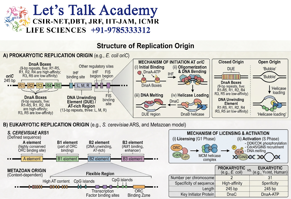
The exact structure of an origin differs between organisms.
Generally, an origin contains DNA sequences or chromatin features that allow replication-initiation proteins to recognize and activate it.
A simplified organization is:
Replication origin
↓
Initiator recognition
↓
DNA unwinding
↓
Helicase loading
↓
Replication machinery assembly
↓
Replication fork formation
5. Bacterial Replication Origin
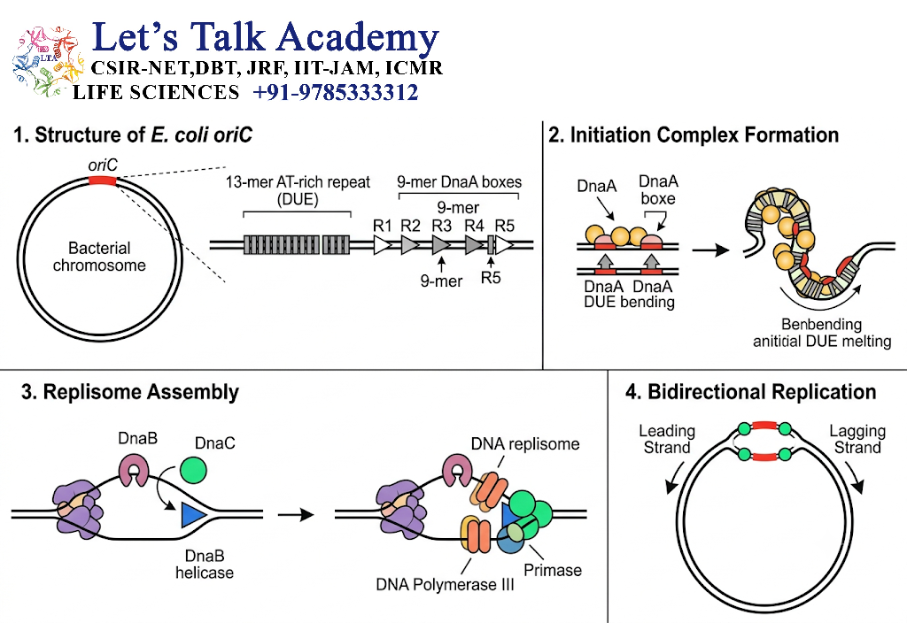
Many bacteria possess a circular chromosome with a major replication origin.
A well-studied example is oriC in Escherichia coli.
The oriC region contains specific DNA sequences that are recognized by the bacterial initiator protein DnaA.
5.1 Initiation at oriC
The general process is:
DnaA binds oriC
↓
DNA locally unwinds
↓
Helicase is loaded
↓
Primase becomes associated
↓
RNA primers are synthesized
↓
DNA polymerase begins DNA synthesis
↓
Replication forks move away from the origin
6. Eukaryotic Replication Origins
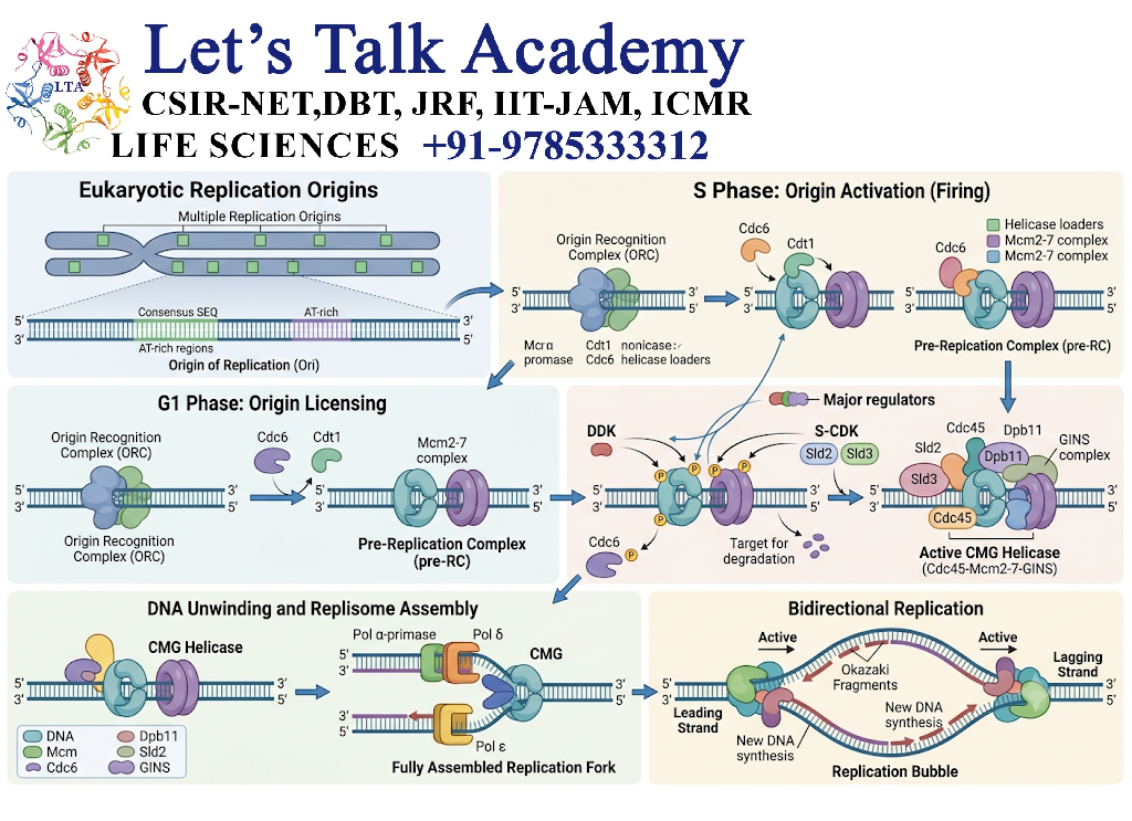
Eukaryotic chromosomes are generally much larger than bacterial chromosomes and therefore contain many replication origins.
Multiple origins allow different regions of a chromosome to be replicated simultaneously.
A simplified chromosome can be represented as:
Origin 1 → Origin 2 → Origin 3 → Origin 4 → Origin 5
Each origin can initiate replication and establish a replicating region.
This organization allows the entire genome to be copied efficiently during S phase of the cell cycle.
7. Origin Recognition

Replication initiation requires specific proteins to recognize the origin.
In bacteria, proteins such as DnaA recognize the origin.
In eukaryotes, the Origin Recognition Complex (ORC) participates in recognizing replication origins.
After origin recognition, additional proteins assemble to establish an active replication machinery.
8. Replication Initiation
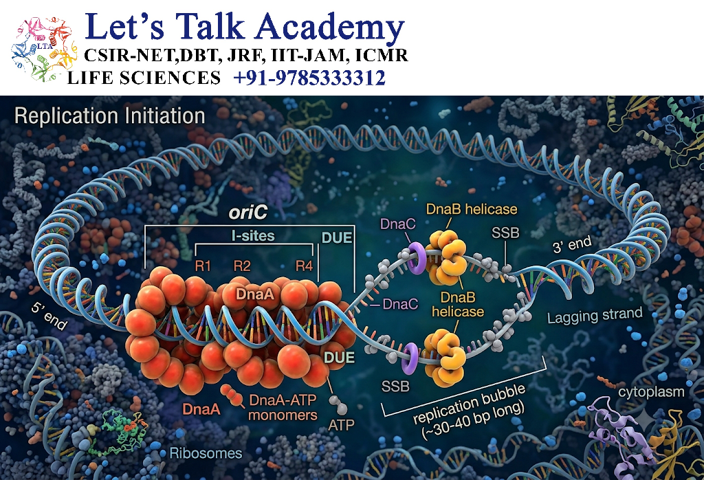
Replication initiation can be divided into several steps:
Step 1: Origin Recognition
Initiator proteins identify the replication origin.
Step 2: DNA Unwinding
The DNA double helix begins to open.
Step 3: Helicase Loading
The replicative helicase is positioned onto DNA.
Step 4: Primer Formation
Primase synthesizes RNA primers.
Step 5: DNA Polymerase Recruitment
DNA polymerases begin synthesizing the new DNA strands.
Step 6: Replication Fork Formation
One or more replication forks become established.
9. Replication Bubble
When DNA is unwound around an origin, a region called a replication bubble can form.
A replication bubble contains:
- The origin
- Unwound parental DNA
- Two replication forks
- Newly synthesized DNA
In bidirectional replication, the two forks move in opposite directions.
Fork ← Origin → Fork
This allows DNA to be replicated on both sides of the origin simultaneously.
10. Replication Fork
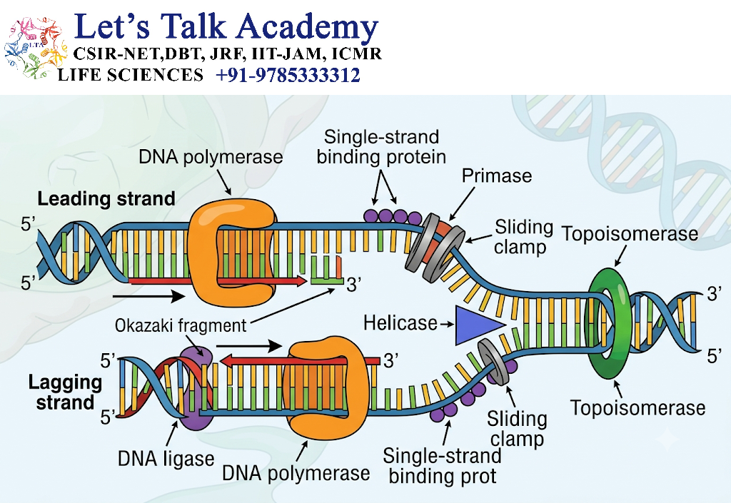
A replication fork is a Y-shaped structure formed when the parental DNA double helix is unwound during DNA replication.
It is the major site at which:
- DNA strands separate
- RNA primers are synthesized
- New DNA is synthesized
- Leading and lagging strands are produced
The replication fork therefore represents the active region of DNA synthesis.
11. Formation of Replication Fork
The replication fork develops after the DNA double helix is opened at the replication origin.
The process is:
Replication origin
↓
Helicase binds
↓
DNA strands separate
↓
Single-stranded DNA exposed
↓
Replication proteins assemble
↓
Replication fork forms
12. Structure of Replication Fork
The replication fork contains several important components.
Major Components
- Parental DNA
- Leading-strand template
- Lagging-strand template
- Helicase
- Primase
- DNA polymerase
- Single-strand DNA-binding proteins
- Sliding clamp
- Topoisomerase
- Newly synthesized DNA
A simplified representation is:
Parental DNA
↙ ↘
Leading strand Lagging strand
↓ ↓
Continuous synthesis Okazaki fragments
13. Leading Strand at the Replication Fork
The leading strand is synthesized continuously in the direction of replication-fork movement.
Because DNA polymerase synthesizes DNA only in the 5′ → 3′ direction, the orientation of the template permits continuous synthesis.
The basic process is:
Primer
↓
DNA polymerase
↓
Continuous DNA synthesis
↓
Leading strand
14. Lagging Strand at the Replication Fork
The lagging strand is synthesized discontinuously.
This occurs because DNA polymerase can synthesize DNA only in the 5′ → 3′ direction.
Short DNA segments called Okazaki fragments are therefore produced.
The process is:
Primer synthesis
↓
DNA synthesis
↓
Okazaki fragment
↓
Primer removal
↓
Gap filling
↓
DNA ligase
↓
Continuous DNA strand
15. Enzymes at the Replication Fork
Several enzymes work together at the replication fork.
| Enzyme/Protein | Function |
|---|---|
| Helicase | Unwinds parental DNA |
| Primase | Synthesizes RNA primers |
| DNA Polymerase | Synthesizes DNA |
| Topoisomerase | Relieves torsional stress |
| DNA Gyrase | Controls DNA supercoiling in bacteria |
| RNase H / nucleases | Participate in primer processing |
| DNA Ligase | Joins DNA fragments |
| Sliding Clamp | Increases DNA polymerase processivity |
| Single-Strand Binding Proteins | Stabilize separated DNA strands |
16. Helicase at the Replication Fork
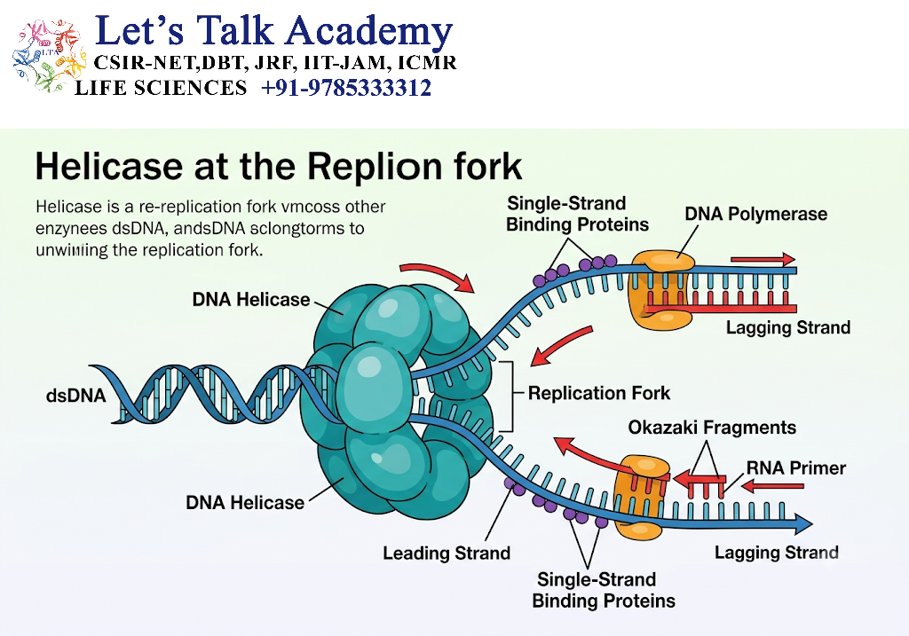
Helicase is one of the central enzymes at the replication fork.
It moves along DNA and separates the parental strands by disrupting interactions that hold the duplex together.
This produces two single-stranded DNA templates.
Double-stranded DNA
↓
Helicase
↓
Two separated DNA strands
↓
Two templates available for DNA synthesis
17. Topoisomerase at the Replication Fork
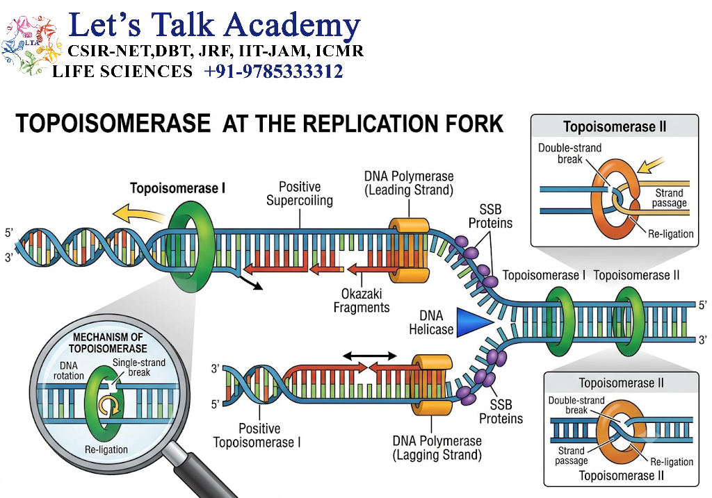
DNA unwinding generates torsional stress ahead of the replication fork.
Topoisomerases reduce this stress by temporarily breaking and resealing DNA.
Without topoisomerase activity, excessive DNA supercoiling could interfere with fork progression.
In bacteria, DNA gyrase is an important topoisomerase involved in this process.
18. Single-Strand Binding Proteins
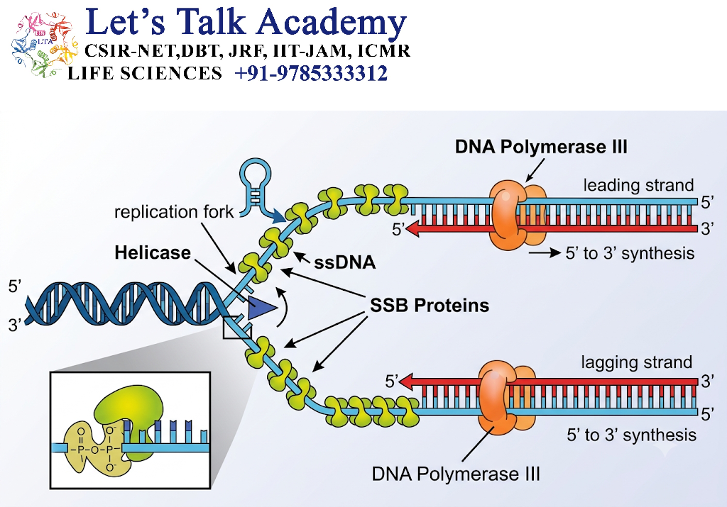
After helicase separates the DNA strands, the exposed single-stranded DNA can potentially reanneal.
Single-strand binding proteins prevent this.
In bacteria, SSB proteins stabilize single-stranded DNA.
In eukaryotes, RPA (Replication Protein A) performs a similar role.
Their functions include:
- Stabilizing single-stranded DNA
- Preventing premature reannealing
- Protecting exposed DNA
- Facilitating replication machinery function
19. Primase at the Replication Fork
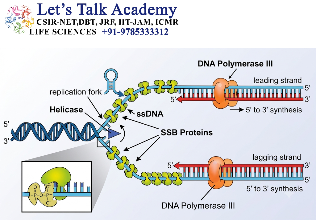
Primase synthesizes short RNA primers on the exposed DNA template.
These primers provide the 3′-OH group required by DNA polymerase.
On the lagging strand, primase repeatedly produces primers for successive Okazaki fragments.
20. DNA Polymerase at the Replication Fork
DNA polymerase extends the RNA primers by adding complementary deoxyribonucleotides.
It synthesizes DNA only in the:
5′ → 3′ direction
DNA polymerase therefore works differently on the two templates:
Leading strand → continuous synthesis
Lagging strand → discontinuous synthesis
21. Replication Fork and Okazaki Fragments
Okazaki fragments are produced on the lagging strand.
Each fragment begins with an RNA primer and is extended by DNA polymerase.
Later:
RNA primer removal
↓
Gap filling
↓
DNA ligase
↓
Continuous DNA
This process is essential for completing lagging-strand synthesis.
22. Bidirectional Replication
Replication frequently proceeds in two directions from an origin.
Two replication forks move away from the origin:
Replication fork ← Origin → Replication fork
This is called bidirectional replication.
Advantages
Bidirectional replication:
- Speeds up genome duplication.
- Allows simultaneous replication of both sides of an origin.
- Helps large DNA molecules replicate efficiently.
23. Unidirectional Replication
Some DNA molecules can replicate primarily in one direction from an origin.
In such cases:
Origin → Replication fork → DNA synthesis
However, bidirectional replication is a common strategy for many cellular chromosomes.
24. Replication Origin vs Replication Fork
| Feature | Replication Origin | Replication Fork |
|---|---|---|
| Definition | Site where replication begins | Site where DNA is actively replicated |
| Location | Specific DNA region | Moving structure along DNA |
| Major role | Initiates replication | Carries out DNA synthesis |
| Main event | Origin recognition and unwinding | Strand separation and synthesis |
| Important proteins | Initiators, helicase-loading factors | Helicase, primase, DNA polymerase, etc. |
| Movement | Fixed DNA location | Moves along DNA |
| Number | One or many depending on genome | Usually two per bidirectionally activated origin |
25. Replication Origin and Replicon
A replicon is the functional unit of DNA replication associated with a particular origin.
The relationship is:
Replication Origin
↓
Initiation
↓
Replication Fork(s)
↓
DNA synthesis
↓
Termination
The DNA replicated from one origin constitutes a replicating unit or replicon.
26. Replication Origin and Cell Cycle
In eukaryotic cells, replication origins are closely associated with cell-cycle control.
During the appropriate stage:
G1 phase
↓
Origin licensing
↓
S phase
↓
Origin activation
↓
DNA replication
↓
G2 phase
↓
M phase
This system helps ensure that DNA is not replicated repeatedly within the same cell cycle.
27. Replication Fork and Cell-Cycle Checkpoints
Replication forks can encounter problems such as:
- DNA damage
- Lack of nucleotides
- Difficult DNA sequences
- DNA-protein complexes
- Transcription-replication conflicts
Cells have checkpoint mechanisms that can slow or stop replication when serious problems occur.
This helps prevent the accumulation of DNA damage and genomic instability.
28. Replication Fork Protection
A normal replication fork must be maintained in a stable configuration.
If the fork stalls, cells can activate:
- DNA damage signaling
- Fork stabilization mechanisms
- DNA repair pathways
- Cell-cycle checkpoints
Proper fork protection is important for maintaining genome integrity.
29. Replication Fork and DNA Damage
When a replication fork encounters damaged DNA, DNA synthesis may slow or stop.
A simplified response is:
Replication fork encounters damage
↓
Replication slows/stalls
↓
Damage sensing
↓
Checkpoint activation
↓
DNA repair / fork restart
↓
Replication resumes
If damage cannot be properly handled, persistent replication stress may contribute to chromosome abnormalities.
30. Replication Fork and Genomic Stability
Proper replication-fork function is essential for maintaining genomic stability.
Defects in replication can result in:
- DNA breaks
- Mutations
- Chromosomal rearrangements
- Copy-number abnormalities
- Replication stress
- Genomic instability
Therefore, replication-fork regulation is closely connected with DNA repair and cell-cycle control.


