
1. Steps in the Cell Cycle
The cell cycle is a highly organized and precisely regulated series of events through which a cell grows, duplicates its genetic material, and divides to produce daughter cells. It is one of the fundamental processes responsible for the growth, development, repair, and reproduction of living organisms.
Before a cell divides, it must accomplish several important tasks. It needs to increase its cellular mass, synthesize proteins and other cellular components, duplicate its DNA accurately, check the integrity of the newly copied DNA, and finally distribute the duplicated chromosomes equally between daughter cells.
The cell cycle can therefore be understood as a carefully coordinated sequence rather than simply a process of cell division.
In eukaryotic cells, the cell cycle is broadly divided into two major phases:
- Interphase
- M phase (Mitotic phase)
Interphase consists of:
- G₁ phase (Gap 1)
- S phase (Synthesis)
- G₂ phase (Gap 2)
The M phase consists mainly of:
- Mitosis
- Cytokinesis
A simplified sequence is:
G₁ → S → G₂ → Mitosis → Cytokinesis → G₁
The cell may also temporarily or permanently leave the cycle and enter a specialized state called the G₀ phase.
2. Overview of the Cell Cycle
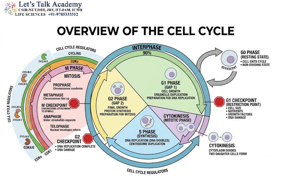
The cell cycle is a continuous process, but it is traditionally divided into distinct stages for convenient study.
2.1 Major Stages of the Cell Cycle
The major stages are:
| Phase | Major Event |
|---|---|
| G₁ phase | Cell growth and preparation for DNA replication |
| S phase | DNA replication and chromosome duplication |
| G₂ phase | Further growth and preparation for mitosis |
| M phase | Nuclear division through mitosis |
| Cytokinesis | Division of cytoplasm |
| G₀ phase | Non-dividing or quiescent state |
The duration of each phase varies among cell types and organisms. Some rapidly dividing cells complete the cycle relatively quickly, whereas differentiated cells may remain outside the active cycle for long periods.
3. Interphase
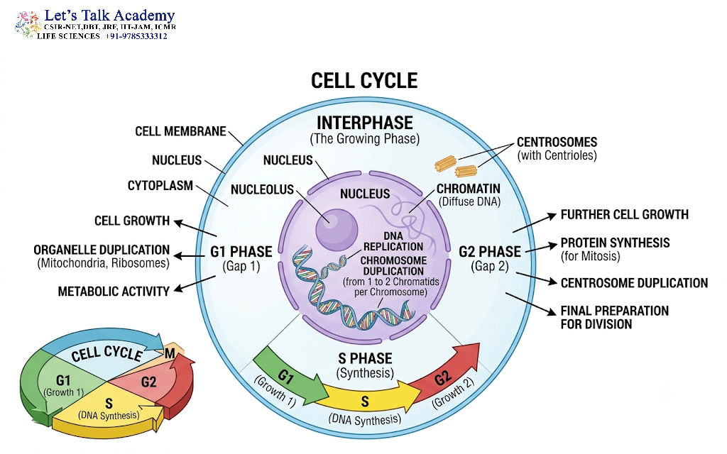
Interphase is the period between two successive cell divisions. It is often incorrectly considered a period of cellular inactivity. In reality, interphase is a highly active stage during which the cell grows, performs its normal functions, duplicates its DNA, synthesizes proteins, and prepares for division.
Interphase is divided into three stages:
- G₁ phase
- S phase
- G₂ phase
3.1 G₁ Phase: First Gap Phase
The G₁ phase is the first major phase after cytokinesis and before DNA synthesis.
During G₁, the newly formed daughter cell grows and resumes its normal metabolic activities.
Major events of G₁ phase
During this stage:
- Cell size increases.
- RNA synthesis occurs.
- Protein synthesis increases.
- Organelles are produced or increased in number.
- Cellular metabolism is highly active.
- Components required for DNA replication are synthesized.
- The cell evaluates whether environmental conditions are suitable for division.
The G₁ phase is particularly important because the cell makes a major decision about whether it should continue toward DNA replication and division.
G₁ restriction point
In many animal cells, cell-cycle progression through G₁ is controlled by a critical regulatory point called the restriction point.
Before passing this point, cell-cycle progression depends strongly on external signals such as:
- Growth factors
- Nutrient availability
- Cell size
- Energy status
- Signals from neighboring cells
After passing the restriction point, the cell is generally committed to completing the cell cycle, provided that major problems do not arise.
3.2 S Phase: DNA Synthesis Phase
The S phase is the stage during which the cell duplicates its DNA.
DNA replication is one of the most important events of the entire cell cycle because each daughter cell must receive a complete genome.
During S phase:
- DNA is replicated.
- Each chromosome produces two identical DNA molecules.
- Histone proteins are synthesized.
- Chromatin is duplicated.
- Centrosomes are duplicated in animal cells.
Following DNA replication, each chromosome consists of two identical sister chromatids connected primarily at the centromeric region.
Important Point
DNA replication does not double the number of chromosomes.
Instead:
- The amount of DNA doubles.
- Each chromosome becomes composed of two sister chromatids.
- The chromosome number remains unchanged.
For example, a diploid cell with 2n chromosomes remains 2n chromosomes after S phase, but its DNA content has doubled.
3.3 Mechanism of DNA Replication During S Phase
DNA replication is semiconservative, meaning that each newly formed DNA molecule contains:
- One parental strand
- One newly synthesized strand
Replication begins at specific regions called origins of replication.
Several proteins and enzymes participate in this process.
Important components include:
- Helicase
- Primase
- DNA polymerases
- DNA ligase
- Single-strand DNA-binding proteins
- Topoisomerases
Helicase
Helicase separates the two DNA strands by disrupting hydrogen bonds between complementary bases.
Primase
Primase synthesizes short RNA primers required to initiate DNA synthesis.
DNA Polymerase
DNA polymerase adds nucleotides to the growing DNA strand in the 5′ → 3′ direction.
DNA Ligase
DNA ligase joins discontinuous DNA fragments, particularly the Okazaki fragments formed on the lagging strand.
The accurate duplication of DNA is essential because errors introduced during replication can become permanent mutations if they are not repaired.
4. G₂ Phase: Second Gap Phase
After DNA replication is completed, the cell enters the G₂ phase.
This stage represents the final preparation period before mitosis.
During G₂:
- The cell continues to grow.
- Protein synthesis continues.
- Microtubule components are produced.
- Energy reserves are maintained.
- DNA replication is checked.
- DNA damage is detected and repaired.
- Proteins required for mitosis are synthesized.
The cell also ensures that DNA replication has been completed properly before entering M phase.
4.1 G₂ Checkpoint
The G₂ checkpoint prevents a cell from entering mitosis when DNA replication is incomplete or when significant DNA damage remains unrepaired.
If damage is detected, cell-cycle progression can be temporarily stopped to allow repair.
If the damage is too severe to repair, the cell may undergo programmed cell death, known as apoptosis.
This checkpoint therefore contributes to genomic stability.
5. M Phase
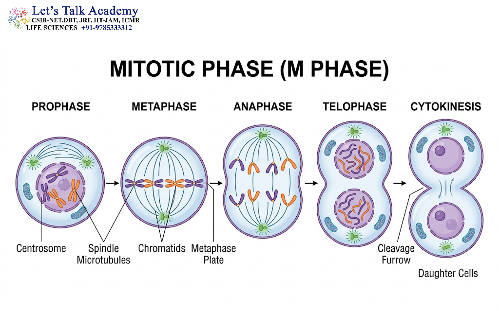
The M phase is the stage in which the duplicated genetic material is distributed between daughter cells.
M phase includes:
- Mitosis
- Cytokinesis
Mitosis is nuclear division, whereas cytokinesis is division of the cytoplasm.
Mitosis is traditionally divided into:
- Prophase
- Prometaphase
- Metaphase
- Anaphase
- Telophase
Some descriptions combine prometaphase with prophase.
6. Prophase
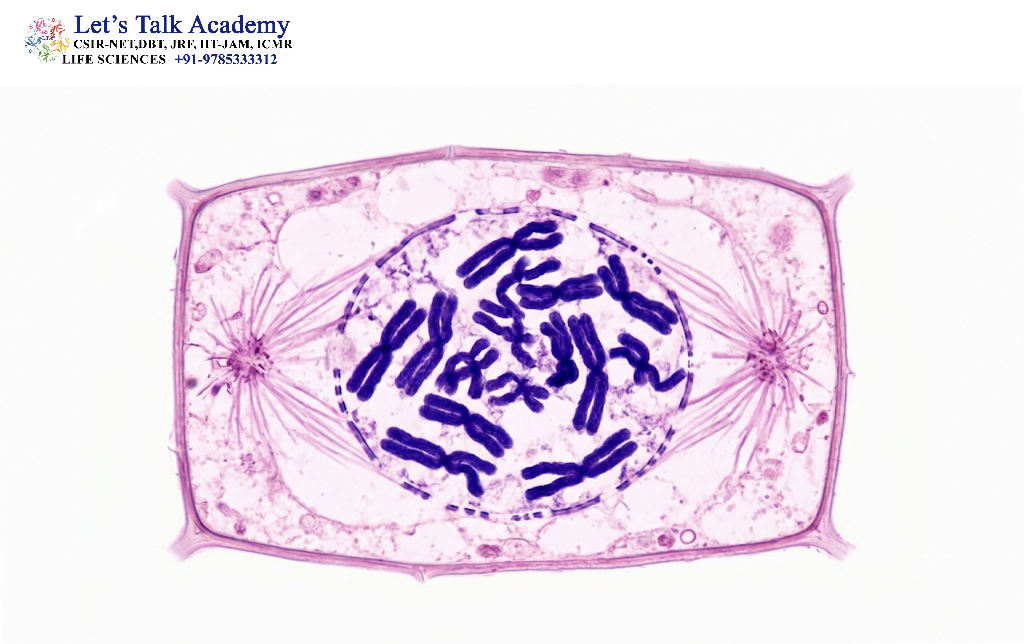
Prophase is the first major stage of mitosis.
At the beginning of prophase, the chromatin becomes progressively condensed.
Major Events
During prophase:
- Chromatin condenses into visible chromosomes.
- Each chromosome consists of two sister chromatids.
- The nucleolus becomes less distinct and eventually disappears.
- The mitotic spindle begins to form.
- Centrosomes move toward opposite poles of the cell.
Chromosome condensation is important because highly compact chromosomes can be moved more efficiently without becoming excessively tangled.
7. Prometaphase
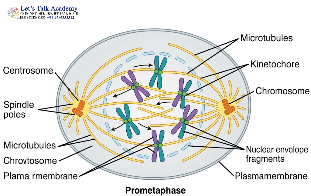
Prometaphase begins with the breakdown of the nuclear envelope.
This allows spindle microtubules to interact directly with chromosomes.
Major Events
During prometaphase:
- Nuclear envelope disintegrates.
- Spindle microtubules enter the former nuclear region.
- Microtubules attach to chromosomes through specialized protein structures called kinetochores.
- Chromosomes begin moving toward the central region of the cell.
Kinetochore
A kinetochore is a multiprotein structure assembled on the centromeric region of a chromosome.
It provides a site for attachment of spindle microtubules.
Proper kinetochore–microtubule attachment is essential for accurate chromosome segregation.
8. Metaphase
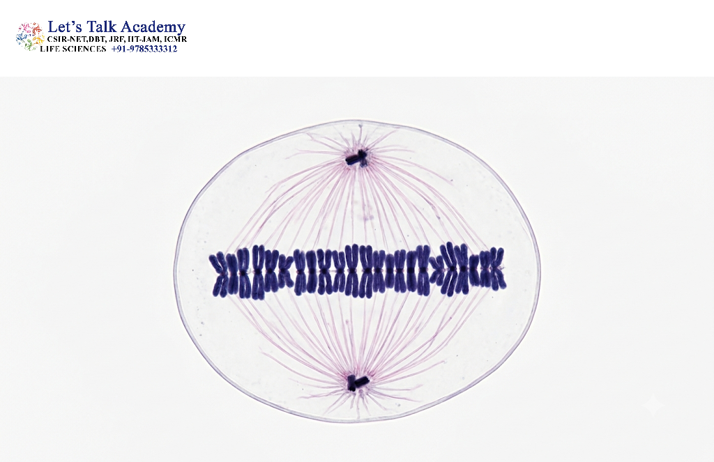
During metaphase, chromosomes become aligned approximately at the center of the cell.
This region is often called the metaphase plate.
Major Events
- Chromosomes are highly condensed.
- Chromosomes align at the metaphase plate.
- Sister chromatids remain attached to one another.
- Spindle microtubules attach to kinetochores.
- Sister kinetochores are connected to microtubules from opposite spindle poles.
The cell carefully checks whether all chromosomes are correctly attached before proceeding to anaphase.
9. Metaphase Checkpoint
The spindle assembly checkpoint is an important surveillance mechanism operating around metaphase.
It ensures that chromosomes are properly attached to the mitotic spindle before sister chromatids separate.
If even one chromosome is incorrectly attached, progression into anaphase can be delayed.
This checkpoint helps prevent:
- Chromosome loss
- Chromosome gain
- Unequal chromosome distribution
- Aneuploidy
The spindle assembly checkpoint therefore plays a major role in maintaining chromosome stability.
10. Anaphase
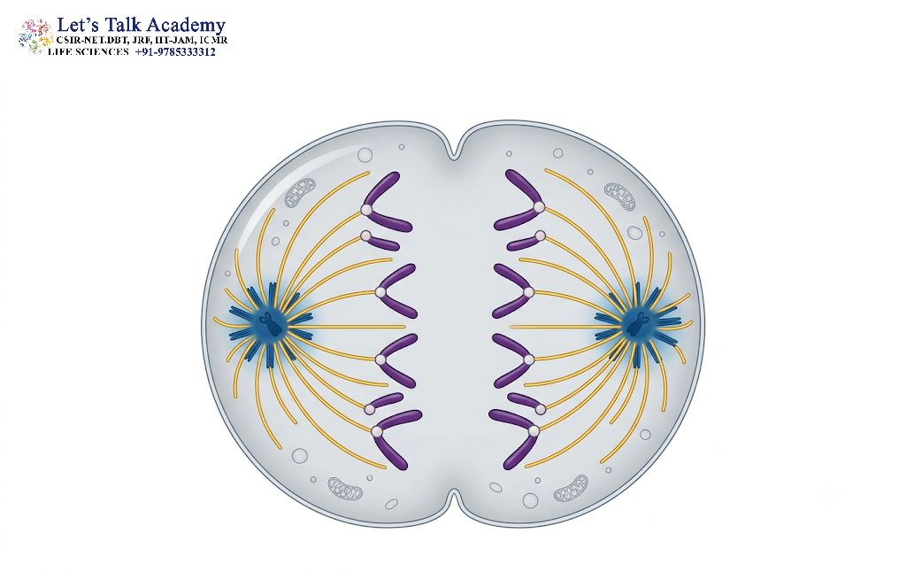
Anaphase begins when the connections holding sister chromatids together are released.
The sister chromatids then move toward opposite poles of the cell.
The protein complex responsible for holding sister chromatids together is known as cohesin.
At the onset of anaphase, cohesin is cleaved by the protease separase, allowing sister chromatids to separate.
Major Events
- Sister chromatids separate.
- Each separated chromatid is now considered an individual chromosome.
- Chromosomes move toward opposite poles.
- The cell begins to elongate.
Anaphase can be described in two components:
Anaphase A
Chromosomes move toward spindle poles primarily through shortening of kinetochore microtubules.
Anaphase B
The two spindle poles move farther apart, contributing to elongation of the cell.
11. Telophase
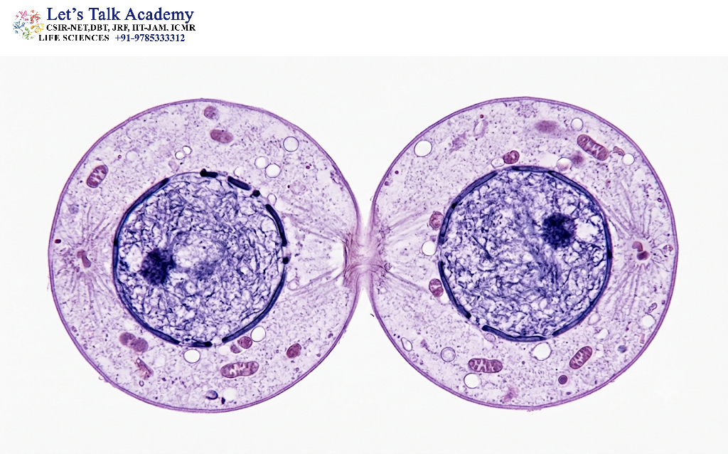
Telophase occurs after chromosomes reach opposite poles.
During this stage, the cell begins reconstructing two separate nuclei.
Major Events
- Chromosomes arrive at opposite poles.
- Chromosomes begin to decondense.
- Nuclear envelopes reform around each chromosome set.
- Nucleoli reappear.
- Mitotic spindle components are disassembled.
At the end of telophase, two genetically equivalent nuclei have generally been established.
12. Cytokinesis
Cytokinesis is the physical division of the cytoplasm into two daughter cells.
It usually overlaps with the later stages of mitosis.
The mechanism of cytokinesis differs between animal and plant cells.
12.1 Cytokinesis in Animal Cells
Animal cells undergo cytokinesis through the formation of a cleavage furrow.
The cleavage furrow develops because of contraction of an actin–myosin contractile ring located beneath the plasma membrane.
The contractile ring tightens progressively, causing the plasma membrane to constrict until two daughter cells are formed.
12.2 Cytokinesis in Plant Cells
Plant cells cannot simply form a cleavage furrow because their rigid cell wall prevents this type of constriction.
Instead, they form a cell plate.
Vesicles derived largely from the Golgi apparatus accumulate at the center of the dividing cell and fuse to form the developing cell plate.
The cell plate expands outward and eventually becomes part of the new cell wall separating the daughter cells.
13. G₀ Phase: A Non-Dividing State
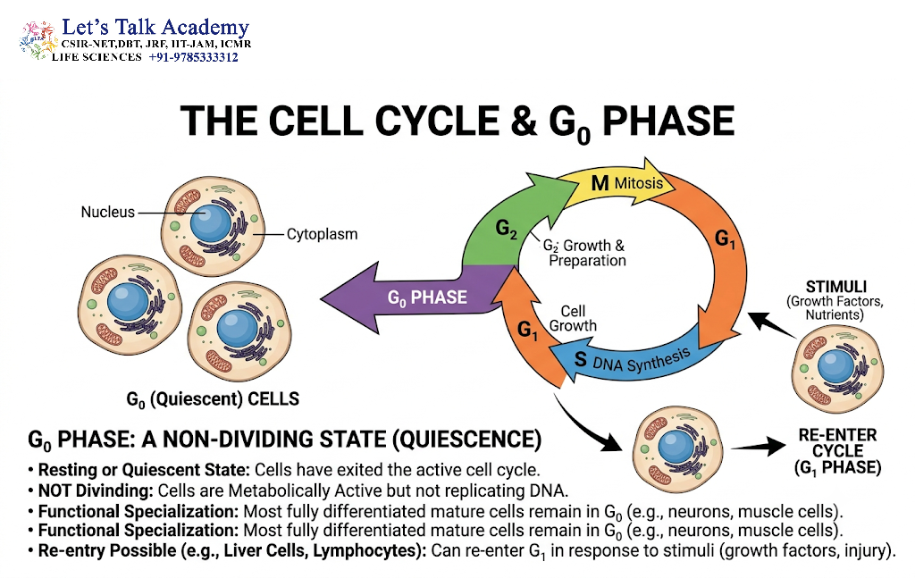
Not every cell continuously progresses through the cell cycle.
Some cells enter a state known as G₀.
G₀ is generally considered a state outside the active proliferative cycle.
Cells in G₀ may be:
- Temporarily quiescent
- Differentiated and highly specialized
- Reversibly capable of re-entering the cell cycle
- Permanently withdrawn from cell division
The nature of G₀ differs among cell types.
Some cells can re-enter G₁ following appropriate stimulation, whereas highly differentiated cells may remain in a non-dividing state for very long periods.
14. Cell-Cycle Checkpoints
Cell-cycle progression is controlled by several checkpoints. These checkpoints function as surveillance systems that ensure the cell does not proceed to the next stage when important conditions have not been satisfied.
The major checkpoints include:
- G₁ checkpoint
- G₂ checkpoint
- Spindle assembly checkpoint
14.1 G₁/S Checkpoint
The G₁/S checkpoint determines whether the cell is ready to initiate DNA replication.
It evaluates factors such as:
- Cell size
- Nutrient availability
- Growth signals
- DNA integrity
- Cellular energy status
If DNA damage is detected, progression may be stopped.
14.2 G₂/M Checkpoint
The G₂/M checkpoint ensures that the cell does not enter mitosis until:
- DNA replication is complete.
- Major DNA damage has been repaired.
- The cell has adequate resources for division.
This checkpoint is particularly important for preventing the transmission of damaged or incompletely replicated DNA.
14.3 Spindle Assembly Checkpoint
The spindle assembly checkpoint operates during mitosis.
It ensures that chromosomes are properly attached to spindle microtubules before sister chromatids separate.
This mechanism helps ensure that each daughter cell receives the correct chromosome complement.
15. Molecular Regulation of the Cell Cycle
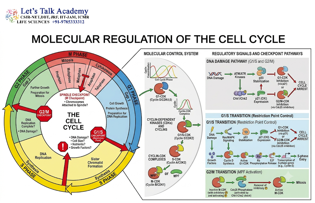
The cell cycle is controlled by a complex network of proteins.
Two major groups of regulatory proteins are:
- Cyclins
- Cyclin-dependent kinases (CDKs)
Cyclins are regulatory proteins whose concentrations change during the cell cycle.
CDKs are protein kinases that become active when associated with specific cyclins.
Together, cyclin–CDK complexes regulate progression through different stages of the cell cycle.
15.1 Cyclins
Cyclins are synthesized and degraded at specific stages of the cell cycle.
Important cyclin classes include:
- G₁ cyclins
- G₁/S cyclins
- S-phase cyclins
- M-phase cyclins
Their changing abundance helps determine when specific CDKs become active.
15.2 Cyclin-Dependent Kinases
CDKs phosphorylate specific target proteins.
This phosphorylation changes the activity, localization, or stability of target proteins involved in cell-cycle progression.
Examples include:
- Cyclin D–CDK4/6
- Cyclin E–CDK2
- Cyclin A–CDK2
- Cyclin A/B–CDK1
These complexes coordinate progression through G₁, S, G₂, and M phases.
16. Role of the Retinoblastoma Protein
The retinoblastoma protein (Rb) is an important regulator of the G₁-to-S transition.
Rb can bind and inhibit E2F transcription factors.
When appropriate growth signals activate cyclin–CDK complexes, Rb becomes progressively phosphorylated.
This reduces its inhibitory effect on E2F.
Activated E2F promotes transcription of genes required for DNA synthesis and S-phase progression.
Thus, the Rb–E2F regulatory pathway is an important mechanism controlling entry into S phase.
17. Role of p53 in Cell-Cycle Regulation
p53 is an important tumor-suppressor protein that responds to cellular stress, particularly DNA damage.
When DNA damage occurs, p53 can become stabilized and activate genes that inhibit cell-cycle progression.
One important target is p21.
p21 inhibits certain cyclin–CDK complexes and can cause cell-cycle arrest.
This gives the cell time to repair damaged DNA.
If damage cannot be successfully repaired, p53-dependent pathways can contribute to apoptosis or other forms of cellular elimination.
18. Regulation of the Cell Cycle by External Signals
Cell division is not controlled only by internal mechanisms.
Cells also respond to external signals.
Important signals include:
- Growth factors
- Hormones
- Nutrient availability
- Cell–cell interactions
- Extracellular matrix signals
For example, growth factors can activate intracellular signaling pathways that increase cyclin production and promote cell-cycle progression.
This coordination ensures that cells divide only when proliferation is biologically appropriate.
19. DNA Damage and Cell-Cycle Arrest
DNA damage can arise from several sources, including:
- Replication errors
- Reactive oxygen species
- Radiation
- Chemical agents
- Environmental stress
Cells possess DNA damage-response systems that detect and respond to such damage.
Depending on the severity of damage, the cell may:
- Pause the cell cycle.
- Repair the DNA.
- Resume proliferation.
- Enter senescence.
- Undergo apoptosis.
This system is essential for preventing damaged genetic information from being propagated through successive generations of cells.
20. Importance of Accurate Chromosome Segregation
Correct chromosome segregation is essential during mitosis.
Each daughter cell should normally receive an equivalent set of chromosomes.
Errors in chromosome segregation can result in:
- Aneuploidy
- Chromosomal instability
- Abnormal cellular function
- Developmental abnormalities
- Contribution to disease processes
The spindle assembly checkpoint, kinetochore attachment, cohesin, separase, and spindle dynamics all contribute to accurate segregation.
21. Cell Cycle and Cancer
Uncontrolled cell proliferation is one of the fundamental characteristics of cancer.
Normal cells possess multiple mechanisms that prevent inappropriate proliferation.
Cancer can arise when mutations disrupt genes involved in:
- Cell-cycle progression
- DNA repair
- Checkpoint control
- Apoptosis
- Growth-factor signaling
Important regulatory proteins involved in cancer biology include:
- p53
- Rb
- Cyclins
- CDKs
- CDK inhibitors
Loss of checkpoint control can allow cells carrying damaged DNA to continue dividing.
As mutations accumulate, abnormal proliferation and genomic instability may increase.
22. Cell Cycle Versus Cell Division
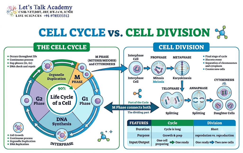
The terms cell cycle and cell division are related but are not identical.
The cell cycle includes the complete sequence of events from one cell division to the next.
Cell division is only one part of this overall process.
Cell Cycle Includes:
G₁ + S + G₂ + M
Whereas M phase includes:
Mitosis + Cytokinesis
Therefore, most of the cell cycle is actually spent in interphase rather than in mitosis.


