
Structure of Molecules
The four major classes of biological molecules traditionally studied in biochemistry are carbohydrates, lipids, proteins, and nucleic acids. Each class has its own characteristic chemical structures and biological functions, but there is extensive interaction among them. Carbohydrates can provide carbon skeletons for metabolic pathways, lipids form biological membranes and serve as energy stores, proteins perform most of the catalytic and regulatory functions of cells, and nucleic acids store and express hereditary information.
A central principle of molecular biology is that structure determines function. The precise arrangement of atoms within a molecule determines its shape, charge distribution, polarity, flexibility, stability, and ability to interact with other molecules. Even a small structural alteration can sometimes produce a major biological consequence. Therefore, when studying biomolecules, it is not sufficient to memorize their names or structures. A strong understanding requires connecting the molecular structure with its chemical properties and ultimately with its biological function.
Chemical Foundation of Biomolecules
The chemical complexity of living organisms is built primarily from a relatively small collection of elements. The most important elements found in biological molecules are carbon, hydrogen, oxygen, nitrogen, phosphorus, and sulfur. These are often collectively remembered as CHNOPS.
Among these elements, carbon occupies a particularly important position because of its ability to form four stable covalent bonds. Carbon atoms can bond with one another to produce long chains, branched structures, rings, and complex three-dimensional frameworks. They can also form stable bonds with hydrogen, oxygen, nitrogen, sulfur, and phosphorus. This exceptional versatility of carbon is one of the fundamental reasons why carbon-based chemistry can generate the enormous structural diversity observed in living organisms.
Hydrogen and oxygen are especially abundant in carbohydrates and water. Nitrogen is an important component of amino acids, proteins, nucleotides, and many biologically active compounds. Phosphorus is essential in nucleic acids, phospholipids, ATP, and many regulatory phosphorylation reactions. Sulfur occurs in specific amino acids such as cysteine and methionine and consequently contributes to the structure and function of many proteins.
Although these elements are individually simple, their different combinations and bonding arrangements produce molecules with completely different properties. For example, glucose and fructose have the same molecular formula, C6H12O6, but their atoms are arranged differently. Consequently, they have different structures and biochemical properties. This illustrates a fundamental concept of biological chemistry: molecular composition alone does not completely determine molecular behavior; molecular arrangement is equally important.
Why Carbon Is the Central Element of Biological Chemistry
Carbon has four valence electrons and can therefore participate in four covalent bonds. More importantly, carbon-carbon bonds are sufficiently stable to allow the formation of large biological molecules while still permitting the chemical reactions required for metabolism.
Carbon atoms can form single, double, and triple bonds. They can also produce linear chains, branched chains, and ring structures. This ability creates an extraordinary number of possible molecular configurations.
For example, a simple hydrocarbon can contain only carbon and hydrogen, whereas the addition of oxygen, nitrogen, phosphorus, or sulfur creates molecules with entirely new chemical properties. The introduction of a hydroxyl group can increase polarity, a carboxyl group can provide acidic behavior, an amino group can provide basic properties, and a phosphate group can introduce negative charge and participate in energy-transfer reactions.
Therefore, the biological importance of carbon does not simply arise from its abundance. It arises from the combination of bonding flexibility, structural stability, and chemical diversity.
The carbon skeleton of a biomolecule is often referred to as its carbon backbone. Functional groups attached to this backbone determine many of its chemical properties. Consequently, understanding carbon skeletons and functional groups is one of the first steps toward understanding biomolecular structure.
Functional Groups
A functional group is a specific arrangement of atoms within an organic molecule that gives the molecule characteristic chemical properties. Functional groups are particularly important in biology because they determine how biomolecules interact with water, acids, bases, enzymes, membranes, and other biological molecules.
Two molecules may have similar carbon skeletons but behave very differently because their functional groups differ. Conversely, molecules belonging to very different biological classes may contain the same functional group and therefore share certain chemical characteristics.
The major functional groups encountered in biological chemistry include hydroxyl, carbonyl, carboxyl, amino, sulfhydryl, phosphate, and methyl groups.
1. Hydroxyl Group
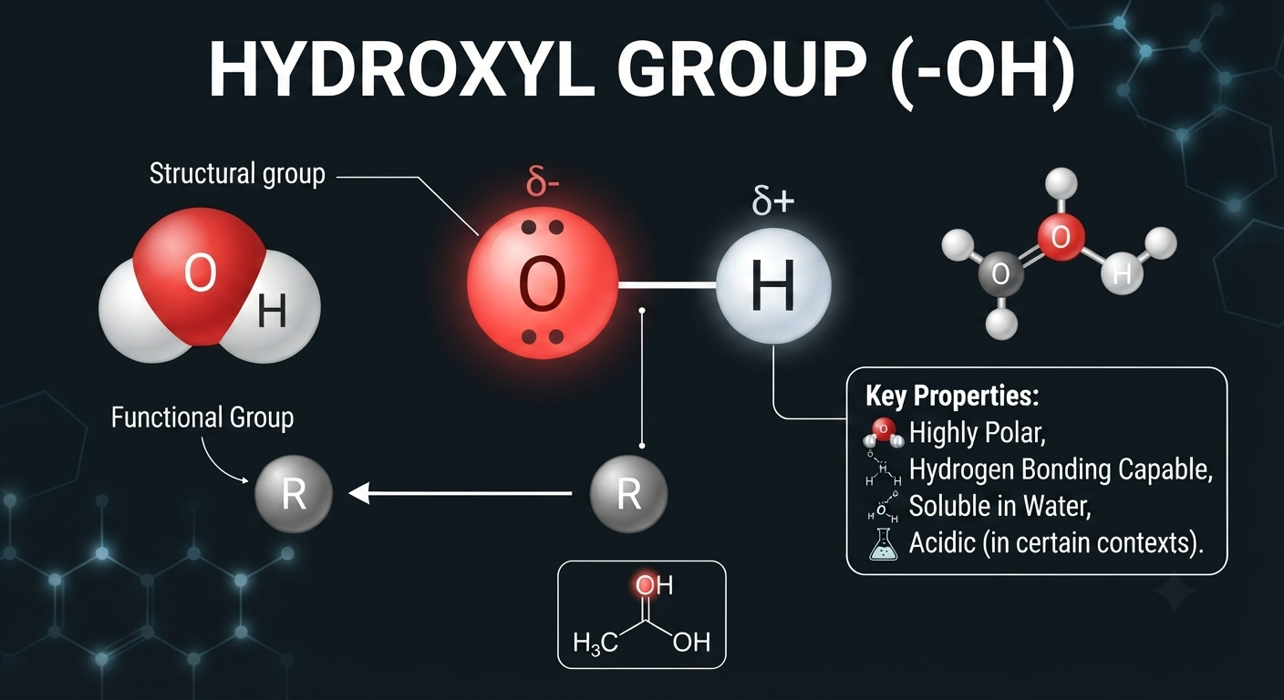
The hydroxyl group is written as –OH. It is commonly found in carbohydrates, alcohols, and several other biological compounds.
The oxygen atom in a hydroxyl group is electronegative and participates in hydrogen bonding. Consequently, the presence of several hydroxyl groups usually increases the polarity and water solubility of a molecule.
Glucose is a good example. It contains multiple hydroxyl groups, which contribute substantially to its interaction with water. This property is biologically important because glucose is transported and metabolized in aqueous cellular environments.
2. Carbonyl Group

The carbonyl group contains a carbon atom double-bonded to oxygen:
C=O
Carbonyl groups are particularly important in carbohydrate chemistry.
When the carbonyl carbon occurs at the end of a carbon chain, the molecule contains an aldehyde group. When the carbonyl group occurs within the carbon chain, the molecule contains a ketone group.
Glucose is classified as an aldose, whereas fructose is classified as a ketose.
This distinction is not merely a naming convention. The position of the carbonyl group influences the chemical reactions that these sugars undergo and contributes to differences in their structures.
3. Carboxyl Group
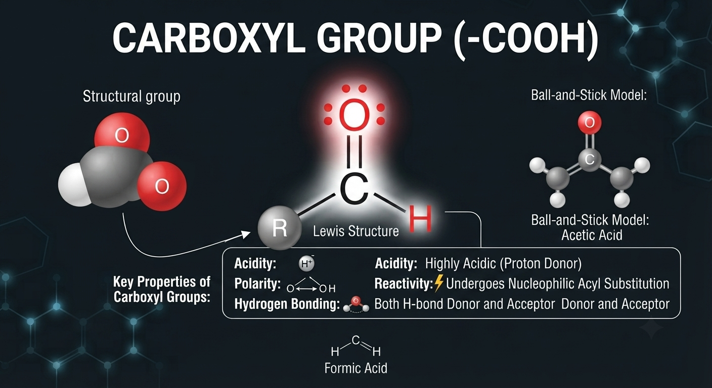
The carboxyl group is represented as:
–COOH
It is characteristic of carboxylic acids and is present in both amino acids and fatty acids.
The carboxyl group can donate a proton:
–COOH ⇌ –COO− + H+
At physiological pH, many carboxyl groups exist predominantly in their negatively charged carboxylate form. This change in charge has important consequences for molecular interactions, solubility, protein structure, and enzyme activity.
The acidic nature of amino acid side chains such as aspartate and glutamate is largely associated with their carboxyl-containing side chains.
4. Amino Group
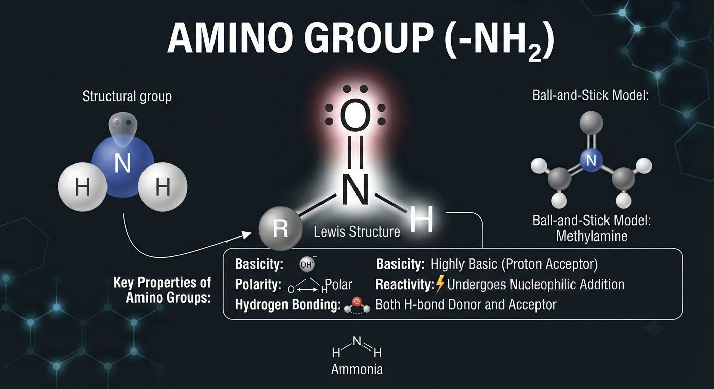
The amino group is represented as:
–NH2
It is a defining functional group of amino acids.
The nitrogen atom can accept a proton, resulting in:
–NH2 + H+ ⇌ –NH3+
At physiological pH, amino groups are frequently protonated and therefore positively charged.
The simultaneous presence of acidic carboxyl groups and basic amino groups allows amino acids to exist in different ionic forms depending on the pH of their environment.
This behavior is fundamental to understanding amino acid titration curves, isoelectric points, protein charge, and electrophoretic separation.
5. Sulfhydryl Group

The sulfhydryl group is written as –SH. It is especially important in the amino acid cysteine.
Two cysteine residues can undergo oxidation to form a covalent disulfide bond:
Cys–SH + HS–Cys → Cys–S–S–Cys
Disulfide bonds can stabilize the three-dimensional structure of proteins, particularly proteins that function in extracellular environments.
This is an important example of how a small chemical group can have a major structural role in a macromolecule.
6. Phosphate Group
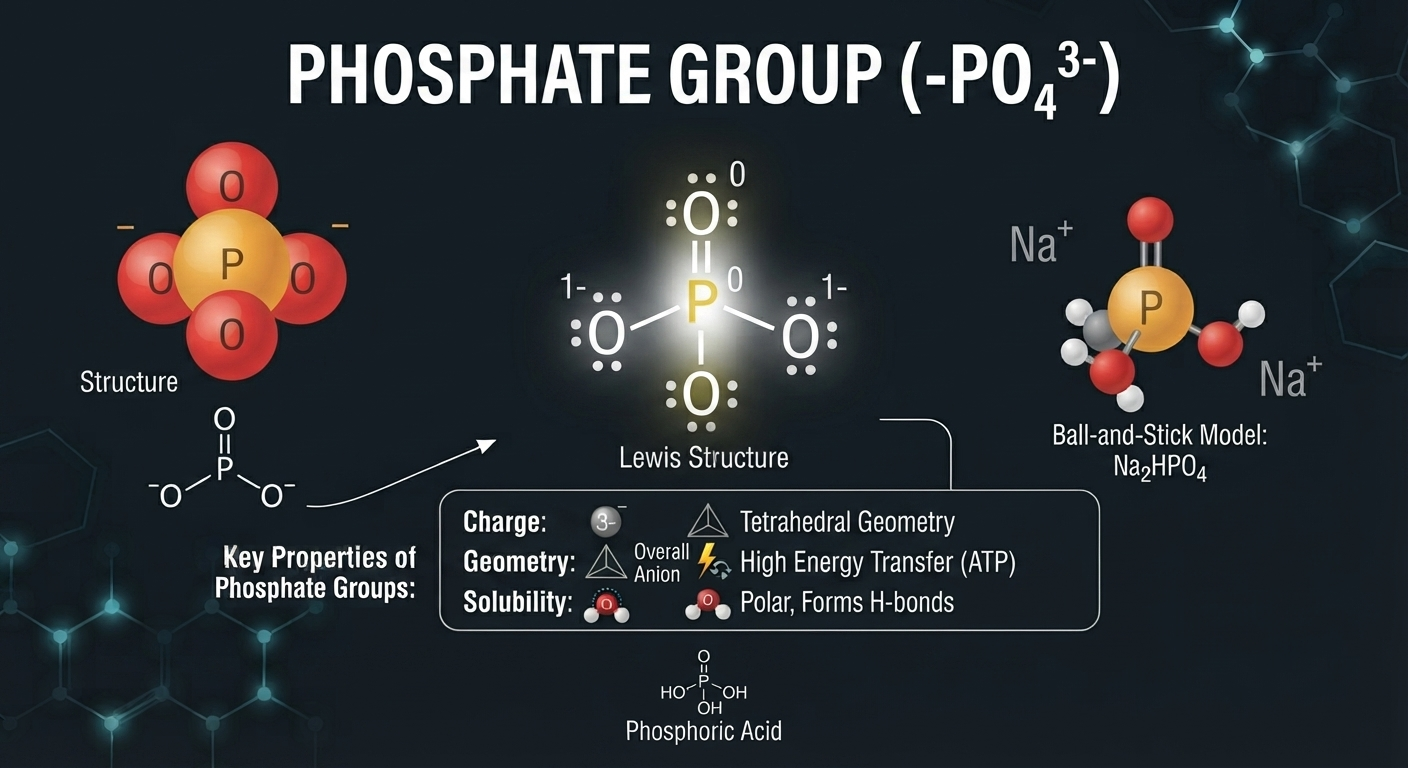
Phosphate groups are among the most important functional groups in biological systems.
They occur in ATP, ADP, DNA, RNA, phospholipids, and phosphorylated proteins. Because phosphate groups can carry substantial negative charge, they strongly influence the interactions and solubility of biomolecules.
Phosphorylation is also a major regulatory mechanism in cells. The addition or removal of phosphate groups can change protein activity, localization, stability, or interactions with other molecules.
Therefore, phosphate chemistry connects the structure of biomolecules with energy metabolism, signal transduction, gene regulation, and cellular communication.
7. Methyl Group

The methyl group is represented as –CH3.
Although it is chemically simple and relatively nonpolar, methyl groups have important biological functions.
Methylation occurs in DNA, proteins, lipids, and small metabolites. In DNA, methylation can influence chromatin organization and gene expression. In proteins, methylation can modify interactions with other molecules and contribute to cellular regulation.
This demonstrates that even a small chemical modification can influence biological function.
Chemical Bonds and Molecular Interactions
The stability and organization of biomolecules depend on several types of chemical interactions. Some interactions are strong covalent bonds that form the primary framework of a molecule. Others are weaker non-covalent interactions that collectively determine molecular folding, recognition, association, and assembly.
The distinction between covalent and non-covalent interactions is particularly important in structural biology.
Covalent bonds generally provide the basic chemical connectivity of biomolecules, whereas hydrogen bonds, ionic interactions, hydrophobic interactions, and van der Waals forces contribute extensively to the higher-order organization of biological molecules.
1. Covalent Bonds
A covalent bond forms when atoms share electrons. Covalent bonds constitute the fundamental structural framework of most biomolecules.
The carbon-carbon bonds of carbohydrates and lipids, peptide bonds of proteins, and phosphodiester bonds of nucleic acids are all covalent bonds.
The strength and directionality of covalent bonds allow biomolecules to maintain stable chemical structures while still participating in controlled biochemical reactions.
Three particularly important covalent linkages in biological macromolecules are peptide bonds, glycosidic bonds, and phosphodiester bonds.
Amino acids are joined by peptide bonds to form proteins. Monosaccharides can be connected through glycosidic bonds to form oligo- and polysaccharides. Nucleotides are linked through phosphodiester bonds to form DNA and RNA.
2. Non-Covalent Interactions
Although non-covalent interactions are individually weaker than covalent bonds, their combined effects are enormous in biological systems.
A protein may contain hundreds or thousands of weak interactions that collectively stabilize its folded structure. DNA base pairing, enzyme-substrate recognition, receptor-ligand binding, antigen-antibody recognition, and protein-protein interactions all depend heavily on non-covalent forces.
The major non-covalent interactions include hydrogen bonding, ionic interactions, van der Waals interactions, and hydrophobic interactions.
An important principle is that biological recognition usually does not depend on a single interaction. Instead, several weak interactions act simultaneously to generate highly specific and reversible molecular associations.
3. Hydrogen Bonding
A hydrogen bond occurs when a hydrogen atom covalently attached to an electronegative atom interacts with another electronegative atom.
Hydrogen bonding is fundamental to biological structure.
Water molecules form extensive hydrogen-bond networks. Proteins use hydrogen bonds to stabilize α-helices and β-sheets. DNA uses hydrogen bonding for complementary base pairing. RNA uses hydrogen bonding to generate complex secondary structures.
In double-stranded DNA, adenine pairs with thymine, while guanine pairs with cytosine. Adenine-thymine pairing involves two hydrogen bonds, whereas guanine-cytosine pairing involves three.
However, it is important not to conclude that DNA stability depends only on the number of hydrogen bonds. Base stacking interactions between adjacent nucleotides are also major contributors to nucleic acid stability.
This type of conceptual distinction is highly relevant to CSIR-NET because examination questions often test whether students can distinguish between memorized facts and mechanistic understanding.
4. Ionic Interactions and Salt Bridges
Ionic interactions occur between groups carrying opposite charges.
For example, a positively charged amino group can interact with a negatively charged carboxylate group. In proteins, such interactions can contribute to tertiary and quaternary structure and are often described as salt bridges.
The strength of an ionic interaction depends on the surrounding environment. Water can weaken electrostatic interactions because it stabilizes charged groups and has a high dielectric constant.
Therefore, the same ionic interaction may behave differently inside the hydrophobic interior of a protein compared with an exposed aqueous environment.
This illustrates an important structural biology principle: the biological environment strongly influences molecular interactions.
5. Van der Waals Forces
Van der Waals forces arise from transient changes in electron distribution that generate temporary dipoles.
Each individual interaction is weak, but a large number of closely packed atoms can generate substantial stabilization.
Van der Waals interactions become especially important when molecular surfaces fit closely together. They therefore contribute to protein packing, ligand binding, and molecular recognition.
The importance of these interactions emphasizes why molecular shape matters. A molecule must have an appropriate three-dimensional arrangement for many weak interactions to occur simultaneously.
6. Hydrophobic Interactions
Hydrophobic interactions are among the most important forces influencing biological organization.
Nonpolar groups do not form favorable interactions with water. Consequently, in an aqueous environment, nonpolar groups tend to cluster together and reduce their exposure to water.
This phenomenon contributes strongly to protein folding, formation of lipid bilayers, membrane organization, and association of hydrophobic molecules.
In globular proteins, many nonpolar amino acid side chains become buried within the protein interior, producing a hydrophobic core. In membranes, hydrophobic lipid tails become oriented away from the surrounding water.
Thus, hydrophobic interactions help explain both protein architecture and membrane formation.
Water: The Most Important Biological Solvent
Water is not simply an inert medium in which biochemical reactions occur. It actively influences the structure, stability, and behavior of biomolecules.
A water molecule contains one oxygen atom covalently bonded to two hydrogen atoms. Because oxygen is more electronegative than hydrogen, the electrons are distributed unequally. Water therefore has a partial negative charge near oxygen and partial positive charges near hydrogen.
This makes water a polar molecule.
The polarity of water allows it to form hydrogen bonds with other water molecules and with polar groups present in biomolecules.
Water can therefore interact strongly with ions and polar molecules while also driving the organization of nonpolar molecules through the hydrophobic effect.
Biological Importance of Water
The properties of water are essential for life.
Water acts as the major solvent in cells and allows ions, metabolites, proteins, nucleic acids, and other substances to interact in a controlled chemical environment.
Its high heat capacity helps organisms resist rapid temperature changes. Its high heat of vaporization makes evaporation an effective cooling mechanism. Its cohesive properties contribute to the movement of water through biological systems, particularly in plants.
One of the unusual properties of water is that solid ice is less dense than liquid water. The open structure created by hydrogen bonding causes ice to occupy more volume than the corresponding liquid. Consequently, ice floats, allowing aquatic environments to remain liquid beneath an insulating layer.
pH and Biomolecular Structure
The pH of a solution is defined as:
pH = −log[H+]
The concentration of hydrogen ions influences the protonation state of functional groups.
For biomolecules such as proteins, this can have profound consequences. Changing pH can alter the charge of amino acid side chains, disrupt ionic interactions, modify hydrogen bonding, and eventually affect protein folding and activity.
Enzymes are particularly sensitive to pH because catalytic activity depends on the precise chemical environment of the active site.
Therefore, pH is not simply a numerical measurement. It is a factor capable of changing the molecular structure and function of biological systems.
Acids, Bases, and pKa
An acid is a substance that can donate a proton, whereas a base can accept a proton.
A generalized acid dissociation reaction can be written as:
HA ⇌ H+ + A−
The equilibrium constant for this reaction is related to the acid dissociation constant, Ka.
The pKa is defined as:
pKa = −log Ka
A lower pKa generally indicates a stronger acid because the protonated form dissociates more readily.
In biological systems, the relationship between pH, pKa, and protonation state is extremely important because it determines the charge of many functional groups.
Henderson-Hasselbalch Equation
The Henderson-Hasselbalch equation is:
pH = pKa + log([A−]/[HA])
This equation provides a quantitative relationship between the pH of a solution and the ratio of protonated and deprotonated forms of a weak acid.
It is particularly useful for understanding biological buffering systems.
At pH = pKa, the concentrations of the protonated and deprotonated forms are equal.
This concept is frequently useful in questions involving amino acid titration, buffer systems, and protein charge.
Biomolecular Buffers
A buffer resists changes in pH when relatively small amounts of acid or base are introduced.
Cells require effective buffering because biochemical reactions continuously produce or consume protons.
Important biological buffering systems include phosphate, bicarbonate, and protein-based buffers.
The bicarbonate system is especially important in maintaining blood pH:
CO2 + H2O ⇌ H2CO3 ⇌ H+ + HCO3−
Carbonic anhydrase catalyzes the interconversion between carbon dioxide and carbonic acid extremely efficiently.
The ability of biological systems to regulate pH is essential because protein structure and enzyme activity depend strongly on the protonation state of functional groups.
Major Classes of Biomolecules
The major classes of biomolecules are carbohydrates, lipids, proteins, and nucleic acids.
Although these groups are often studied separately, they are deeply interconnected.
For example, carbohydrates can be converted into metabolic intermediates that contribute to lipid and amino acid synthesis. Amino acids can enter metabolic pathways and contribute to energy production. Lipids provide components for membranes and signaling molecules. Nucleotides participate not only in genetic information storage but also in energy transfer and cellular regulation.
Thus, biomolecules should be viewed as an interconnected biochemical system rather than four completely independent categories.
1. Carbohydrates
Carbohydrates are organic molecules composed mainly of carbon, hydrogen, and oxygen. They are traditionally represented by the approximate empirical relationship Cn(H2O)n for many simple carbohydrates, although this formula does not apply universally to all carbohydrates.
Carbohydrates perform diverse biological functions. Glucose is an important metabolic fuel, glycogen and starch serve as storage materials, while cellulose and chitin provide structural support.
Carbohydrates also participate in cell recognition and molecular signaling. Complex carbohydrates attached to proteins and lipids at the cell surface can act as biological identity markers and recognition molecules.
(i) Monosaccharides
Monosaccharides are the simplest carbohydrate units and cannot be hydrolyzed into smaller carbohydrate molecules.
Glucose, fructose, galactose, ribose, and deoxyribose are important biological monosaccharides.
Monosaccharides can be classified according to the number of carbon atoms present in their structure. They may also be classified as aldoses or ketoses depending on the nature and position of their carbonyl group.
In aqueous solution, many monosaccharides do not remain exclusively in their open-chain form. They can undergo intramolecular reactions that produce cyclic structures.
This is particularly important for glucose, which predominantly exists in cyclic forms under physiological conditions.
(ii) Stereochemistry of Carbohydrates
Carbohydrates provide an excellent example of the importance of three-dimensional molecular structure.
Two molecules may contain exactly the same atoms and chemical bonds but differ in the spatial arrangement of those atoms. Such molecules are known as stereoisomers.
Biological systems are highly sensitive to stereochemistry because enzymes and receptors are themselves three-dimensional structures.
Therefore, two stereoisomers may interact very differently with the same enzyme.
Concepts such as D/L configuration, epimers, anomers, and optical activity are therefore important for advanced understanding of carbohydrate chemistry.
(iii) Disaccharides
Disaccharides are formed when two monosaccharides become linked through a glycosidic bond.
The most familiar examples are sucrose, lactose, and maltose.
Sucrose consists of glucose and fructose. Lactose consists of glucose and galactose. Maltose consists of two glucose units.
The exact position and configuration of the glycosidic bond determine the chemical properties of the resulting disaccharide.
This is why structurally related carbohydrates can behave differently in biological systems.
(iv) Polysaccharides
Polysaccharides are large carbohydrate molecules composed of many monosaccharide residues.
Starch is a major storage polysaccharide in plants, whereas glycogen is an important storage polysaccharide in animals and fungi.
Cellulose is a structural polysaccharide that forms a major component of plant cell walls. Chitin is another important structural polysaccharide and occurs in fungal cell walls and the exoskeletons of arthropods.
The different biological properties of these polysaccharides arise largely from differences in glycosidic linkage, branching, and three-dimensional organization.
A particularly important conceptual example is the difference between starch and cellulose. Both contain glucose as their fundamental building block, yet their linkage patterns result in dramatically different structures and biological properties.
Lipids
Lipids are a chemically diverse group of molecules characterized primarily by their hydrophobic or amphipathic properties.
Unlike proteins and nucleic acids, lipids do not form a single class of repeating polymers. Instead, the term lipid encompasses several structurally distinct molecules.
Lipids include fatty acids, triacylglycerols, phospholipids, sphingolipids, sterols, and many signaling molecules.
Their biological roles extend far beyond energy storage. Lipids form the structural basis of biological membranes, regulate membrane properties, act as signaling molecules, and serve as precursors for hormones and other biologically active compounds.
Fatty Acids
Fatty acids generally consist of a hydrocarbon chain terminating in a carboxyl group.
The hydrocarbon chain may contain no double bonds or one or more double bonds.
Saturated fatty acids contain no carbon-carbon double bonds. Unsaturated fatty acids contain one or more carbon-carbon double bonds.
The presence and position of double bonds influence the physical properties of fatty acids and membrane lipids.
In many naturally occurring unsaturated fatty acids, cis double bonds introduce bends into the hydrocarbon chain. These structural changes can affect how closely lipid molecules pack together and consequently influence membrane fluidity.
Triacylglycerols
Triacylglycerols consist of glycerol esterified with three fatty acids.
They are major energy-storage molecules.
Their high energy density makes them particularly effective for long-term energy storage. Compared with carbohydrates, fats can store large amounts of energy in a relatively compact form.
The hydrophobic nature of triacylglycerols also means that they can be stored with relatively little associated water.
Phospholipids and Membrane Structure
Phospholipids are fundamental components of biological membranes.
They possess both hydrophilic and hydrophobic regions and are therefore described as amphipathic molecules.
When amphipathic lipids are placed in an aqueous environment, their hydrophobic regions tend to avoid water while their hydrophilic regions interact with the surrounding aqueous phase. This drives the spontaneous formation of organized structures such as lipid bilayers.
The lipid bilayer provides the basic structural framework of the plasma membrane and intracellular membranes.
Understanding phospholipid structure therefore provides the chemical foundation for understanding membrane transport, receptors, membrane proteins, signaling, and cellular compartmentalization.
Steroids and Sterols
Steroids possess a characteristic four-ring carbon framework.
Cholesterol is the most familiar biological sterol in animals. It contributes to membrane organization and serves as a precursor for steroid hormones and other biologically important molecules.
Steroid hormones such as cortisol, aldosterone, testosterone, estrogen, and progesterone are derived from cholesterol.
Because steroid molecules are relatively hydrophobic, they can cross lipid bilayers more readily than many polar signaling molecules and can interact with intracellular receptors.
Proteins
Proteins are among the most functionally diverse biomolecules found in living organisms.
They act as enzymes, receptors, channels, transporters, structural components, molecular motors, antibodies, transcription regulators, and signaling molecules.
The extraordinary diversity of proteins arises from the ability of cells to construct polypeptide chains using combinations of 20 standard amino acids.
The sequence of amino acids determines how the polypeptide folds, and the resulting three-dimensional structure determines its biochemical properties.
This relationship can be represented conceptually as:
Amino acid sequence → Molecular folding → Three-dimensional structure → Biological function
Although this relationship is fundamental, it is not always simple. Protein folding depends on the sequence as well as the cellular environment, molecular interactions, cofactors, chaperones, and post-translational modifications.
Amino Acids: Building Blocks of Proteins
A standard α-amino acid contains an amino group, a carboxyl group, a hydrogen atom, and a variable side chain attached to the α-carbon.
The general structure can be represented as:
NH2–CH(R)–COOH
The R group determines the chemical identity of the amino acid.
The side chains vary widely in size, polarity, charge, aromaticity, flexibility, and chemical reactivity.
This diversity allows proteins to generate highly specialized chemical environments.
For example, hydrophobic amino acids can contribute to the formation of protein interiors, while charged amino acids can participate in electrostatic interactions. Cysteine can form disulfide bonds, histidine can participate in acid-base catalysis, and aromatic residues can contribute to stacking and hydrophobic interactions.
Zwitterionic Nature of Amino Acids

At physiological pH, amino acids often exist predominantly as zwitterions, containing both positive and negative charges.
The amino group is commonly protonated:
–NH3+
while the carboxyl group is commonly deprotonated:
–COO−
Thus, an amino acid can possess an overall neutral charge while still containing charged functional groups.
The charge of an amino acid changes as the pH changes. This property is central to understanding amino acid titration, protein solubility, electrophoresis, and isoelectric points.
Peptide Bond Formation
Amino acids are connected through peptide bonds.
A peptide bond forms between the carboxyl group of one amino acid and the amino group of another amino acid, with the release of water.
The resulting linkage has the form:
–CO–NH–
The peptide bond has partial double-bond character because of resonance. Consequently, rotation around the peptide bond is restricted and the peptide group is approximately planar.
This structural restriction is important because it limits the conformational freedom of proteins and influences how polypeptide chains fold.
1. Primary Structure of Proteins

The primary structure refers to the linear sequence of amino acids in a polypeptide chain.
The sequence is not simply a list of amino acids. It contains the information necessary for the polypeptide to acquire its characteristic three-dimensional structure under appropriate conditions.
A single amino acid substitution can sometimes have profound consequences.
A classical example is sickle-cell disease, in which a mutation in the β-globin gene results in a change in the amino acid sequence of β-globin. The altered protein has different properties, contributing to the formation of abnormal hemoglobin polymers under deoxygenated conditions.
This example demonstrates how a change at the molecular level can ultimately produce a major cellular and physiological phenotype.
2. Secondary Structure of Proteins
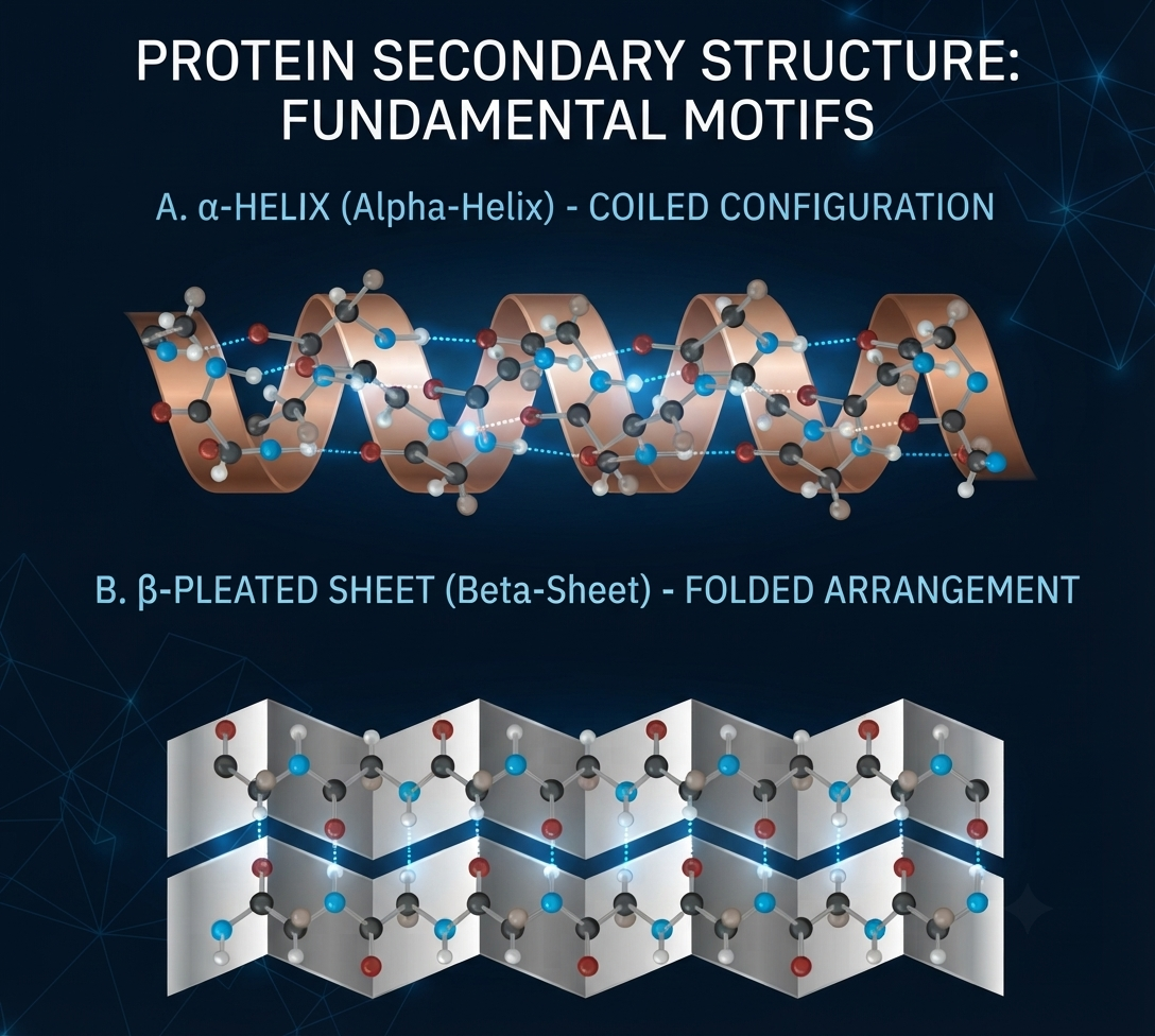
Secondary structure describes local structural patterns formed by the polypeptide backbone.
The two major regular secondary structures are the α-helix and β-sheet.
These structures are stabilized primarily through hydrogen bonding between backbone carbonyl oxygen atoms and amide hydrogen atoms.
In an α-helix, the polypeptide backbone coils into a helical arrangement. In β-sheets, extended polypeptide segments align with one another to form sheet-like structures.
The orientation of β-strands may be parallel, antiparallel, or mixed.
3. Tertiary Structure
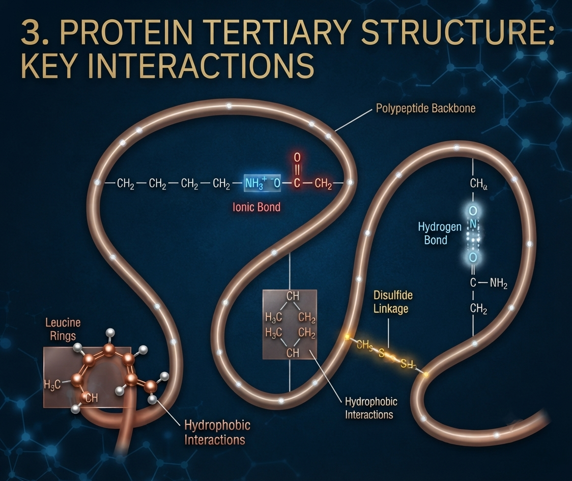
Tertiary structure describes the complete three-dimensional arrangement of a single polypeptide chain.
It results from interactions among amino acid side chains and between side chains and the surrounding environment.
Hydrophobic interactions often drive nonpolar residues toward the interior of globular proteins, while charged and polar residues are frequently exposed to the aqueous environment.
Hydrogen bonds, ionic interactions, van der Waals forces, and disulfide bonds can further stabilize the folded structure.
The final structure creates specific regions such as enzyme active sites, ligand-binding pockets, and protein interaction surfaces.
4. Quaternary Structure

Some proteins contain multiple polypeptide subunits. The arrangement and interactions of these subunits constitute the quaternary structure.
Hemoglobin is a classic example. It contains four polypeptide subunits that interact to produce the functional protein complex.
Quaternary organization can provide important functional advantages, including cooperative binding, regulation, structural stability, and division of functional roles among subunits.
The distinction between tertiary and quaternary structure is therefore important: tertiary structure describes the three-dimensional organization of one polypeptide chain, whereas quaternary structure concerns the organization of multiple polypeptide chains.
Nucleic Acids
Nucleic acids are macromolecules specialized for the storage, transmission, regulation, and expression of genetic information.
DNA and RNA are polymers of nucleotides.
DNA acts as the principal long-term repository of genetic information in most organisms, whereas RNA performs numerous functions in information transfer, translation, catalysis, regulation, and structural organization.
The structure of nucleic acids is therefore closely connected to molecular genetics.
Nucleotides and Nucleosides
A nucleotide contains three basic components:
- Nitrogenous base
- Pentose sugar
- Phosphate group
A nucleoside contains:
Nitrogenous base + Pentose sugar
Therefore, a nucleotide is essentially a phosphorylated nucleoside.
This distinction is extremely important in molecular biology and is frequently tested in competitive examinations.
The sugar in DNA is deoxyribose, whereas RNA contains ribose.
Nitrogenous Bases
Nitrogenous bases are divided into purines and pyrimidines.
Purines possess a fused two-ring structure and include adenine and guanine.
Pyrimidines possess a single six-membered ring and include cytosine, thymine, and uracil.
DNA contains adenine, guanine, cytosine, and thymine.
RNA contains adenine, guanine, cytosine, and uracil.
The difference between thymine and uracil is chemically small but biologically important.
DNA Structure
DNA generally consists of two polynucleotide strands arranged in a double-helical structure.
The two strands are antiparallel. This means that one strand runs from the 5′ end toward the 3′ end, whereas the complementary strand runs from 3′ toward 5′.
The sugar-phosphate backbone forms the external framework of the molecule, while the nitrogenous bases are oriented toward the interior.
Complementary base pairing allows the sequence of one strand to determine the sequence of the other.
Adenine pairs with thymine, while guanine pairs with cytosine.
This complementarity provides the structural basis for accurate DNA replication and contributes to mechanisms of DNA repair and information transfer.
RNA Structure and Diversity
RNA contains ribose sugar and uracil.
Although RNA is often described as a single-stranded molecule, this does not mean that RNA lacks structural complexity. RNA can fold into extensive secondary and tertiary structures through internal base pairing and other molecular interactions.
Different RNA molecules perform distinct functions.
Messenger RNA carries coding information from DNA to ribosomes. Transfer RNA participates in decoding codons during translation. Ribosomal RNA forms an essential structural and catalytic component of ribosomes.
Other RNA molecules, including microRNAs, small interfering RNAs, small nuclear RNAs, small nucleolar RNAs, and long non-coding RNAs, participate in gene regulation, RNA processing, and other cellular processes.
Thus, RNA should be considered a structurally and functionally diverse class of biomolecules rather than simply a temporary copy of DNA.
Bonds Linking Biological Monomers
One of the most important ways to understand biomolecules is to identify the bonds connecting their building blocks.
Proteins contain amino acids connected by peptide bonds.
Polysaccharides contain monosaccharides connected through glycosidic bonds.
DNA and RNA contain nucleotides connected through phosphodiester bonds.
Lipids contain characteristic ester or other linkages depending on the lipid class.
These bonds are chemically different and produce different structural properties.
For example, peptide bonds create a relatively rigid protein backbone, while glycosidic linkages determine the shape and branching pattern of polysaccharides.
Condensation Reactions
Many biological macromolecules are assembled through condensation reactions.
During condensation, two molecules are joined while a small molecule, often water, is released.
Formation of peptide bonds and many glycosidic linkages can be understood in this context.
The reverse process is hydrolysis, in which water participates in bond cleavage.
These reactions are fundamental to digestion and metabolism.
However, in living cells, the actual biochemical mechanisms of polymer synthesis and degradation are usually enzyme-catalyzed and often involve activated intermediates rather than simple uncatalyzed condensation or hydrolysis.
This distinction becomes increasingly important at an advanced CSIR-NET level.
Molecular Complementarity
Biological systems depend extensively on molecular recognition.
A molecule can recognize another molecule because their structures are chemically complementary.
Complementarity involves several characteristics simultaneously, including molecular shape, charge, hydrogen-bonding capacity, hydrophobicity, and flexibility.
For example, an enzyme active site contains amino acid residues arranged in a three-dimensional environment that is complementary to particular substrates. Similarly, receptors recognize signaling molecules, antibodies recognize antigens, and transcription factors recognize specific DNA sequences.
This principle explains why biological interactions are often highly specific.
Structure-Function Relationship
The relationship between molecular structure and biological function represents one of the most important themes in Life Science.
A protein’s amino acid sequence influences its folding pattern. Its folding pattern creates specific surfaces and pockets. These surfaces determine what molecules the protein can bind and what reactions it can perform.
Similarly, lipid structure influences membrane properties. The saturation level and chain length of fatty acids can influence membrane packing and fluidity.
Carbohydrate structure determines whether a polysaccharide behaves as an energy-storage molecule or structural material.
Nucleic acid structure allows genetic information to be stored, copied, and expressed.
Therefore, molecular structure provides the physical basis for biological function.
Molecular Structure and Biological Specificity
Biological molecules operate within highly organized cellular environments. A receptor does not bind every molecule present in the cell. An enzyme does not catalyze every possible chemical reaction. A transcription factor does not recognize every DNA sequence.
Specificity arises because molecular structures are complementary.
This specificity is one reason biological systems can perform thousands of biochemical reactions simultaneously without complete chemical chaos.
Biomolecules and Cellular Organization
Biomolecules are also responsible for the organization of cells into functional compartments.
Phospholipids form membranes. Proteins create transport systems and molecular machines. Carbohydrates contribute to extracellular structures and cell recognition. Nucleic acids organize genetic information.
The cell can therefore be considered a highly organized molecular system in which biomolecules interact continuously.
For example, a plasma membrane is not simply a lipid sheet. It contains proteins, carbohydrates, cholesterol, signaling molecules, and dynamic interactions that allow the cell to communicate with its environment.
Similarly, chromatin is not simply DNA. It is a complex association of DNA with histones and many other proteins and regulatory molecules.
Biomolecular Interactions and Enzymes
Enzymes are proteins or catalytic RNA molecules that accelerate biochemical reactions.
Their catalytic activity depends heavily on molecular structure.
An enzyme contains an active site where substrates bind and chemical reactions occur. The active site possesses a specific arrangement of amino acid side chains that creates a chemically suitable environment.
Substrate recognition depends on multiple interactions, including hydrogen bonds, ionic interactions, hydrophobic interactions, and van der Waals forces.
Therefore, enzyme catalysis provides one of the clearest examples of how molecular structure produces biological function.
Biomolecules in Energy Metabolism
Biomolecules are continuously transformed through metabolic pathways.
Carbohydrates can be oxidized to generate ATP. Lipids can be degraded into fatty acids and other intermediates that contribute to energy production. Amino acids can be used for biosynthesis or enter metabolic pathways under appropriate conditions.
Nucleotides such as ATP and GTP also function as energy-rich molecules and participate in cellular processes requiring controlled energy transfer.
Thus, biomolecules are not static structures. They exist within dynamic networks of synthesis, modification, degradation, and interconversion.
Biomolecules and Genetic Information
Nucleic acids provide the molecular foundation of heredity.
The sequence of nucleotides in DNA contains genetic information. During replication, complementary base pairing allows the information to be copied. During transcription, selected DNA sequences are used as templates for RNA synthesis. During translation, nucleotide information in messenger RNA is converted into the amino acid sequence of proteins.
Therefore, there is a fundamental relationship:
DNA → RNA → Protein
This relationship connects nucleic acid structure directly with protein structure and cellular function.
Structural Changes and Disease
Changes in biomolecular structure can lead to disease.
A mutation may change a protein’s amino acid sequence. A chemical modification may alter enzyme activity. An abnormal lipid composition may affect membrane properties. Improper protein folding may lead to aggregation.
Diseases such as sickle-cell disease illustrate how a molecular change can influence cellular behavior and ultimately produce physiological consequences.
Other disorders involve abnormal protein aggregation, defective nucleic acid processing, altered lipid metabolism, or changes in carbohydrate metabolism.
Therefore, structural biochemistry has direct relevance to medicine, biotechnology, pharmacology, and disease research.
Biomolecules and Biotechnology
Modern biotechnology relies extensively on our understanding of biomolecular structure.
Recombinant DNA technology depends on the complementary base-pairing properties of nucleic acids. PCR depends on DNA structure and strand separation. Protein engineering depends on understanding amino acid sequences and protein folding. Enzyme technology depends on molecular recognition and catalytic mechanisms.
Drug development also relies heavily on structural biology. A drug molecule is designed to interact with a particular protein, enzyme, receptor, or nucleic acid structure.
Thus, the study of biomolecular structure provides the chemical foundation for modern biotechnology and molecular medicine.


