
1. Introduction
The adaptive immune system must distinguish between different molecular structures and communicate information about them to T lymphocytes. Unlike antibodies and B-cell receptors, which can recognize intact antigens, T-cell receptors (TCRs) generally recognize antigen-derived peptide fragments only when they are presented by specialized cell-surface molecules called Major Histocompatibility Complex (MHC) molecules.
MHC molecules are membrane-bound glycoproteins that bind peptide fragments and display them on the surface of cells. T cells recognize the combination of peptide + MHC molecule through their T-cell receptors.
MHC molecules therefore form a central link between antigen processing and T-cell-mediated immunity.
The two major classes are:
- MHC Class I
- MHC Class II
In humans, MHC molecules are encoded by genes within the human leukocyte antigen (HLA) system.
2. Definition of MHC Molecules
Major Histocompatibility Complex (MHC) refers to a group of highly polymorphic genes that encode cell-surface proteins involved primarily in the presentation of peptide antigens to T lymphocytes.
The basic function can be represented as:
Antigen → Processing → Peptide generation → MHC binding → Cell-surface presentation → TCR recognition
MHC molecules therefore act as molecular platforms for displaying antigen-derived peptides to T cells.
3. Human MHC: The HLA System
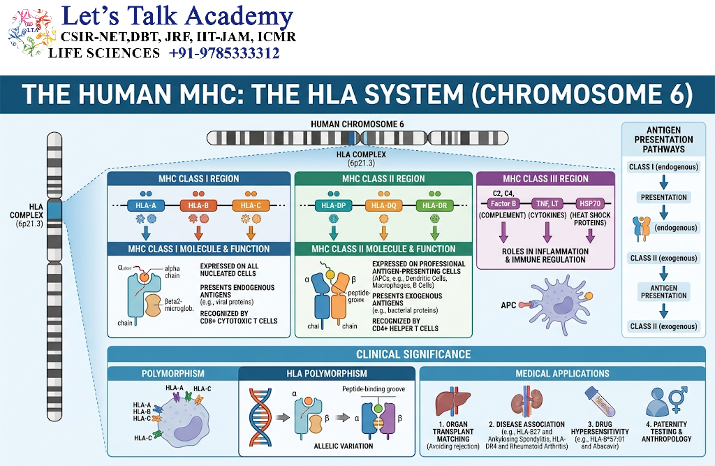
In humans, MHC genes are called Human Leukocyte Antigen (HLA) genes.
The major classical HLA molecules include:
MHC Class I
- HLA-A
- HLA-B
- HLA-C
MHC Class II
- HLA-DP
- HLA-DQ
- HLA-DR
These molecules are encoded by genes located in the MHC region on chromosome 6.
The MHC region is one of the most genetically diverse regions of the human genome.
4. Major Functions of MHC Molecules
MHC molecules perform several important functions.
4.1 Antigen Presentation
MHC molecules bind peptides and present them to T cells.
4.2 T-Cell Recognition
TCRs recognize antigenic peptides in association with MHC molecules.
4.3 Immune Surveillance
MHC molecules allow T cells to monitor cells for signs of:
- Viral infection
- Abnormal protein production
- Intracellular pathogens
- Cellular transformation
4.4 Self-Recognition
MHC molecules participate in distinguishing normal host cells from cells displaying abnormal or foreign peptide patterns.
4.5 Transplantation
Differences in MHC molecules between individuals can contribute strongly to transplant rejection and tissue compatibility.
5. Classification of MHC Molecules
MHC molecules are broadly divided into two major classes.
| Feature | MHC Class I | MHC Class II |
|---|---|---|
| Main molecules | HLA-A, HLA-B, HLA-C | HLA-DP, HLA-DQ, HLA-DR |
| Main expression | Most nucleated cells | Mainly professional APCs |
| Main antigen source | Intracellular | Extracellular |
| Peptide-presenting cells | Most nucleated cells | Dendritic cells, macrophages, B cells |
| Main T cell | CD8⁺ T cells | CD4⁺ T cells |
| Peptide-binding groove | Closed at ends | Open at ends |
| Typical peptide length | About 8–10 amino acids | Commonly about 13–18 or more amino acids |
These are general patterns, and peptide length can vary depending on the MHC molecule and antigen.
6. Structure of MHC Class I Molecules
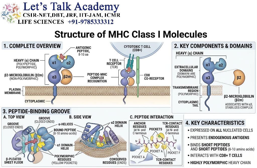
MHC Class I molecules are composed of:
- A large polymorphic α chain
- A smaller β2-microglobulin molecule
The α chain contains three extracellular domains:
- α1
- α2
- α3
The peptide-binding groove is formed mainly by the α1 and α2 domains.
The α3 domain interacts with the CD8 co-receptor.
6.1 Basic Structure
             Peptide
        ───────────────
       /              \
      α1              α2
       \              /
        \____________/
             α3
              |
        Cell membrane
              |
      Cytoplasmic region

        β2-microglobulin
7. MHC Class I Peptide-Binding Groove
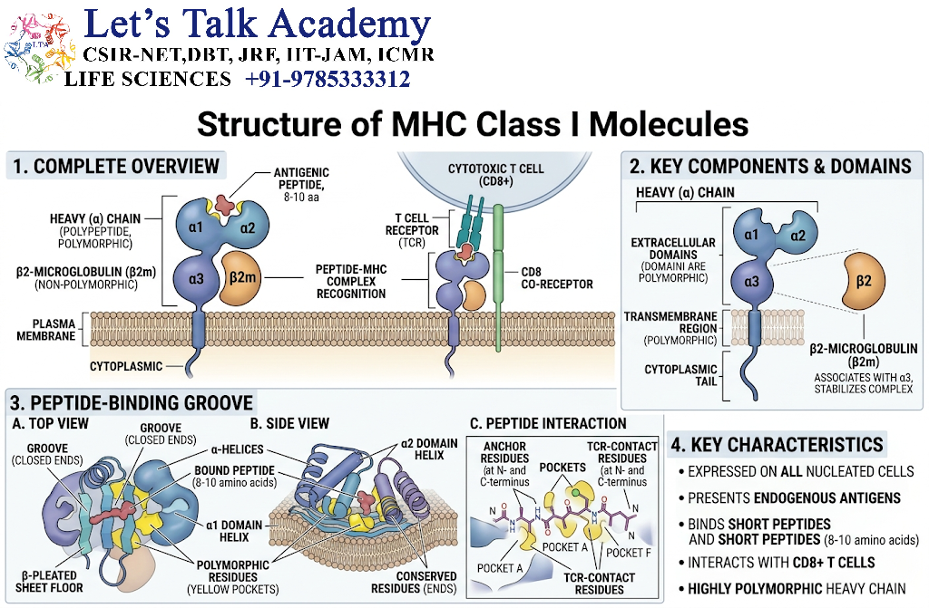
The peptide-binding groove of MHC Class I is formed by the α1 and α2 domains.
It is generally closed at both ends.
Therefore, MHC Class I commonly binds relatively short peptides.
Typical peptides are approximately:
8–10 amino acids long
However, some MHC class I molecules can accommodate peptides of different lengths, especially when longer peptides bulge from the binding groove.
8. Structure of MHC Class II Molecules
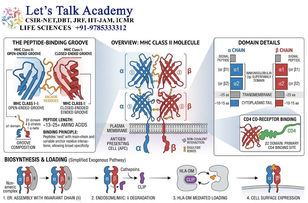
MHC Class II molecules contain two membrane-associated polypeptide chains:
- α chain
- β chain
Each chain has two extracellular domains.
α chain
- α1
- α2
β chain
- β1
- β2
The peptide-binding groove is mainly formed by:
α1 + β1
The β2 domain contributes to interaction with the CD4 co-receptor.
8.1 Basic Structure
             Peptide
      ───────────────────
       α1              β1
        \              /
         \____________/
          α2        β2
             |     |
          Membrane
9. MHC Class II Peptide-Binding Groove
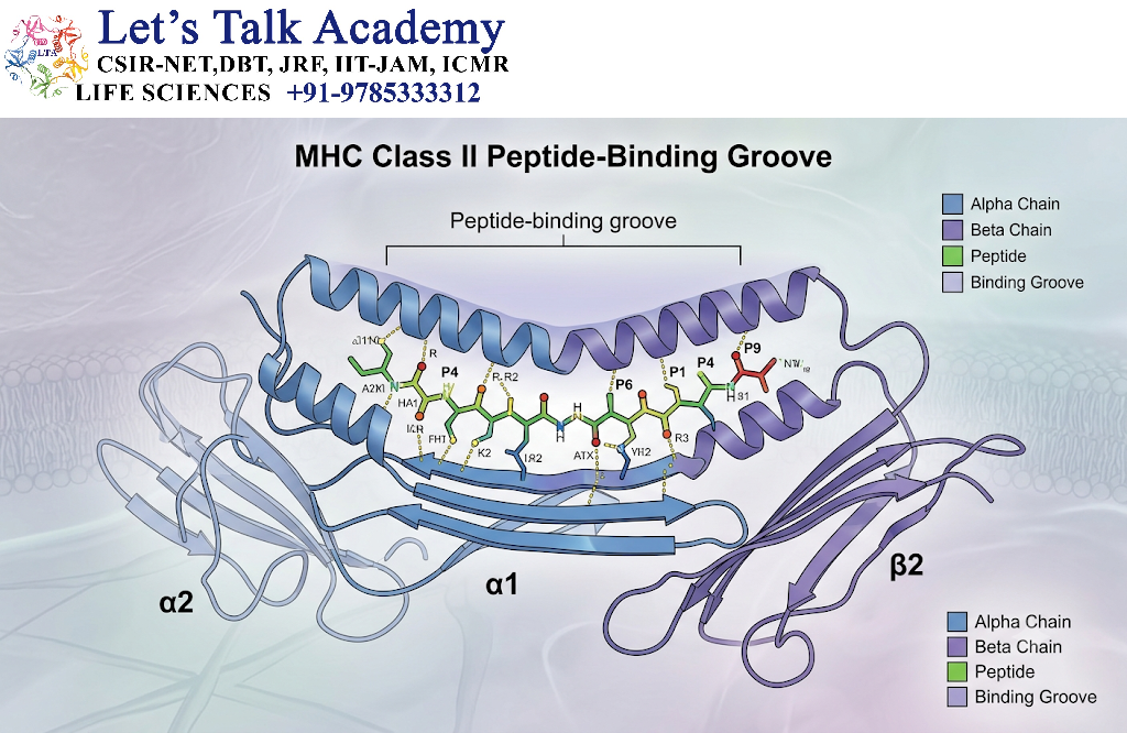
Unlike MHC Class I, the MHC Class II peptide-binding groove is generally open at both ends.
This allows longer peptide fragments to bind.
Peptides commonly contain approximately:
13–18 or more amino acids
The actual peptide can extend beyond the core region that makes the most important contacts with the MHC molecule.
10. Expression of MHC Class I
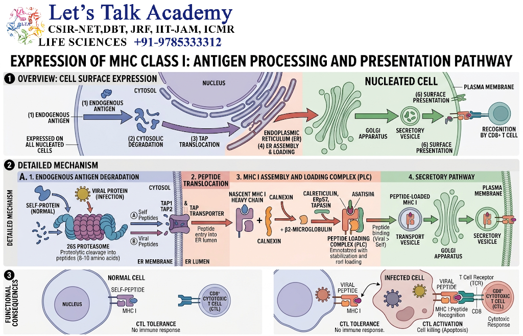
MHC Class I molecules are expressed on most nucleated cells.
They are generally absent from mature red blood cells because mature mammalian erythrocytes lack a nucleus and do not express conventional MHC class I at the cell surface.
MHC Class I expression is particularly important for monitoring intracellular protein production.
11. Expression of MHC Class II
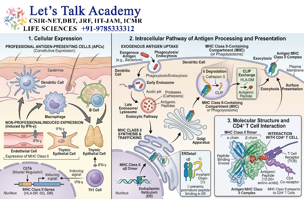
MHC Class II molecules are primarily expressed by professional antigen-presenting cells (APCs).
Major MHC Class II-expressing cells include:
- Dendritic cells
- Macrophages
- B lymphocytes
These cells are specialized for capturing, processing, and presenting antigens to CD4⁺ T cells.
12. MHC Class I and Intracellular Antigens
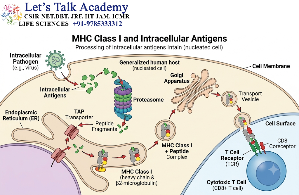
MHC Class I mainly presents peptides derived from proteins located within the cell.
These may originate from:
- Viral proteins
- Abnormal cellular proteins
- Normal cellular proteins
- Tumor-associated proteins
- Intracellular pathogens
The pathway can be summarized as:
Intracellular protein → Proteasomal degradation → Peptides → TAP transport → ER → MHC I loading → Cell surface → CD8⁺ T cell
13. MHC Class II and Extracellular Antigens
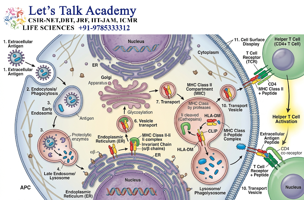
MHC Class II mainly presents peptides derived from extracellular material that has been taken up by antigen-presenting cells.
Examples include proteins from:
- Bacteria
- Extracellular parasites
- Environmental antigens
- Soluble proteins
The general pathway is:
Extracellular antigen → Endocytosis/phagocytosis → Endosomal processing → Peptide generation → MHC II loading → Cell surface → CD4⁺ T cell
14. Antigen Processing for MHC Class I
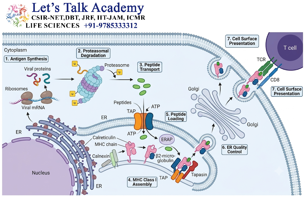
MHC Class I antigen presentation involves several major steps.
Step 1: Protein Generation
Intracellular proteins are produced within the cell.
Step 2: Proteasomal Degradation
Some proteins are degraded into peptide fragments by the proteasome.
Step 3: Peptide Transport
Peptides are transported into the endoplasmic reticulum by TAP, meaning transporter associated with antigen processing.
Step 4: MHC Class I Loading
Peptides are loaded onto newly synthesized MHC Class I molecules.
Step 5: Transport to Cell Surface
The peptide–MHC I complex passes through the secretory pathway and reaches the plasma membrane.
Step 6: T-Cell Recognition
CD8⁺ T cells recognize the peptide–MHC I complex.
15. MHC Class I Processing Flowchart
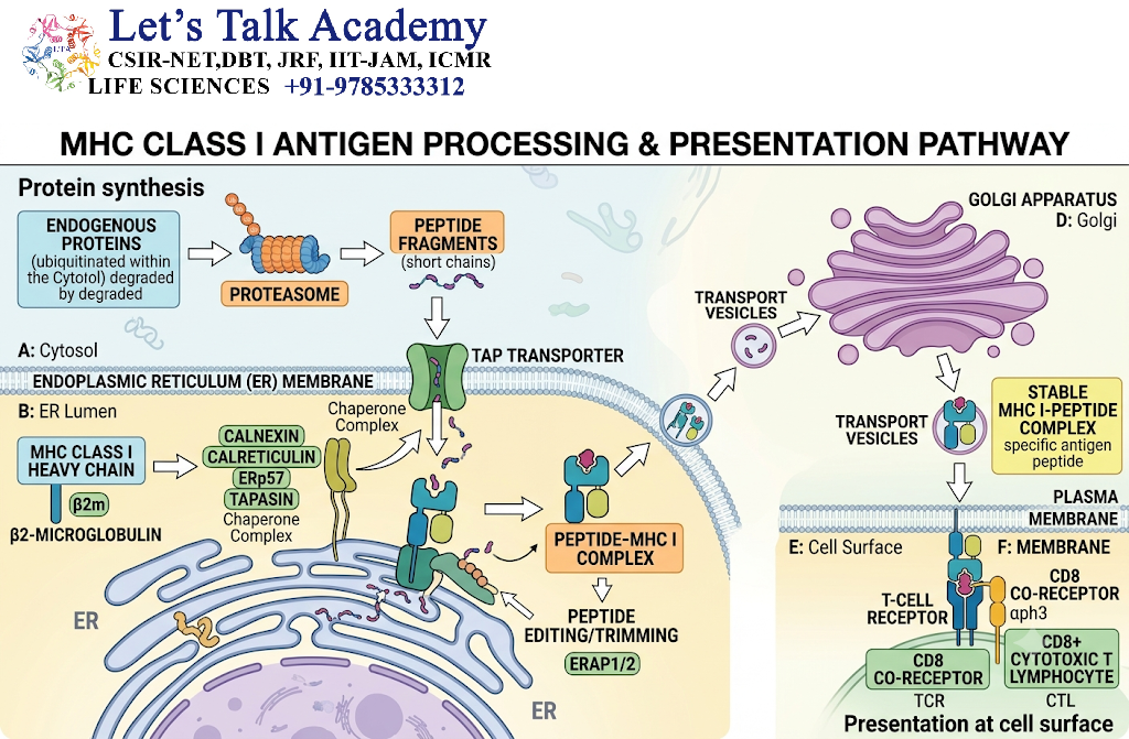
Intracellular Protein
        ↓
Proteasome
        ↓
Peptide Fragments
        ↓
TAP Transporter
        ↓
Endoplasmic Reticulum
        ↓
MHC Class I Peptide Loading
        ↓
Golgi / Secretory Pathway
        ↓
Cell Surface
        ↓
CD8⁺ T Cell Recognition
16. Antigen Processing for MHC Class II
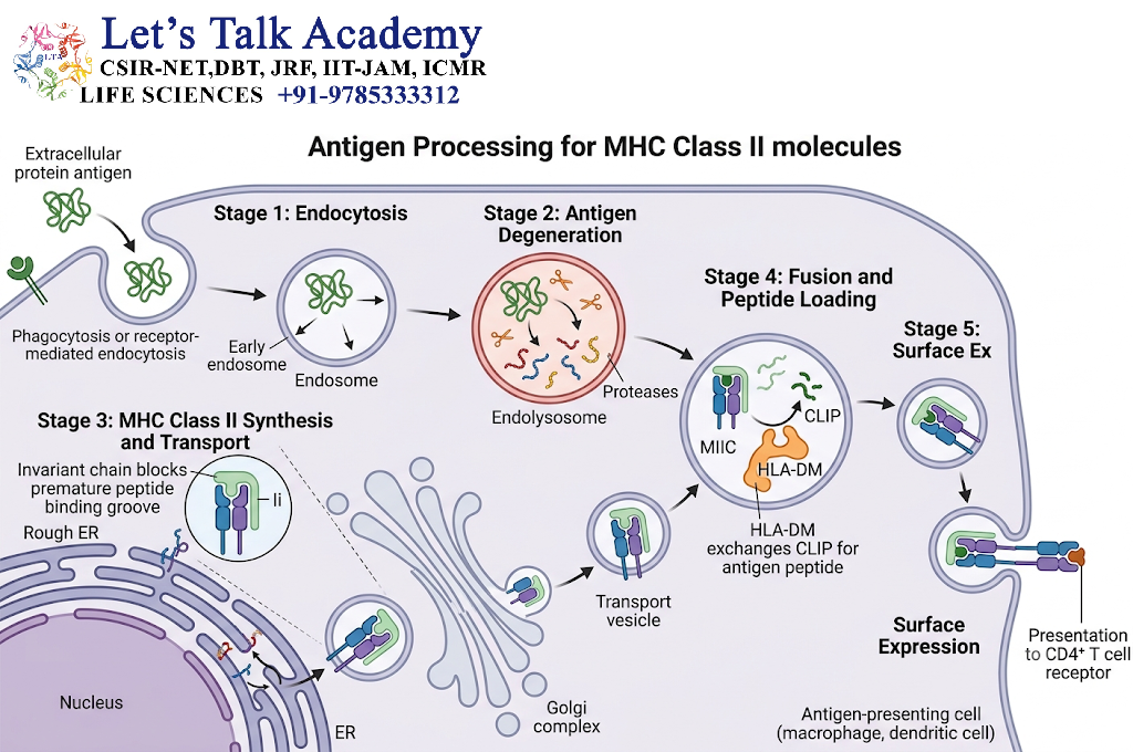
MHC Class II antigen processing follows a different pathway.
Step 1: Antigen Uptake
Extracellular antigen is internalized by an antigen-presenting cell.
Step 2: Endosomal Processing
The antigen enters endosomal or lysosomal compartments.
Step 3: Proteolysis
Proteases break the antigen into peptide fragments.
Step 4: MHC Class II Preparation
MHC Class II molecules are synthesized in the endoplasmic reticulum.
They associate with an invariant chain, which helps prevent premature peptide loading in the ER.
Step 5: CLIP Formation
The invariant chain is progressively degraded, leaving a fragment called CLIP in the peptide-binding groove.
Step 6: Peptide Loading
In specialized endosomal compartments, CLIP is removed and replaced by an antigen-derived peptide.
HLA-DM plays an important role in peptide exchange and loading.
Step 7: Cell-Surface Expression
The peptide–MHC II complex is transported to the cell surface.
Step 8: CD4⁺ T-Cell Recognition
CD4⁺ T cells recognize the peptide–MHC II complex.
17. MHC Class II Processing Flowchart
Extracellular Antigen
        ↓
Endocytosis / Phagocytosis
        ↓
Endosomal / Lysosomal Processing
        ↓
Peptide Fragments
        ↓
MHC Class II Compartment
        ↓
CLIP Removal
        ↓
HLA-DM-Assisted Peptide Loading
        ↓
Peptide–MHC II Complex
        ↓
Cell Surface
        ↓
CD4⁺ T Cell Recognition
18. CD8⁺ T Cells and MHC Class I
CD8⁺ cytotoxic T lymphocytes recognize antigenic peptides presented by MHC Class I molecules.
The CD8 co-receptor interacts with the MHC Class I α3 region.
When an appropriate peptide–MHC I complex is recognized, activated CD8⁺ T cells can kill infected or abnormal cells.
Major mechanisms include:
- Perforin/granzyme pathway
- Fas–FasL-mediated apoptosis
19. CD4⁺ T Cells and MHC Class II
CD4⁺ helper T cells recognize peptides presented by MHC Class II molecules.
The CD4 co-receptor interacts with the MHC Class II molecule.
Activated CD4⁺ T cells can regulate immune responses through:
- Cytokine secretion
- B-cell help
- Macrophage activation
- Support of immune memory
- Coordination of adaptive immunity
20. MHC Restriction
A major principle of T-cell recognition is MHC restriction.
T cells generally recognize antigenic peptides only when those peptides are presented by appropriate MHC molecules.
Therefore, a TCR does not simply recognize:
Peptide
Instead, it recognizes:
Peptide + MHC
This principle is fundamental to T-cell-mediated immunity.
21. MHC Polymorphism
MHC genes are among the most polymorphic genes in humans.
Polymorphism means that many different allelic variants exist within a population.
Different MHC alleles can bind different sets of peptides.
This creates substantial variation in antigen presentation among individuals.
22. Importance of MHC Polymorphism
MHC polymorphism has several consequences.
22.1 Population-Level Protection
Different individuals can present different pathogen-derived peptides.
This increases the diversity of immune responses within a population.
22.2 Transplantation
MHC differences between donor and recipient can cause strong immune responses.
22.3 Disease Associations
Certain MHC variants are statistically associated with increased or decreased susceptibility to particular immune-mediated diseases.
These associations do not necessarily mean that an MHC allele alone causes a disease.
23. MHC Polygeny
Polygeny means that multiple genes encode related MHC molecules.
For example, classical MHC Class I molecules are encoded by:
- HLA-A
- HLA-B
- HLA-C
Class II molecules are encoded by several gene groups, including:
- HLA-DP
- HLA-DQ
- HLA-DR
Together, these genes allow an individual to present a broad variety of peptides.
24. Peptide Binding by MHC Molecules
MHC molecules do not bind all peptides equally.
Binding depends on specific molecular features of the peptide.
Important factors include:
- Peptide sequence
- Anchor residues
- Peptide length
- MHC allele
- Chemical properties of amino acid side chains
24.1 Anchor Residues
Certain peptide residues fit into pockets within the MHC binding groove.
These residues are often called anchor residues.
Their compatibility can strongly influence peptide binding.
25. MHC Peptide-Binding Specificity
MHC molecules have broad but selective peptide-binding preferences.
This means that a single MHC molecule can bind many different peptides, but those peptides usually share certain structural or chemical characteristics.
Thus:
One MHC molecule → Many peptides
but:
Not every peptide → Same MHC molecule
26. Antigen Presentation and Immunodominance
During an immune response, several peptides may potentially be generated from an antigen.
However, some peptides are presented more efficiently than others.
Factors include:
- Protein abundance
- Proteolytic processing
- Transport efficiency
- MHC binding affinity
- Peptide stability
- TCR recognition
- Competition among peptides
Peptides that generate stronger T-cell responses are often described as immunodominant epitopes.
27. Cross-Presentation
Cross-presentation is a specialized mechanism in which certain antigen-presenting cells, particularly dendritic cells, present extracellularly derived antigens on MHC Class I molecules.
This is important for activating CD8⁺ T cells against some antigens that are not produced directly within the dendritic cell.
General mechanism:
Extracellular antigen → Dendritic cell uptake → MHC I presentation → CD8⁺ T-cell activation
This process is particularly important in antiviral immunity and anti-tumor immune responses.
28. MHC and Antigen-Presenting Cells
Professional antigen-presenting cells play a major role in MHC-mediated immunity.
Dendritic Cells
Highly effective at initiating primary T-cell responses.
Macrophages
Present antigens and can receive activating signals from CD4⁺ T cells.
B Cells
Present peptide–MHC II complexes to helper T cells and can receive signals that promote antibody responses.
29. MHC Molecules and B-Cell Activation
MHC Class II is important for cooperation between B cells and helper T cells.
The process can be summarized as:
B Cell binds antigen
        ↓
Antigen internalization
        ↓
Antigen processing
        ↓
Peptide loading onto MHC II
        ↓
Peptide–MHC II displayed
        ↓
CD4⁺ T cell recognizes complex
        ↓
T-cell help
        ↓
B-cell activation
        ↓
Plasma and memory B cells
This interaction supports antibody production and affinity maturation.
30. MHC and Immune Tolerance
MHC molecules also contribute to the development of immune tolerance.
During T-cell development in the thymus, developing T cells undergo selection based partly on their ability to interact appropriately with self-MHC molecules.
Positive Selection
T cells capable of recognizing self-MHC with appropriate affinity receive survival signals.
Negative Selection
T cells with excessively strong recognition of certain self-antigen–MHC complexes can be eliminated or diverted into regulatory pathways.
This helps reduce harmful self-reactivity.
31. MHC and Transplantation
MHC molecules are major determinants of tissue compatibility.
When donor and recipient MHC molecules differ substantially, recipient T cells may recognize donor-derived MHC structures as foreign.
This can contribute to:
- Acute rejection
- Chronic rejection
- Graft-versus-host disease in certain transplant settings
Therefore, HLA typing and compatibility assessment are important in transplantation.
32. MHC and Autoimmunity
Specific HLA variants are associated with susceptibility to several autoimmune diseases.
The mechanisms can involve:
- Presentation of self-derived peptides
- Altered peptide-binding preferences
- Effects on thymic selection
- Changes in immune regulation
However, autoimmune disease is usually multifactorial and involves genetic and environmental influences.
33. MHC and Infectious Diseases
MHC molecules play a major role in immunity against infectious organisms.
For intracellular pathogens:
Intracellular antigen → MHC I → CD8⁺ T-cell response
For extracellular antigens:
Extracellular antigen → MHC II → CD4⁺ T-cell response
Pathogens may evolve mechanisms that interfere with antigen processing or MHC expression.
34. Viral Evasion of MHC Presentation
Some viruses have evolved mechanisms that reduce immune recognition.
They may interfere with:
- Protein degradation
- Peptide transport
- MHC assembly
- MHC trafficking
- Cell-surface MHC expression
Reducing MHC Class I presentation can help infected cells evade CD8⁺ T-cell recognition.
However, reduced MHC I expression can also influence recognition by natural killer (NK) cells, which monitor changes in MHC expression.
35. MHC and Cancer
Tumor cells may contain abnormal proteins that generate peptides capable of being presented by MHC molecules.
These peptides can sometimes be recognized by T cells.
However, tumors may evade immune responses through mechanisms such as:
- Reduced MHC expression
- Altered antigen processing
- Immunosuppressive signaling
- Creation of an immunosuppressive tumor microenvironment
MHC-mediated antigen presentation is therefore important in cancer immunology.
36. MHC Class I vs MHC Class II
| Characteristic | MHC Class I | MHC Class II |
|---|---|---|
| Main role | Presents intracellular peptides | Presents extracellularly derived peptides |
| Main HLA genes | HLA-A, B, C | HLA-DP, DQ, DR |
| Structure | α chain + β2-microglobulin | α chain + β chain |
| Peptide groove | Closed ends | Open ends |
| Typical peptide | ~8–10 aa | ~13–18+ aa |
| Main expression | Most nucleated cells | Professional APCs |
| T cell | CD8⁺ | CD4⁺ |
| Main response | Cytotoxic immunity | Helper/regulatory immunity |
| Processing site | Proteasome/ER | Endosome/lysosome |
| Important transporter | TAP | HLA-DM for peptide exchange |
37. MHC Molecules vs Antibodies
| Feature | MHC Molecules | Antibodies |
|---|---|---|
| Main role | Present peptides to T cells | Directly bind antigens |
| Recognized by | TCR | Antigen |
| Antigen form | Usually processed peptide | Intact molecular structure |
| Location | Cell surface | Secreted or membrane-bound |
| Main immune arm | T-cell immunity | Humoral immunity |
| Specificity | MHC allele + peptide | Antibody variable region |
| Major classes | MHC I and II | IgG, IgA, IgM, IgD, IgE |
38. MHC Molecules and TCR Recognition
TCR recognition is a combined recognition event.
The TCR interacts with:
- The presented peptide
- The MHC molecule
The co-receptor helps stabilize the interaction:
CD8 → MHC I
CD4 → MHC II
This arrangement helps connect antigen recognition with the appropriate T-cell response.
39. MHC Assembly and Quality Control
MHC molecules undergo controlled assembly before reaching the cell surface.
For MHC Class I:
α chain synthesis → β2-microglobulin association → peptide loading → stable complex → cell surface
Only appropriately assembled peptide–MHC complexes are efficiently transported to the cell surface.
MHC Class II assembly similarly involves intracellular chaperoning and controlled peptide loading.
40. MHC and Immune Surveillance
MHC molecules allow immune cells to continuously monitor cellular protein-derived peptides.
This creates a type of molecular surveillance system:
Cellular proteins
      ↓
Antigen processing
      ↓
Peptide generation
      ↓
MHC loading
      ↓
Cell-surface peptide–MHC
      ↓
T-cell surveillance
      ↓
Normal / abnormal recognition
41. Biological Significance of MHC Molecules
MHC molecules are essential for:
- Antigen presentation
- T-cell activation
- Cellular immune responses
- Immune surveillance
- B-cell–T-cell cooperation
- Immune tolerance
- Transplant compatibility
- Host defense
- Tumor immunology
- Vaccine-induced cellular immunity


