
1. Introduction
Cell-cell fusion is a fundamental biological process in which two or more individual cells come together and merge their plasma membranes to form a single cell containing the genetic and cytoplasmic material contributed by the participating cells. Although fusion may initially appear to be a simple membrane-merging event, it is a highly regulated and biologically complex process involving cell recognition, adhesion, membrane rearrangement, cytoskeletal changes, signaling pathways, and precise control of cellular identity.
Cell fusion occurs naturally in many organisms and is essential for several physiological processes. Well-known examples include the fusion of sperm and egg during fertilization, fusion of myoblasts during skeletal muscle formation, and fusion of trophoblast cells during the development of the placenta. In the immune system, certain macrophages can also fuse to form multinucleated giant cells under particular physiological or inflammatory conditions.
However, cell-cell fusion is not restricted to normal physiological conditions. Abnormal or pathological cell fusion can occur during chronic inflammation, infection, tissue injury, cancer progression, and certain degenerative conditions. Such fusion events can produce multinucleated cells, alter genomic organization, modify cellular behavior, and contribute to disease progression.
Understanding cell-cell fusion therefore requires knowledge of membrane biology, cell signaling, developmental biology, immunology, cancer biology, genetics, and molecular mechanisms of cellular communication.
2. Basic Concept of Cell-Cell Fusion
2.1 Definition of Cell-Cell Fusion
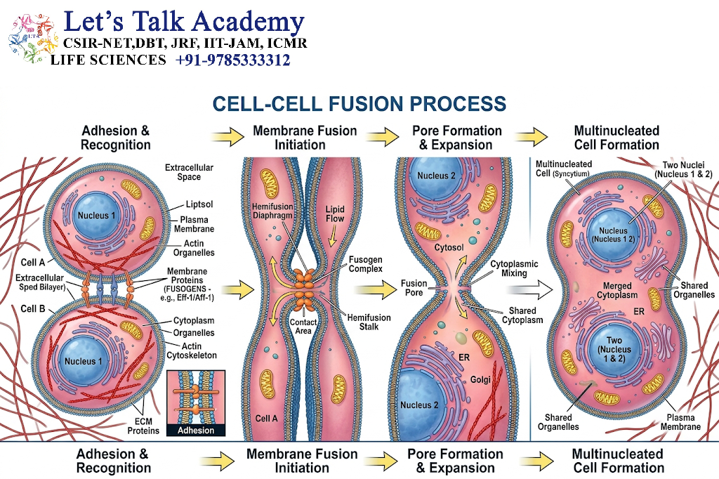
Cell-cell fusion is the process through which two separate cells establish physical contact and ultimately merge their plasma membranes to generate a single cellular entity. Following membrane fusion, the cytoplasmic contents of the participating cells become continuous.
Depending on the biological context, the nuclei may also undergo subsequent fusion. Therefore, cell fusion does not necessarily mean immediate nuclear fusion.
The overall process can be represented conceptually as:
Cell A + Cell B → Cell-cell recognition → Adhesion → Membrane fusion → Cytoplasmic continuity → Nuclear fusion or multinucleation
The final outcome depends on the type of cells involved and the physiological or pathological context.
2.2 Cell Fusion and Cell Division Are Opposite Processes
Cell division increases the number of cells by separating one cell into two or more daughter cells. Cell fusion, in contrast, reduces the number of independent cellular units by combining two or more cells.
During cell division:
One cell → Two daughter cells
During cell fusion:
Two or more cells → One fused cell
Despite being opposite in their immediate outcomes, both processes are tightly regulated and essential for normal development and tissue organization.
2.3 Cytoplasmic Fusion and Nuclear Fusion
An important distinction should be made between cytoplasmic fusion and nuclear fusion.
In cytoplasmic fusion, the plasma membranes of two cells merge and their cytoplasms become continuous. The nuclei may remain separate.
If the nuclei subsequently merge, the process is called karyogamy, or nuclear fusion.
For example, during fertilization, fusion between the sperm and egg plasma membranes is followed by highly coordinated nuclear events that ultimately establish the diploid genome of the zygote.
In other situations, such as skeletal muscle formation, multiple cells fuse but their nuclei remain distinct within a common cytoplasm. This produces a multinucleated structure called a syncytium.
3. Molecular Basis of Cell-Cell Fusion
3.1 Recognition Between Fusion-Competent Cells
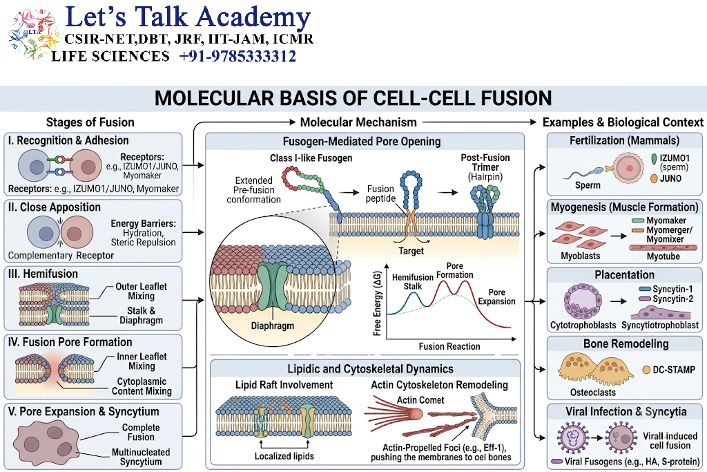
Cell fusion generally begins with the recognition of compatible cells. The participating cells must possess the appropriate surface molecules that allow them to identify one another and establish stable contact.
Cell-surface proteins, adhesion molecules, receptors, and signaling proteins can participate in this recognition process.
Recognition is particularly important because inappropriate fusion could disrupt tissue organization and cellular function.
3.2 Cell Adhesion
After recognition, the cells establish close physical contact through adhesion molecules.
Cell adhesion performs several important functions:
- It brings the two plasma membranes into close proximity.
- It stabilizes the interaction between the cells.
- It initiates intracellular signaling.
- It allows fusion-promoting proteins to act efficiently.
- It helps determine the specificity of the fusion event.
The actin cytoskeleton is frequently involved in organizing the contact region between cells.
3.3 Activation of Fusion Machinery
Following cell recognition and adhesion, specialized proteins and signaling pathways promote membrane fusion.
Different organisms and tissues use different molecular mechanisms. In several systems, proteins known as fusogens play a central role.
Fusogens are membrane-associated proteins that facilitate the physical merger of two lipid bilayers.
The exact molecular components vary considerably among organisms and tissues, and not all cell-cell fusion events depend on the same fusogen.
3.4 Plasma Membrane Remodeling
The plasma membrane is composed primarily of a lipid bilayer containing proteins, cholesterol, and other components. For two membranes to fuse, substantial structural rearrangement must occur.
The process can involve:
Membrane apposition → Hemifusion → Fusion pore formation → Pore expansion → Cytoplasmic continuity
During membrane apposition, the two membranes are brought extremely close together.
In hemifusion, the outer leaflets of the two lipid bilayers become connected while the inner leaflets remain distinct.
A fusion pore can then develop between the cells. Expansion of this pore ultimately allows the cytoplasms to become continuous.
3.5 Cytoskeletal Regulation
The cytoskeleton plays an important role in organizing cellular shape and membrane dynamics during fusion.
Actin filaments can help:
- establish cell-cell contacts,
- organize membrane proteins,
- generate mechanical forces,
- remodel the plasma membrane,
- stabilize fusion sites.
Changes in actin organization can therefore strongly influence the efficiency of cell fusion.
4. Major Types of Cell Fusion
4.1 Homotypic Cell Fusion
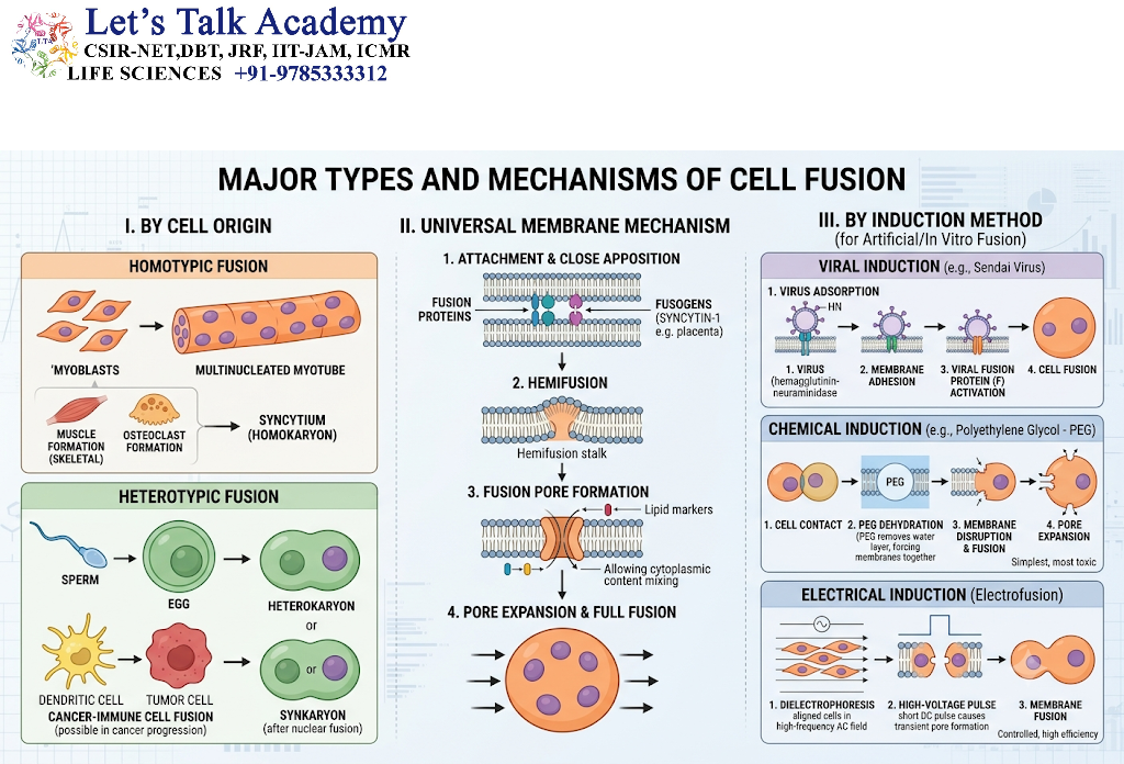
Homotypic fusion occurs between cells of the same general type.
For example, myoblasts can fuse with other myoblasts during skeletal muscle development.
Myoblast + Myoblast → Multinucleated muscle fiber
This process is important for the development and repair of skeletal muscle.
4.2 Heterotypic Cell Fusion
Heterotypic fusion occurs between different cell types.
A classic example is the fusion between a sperm cell and an egg cell during fertilization.
Other heterotypic fusion events can occur under pathological conditions, including interactions between immune cells and other tissue cells.
4.3 Homologous Fusion
Homologous fusion refers broadly to fusion between cells belonging to the same cell lineage or closely related populations.
This type of fusion is common in developmental and regenerative processes.
4.4 Heterologous Fusion
Heterologous fusion involves cells with substantially different cellular identities.
Such events are particularly important when considering pathological cell fusion, cell reprogramming, and interactions between tumor cells and cells from the surrounding tissue.
5. Cell-Cell Fusion in Normal Cells
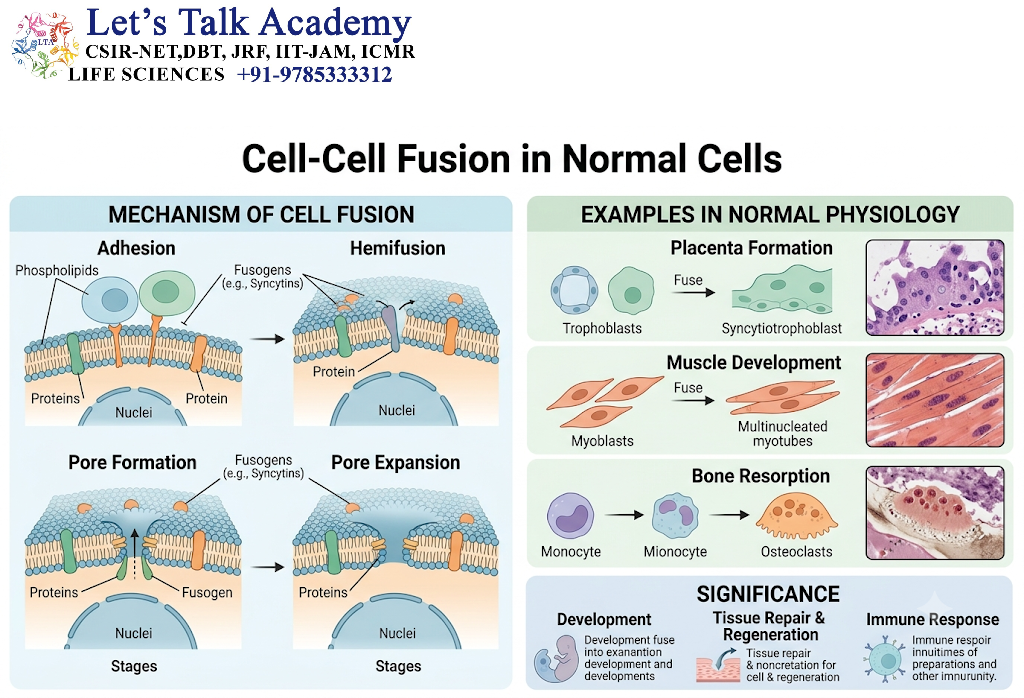
Physiological cell fusion is essential for several normal biological processes.
5.1 Fertilization
Fertilization is one of the best-known examples of cell-cell fusion.
A sperm cell and an egg cell recognize and interact with one another through highly regulated molecular mechanisms. The sperm eventually fuses with the egg plasma membrane.
The fusion event permits the sperm genetic material to enter the egg cytoplasm and contributes to the formation of the zygote.
The major events include:
- Sperm-egg recognition.
- Sperm attachment to the egg.
- Activation of sperm-associated fusion mechanisms.
- Plasma membrane fusion.
- Entry of sperm contents.
- Formation and organization of parental pronuclei.
- Establishment of the embryonic genome.
Fertilization demonstrates that cell fusion can be extremely selective and tightly controlled.
5.2 Myoblast Fusion and Skeletal Muscle Formation
Skeletal muscle provides another major example of physiological cell fusion.
Muscle development begins with precursor cells known as myoblasts. These cells proliferate and differentiate before participating in fusion.
Individual myoblasts fuse with one another to form larger multinucleated structures.
The sequence can be summarized as:
Myoblast proliferation → Differentiation → Alignment → Adhesion → Fusion → Multinucleated myotube → Mature muscle fiber
The resulting muscle fibers contain multiple nuclei within a common cytoplasm.
This arrangement supports the large size and specialized functions of skeletal muscle cells.
5.3 Placental Trophoblast Fusion
Cell fusion is also essential for placental development.
During placentation, trophoblast cells differentiate and fuse to form the syncytiotrophoblast, a multinucleated cellular layer that interfaces with maternal blood.
The syncytiotrophoblast has several important functions, including:
- nutrient exchange,
- hormone production,
- interaction with maternal tissues,
- formation of an effective placental barrier,
- regulation of maternal-fetal communication.
Trophoblast fusion is therefore critical for normal pregnancy and placental function.
5.4 Macrophage Fusion
Macrophages can undergo fusion under specific physiological and pathological conditions.
Fusion of macrophages may result in the formation of multinucleated giant cells.
These cells can occur in response to persistent foreign material, chronic inflammation, or certain tissue environments.
Macrophage fusion demonstrates that the immune system can use cell fusion as part of tissue defense and remodeling.
5.5 Osteoclast Formation
Osteoclasts are specialized multinucleated cells responsible for bone resorption.
They develop through the fusion of mononuclear precursor cells derived from the monocyte/macrophage lineage.
The process can be simplified as:
Mononuclear precursors → Differentiation → Cell-cell adhesion → Fusion → Multinucleated osteoclast
The formation of osteoclasts is essential for normal bone remodeling.
The coordinated actions of osteoblasts and osteoclasts help maintain bone homeostasis.
5.6 Giant Cell Formation
Multinucleated giant cells can form through fusion of macrophage-lineage cells.
Depending on the biological context, these cells can participate in:
- removal of foreign material,
- inflammatory responses,
- tissue remodeling,
- granulomatous reactions.
Their presence is therefore not necessarily abnormal by itself; the biological context determines their significance.
6. Molecular Regulation of Physiological Cell Fusion
6.1 Cell-Surface Adhesion Molecules
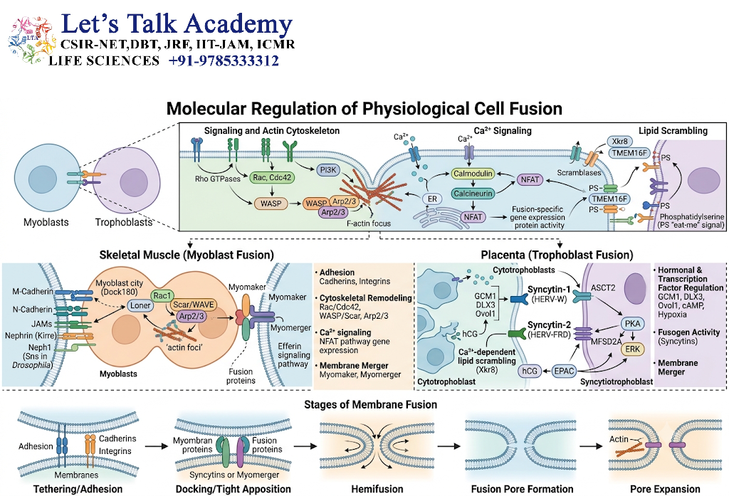
Adhesion molecules help bring fusion-competent cells together.
They can recognize molecules on neighboring cells and establish stable cellular contacts.
These interactions are particularly important during muscle development and immune-cell fusion.
6.2 Fusogenic Proteins
Fusogenic proteins promote the actual membrane fusion process.
Different organisms have evolved different families of fusogens.
In mammals, examples of fusion-associated proteins include syncytins, which are endogenous retrovirus-derived envelope proteins that have been adapted for physiological functions, particularly placental development.
This provides an important example of how viral membrane-fusion machinery can become integrated into host biology.
6.3 Signaling Pathways
Cell fusion is controlled by intracellular signaling pathways that regulate:
- cell differentiation,
- adhesion,
- cytoskeletal organization,
- membrane trafficking,
- expression of fusion-associated proteins.
Signaling pathways involving small GTPases, kinases, transcription factors, and calcium-dependent mechanisms may contribute depending on the cellular system.
6.4 Calcium Signaling
Calcium ions function as important intracellular signaling molecules.
Changes in intracellular calcium concentration can influence cytoskeletal remodeling, membrane dynamics, enzyme activity, and other processes associated with fusion.
The precise role of calcium varies between different fusion systems.
7. Cell-Cell Fusion in Abnormal and Pathological Conditions
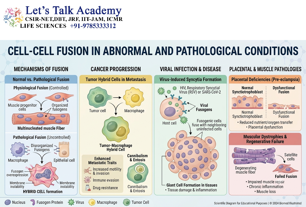
Although physiological fusion is tightly controlled, cell fusion can also occur in pathological environments.
Abnormal fusion has been observed or proposed in conditions involving:
- chronic inflammation,
- viral infection,
- cancer,
- tissue injury,
- fibrosis,
- degenerative diseases,
- foreign-body reactions.
The consequences depend on which cells fuse and how the resulting hybrid cell behaves.
7.1 Cell Fusion During Chronic Inflammation
Persistent inflammation can increase cellular interactions and create an environment favorable to macrophage fusion.
Repeated inflammatory stimulation may result in the formation of multinucleated giant cells.
Such cells can surround and interact with persistent foreign material or tissue components that are difficult for individual macrophages to eliminate.
7.2 Foreign-Body Giant Cells
When macrophages encounter large foreign particles or materials that cannot be engulfed efficiently by a single cell, macrophages may fuse to form foreign-body giant cells.
These cells can contain many nuclei and remain associated with the foreign material for prolonged periods.
They are frequently observed in chronic foreign-body reactions.
7.3 Viral-Induced Cell Fusion
Some viruses possess proteins that promote fusion between infected cells and neighboring cells.
This can generate large multinucleated cells called syncytia.
Viral fusion proteins normally evolved to facilitate viral entry into host cells, but their expression at the surface of infected cells can sometimes cause neighboring host cells to fuse.
The formation of syncytia can therefore serve as an important cytopathic effect of viral infection.
7.4 Cancer and Cell Fusion
Cell fusion has attracted considerable attention in cancer biology.
Tumor cells may encounter stromal cells, immune cells, endothelial cells, or other cell populations within the tumor microenvironment.
Under certain circumstances, fusion between tumor cells and non-tumor cells may produce hybrid cells with altered characteristics.
These hybrid cells can exhibit changes in:
- chromosome number,
- gene expression,
- cellular metabolism,
- proliferation,
- migration,
- differentiation,
- interaction with the immune system.
However, the biological significance of tumor cell fusion is context-dependent, and the extent to which particular fusion events contribute directly to human cancer progression remains an active area of research.
7.5 Fusion and Genomic Instability
Fusion can bring together two different genomes within a single cell.
If the resulting cell undergoes abnormal cell division, chromosome segregation errors may occur.
This can produce:
- aneuploidy,
- polyploidy,
- chromosome rearrangements,
- genomic instability.
These changes may alter cellular behavior.
Thus, cell fusion can create a connection between membrane biology and genome organization.
8. Cell Fusion and Hybrid Cells
8.1 Formation of Hybrid Cells
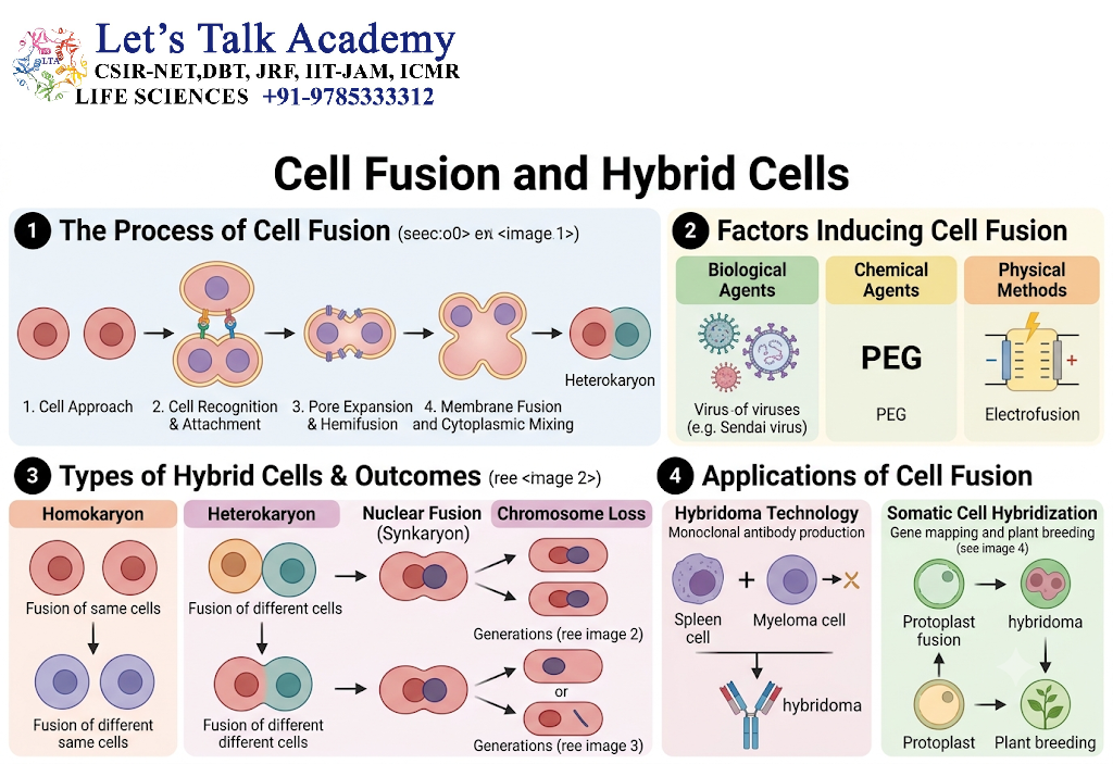
When two genetically distinct cells fuse, the resulting cell may contain genetic material from both parental cells.
Such a cell is commonly referred to as a hybrid cell.
Hybrid cells can display characteristics derived from both parental cells, although the final phenotype is determined by complex interactions among the genomes, epigenetic states, cytoplasmic components, and environmental signals.
8.2 Nuclear Fusion in Hybrid Cells
Following cytoplasmic fusion, the parental nuclei may remain separate for some time.
In some circumstances, the nuclei subsequently fuse.
This can produce a single nucleus containing genetic contributions from both cells.
In other cases, nuclear fusion does not occur, and the cell remains multinucleated.
8.3 Polyploidy
Cell fusion can increase the amount of DNA present in a cell.
For example, fusion of two diploid cells can initially generate a cell containing approximately twice the normal diploid DNA complement.
Depending on subsequent cell-cycle events, this can result in polyploid or aneuploid cellular states.
9. Cell-Cell Fusion and the Cell Cycle
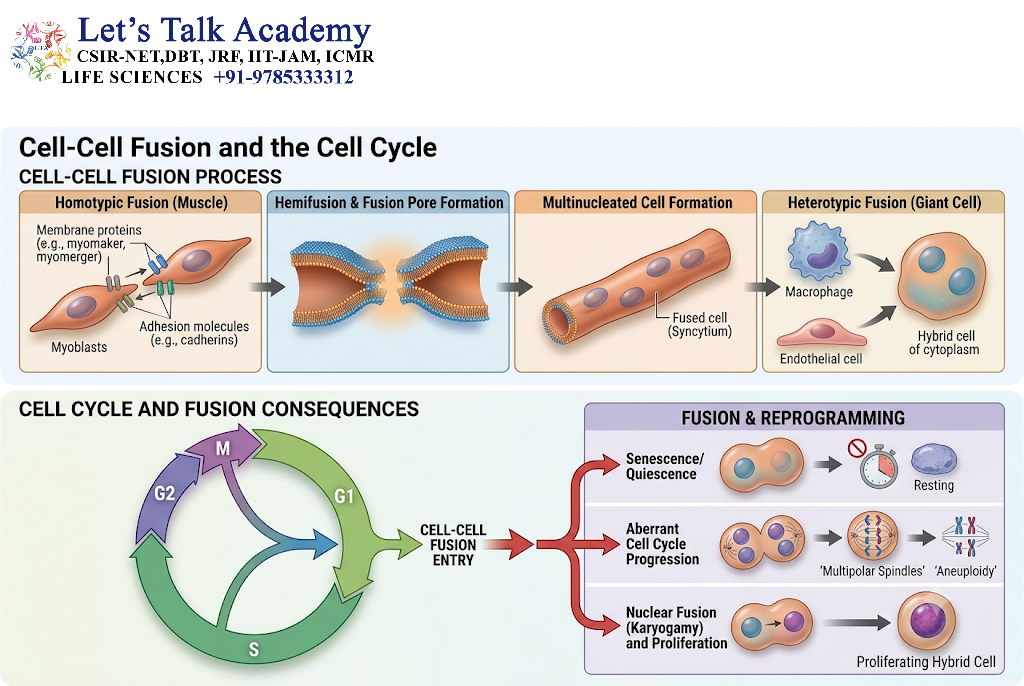
Cell fusion can strongly influence cell-cycle regulation.
A fused cell may contain two nuclei or a single nucleus with an altered chromosome complement.
If cells at different stages of the cell cycle fuse, abnormal interactions between their cell-cycle programs may occur.
This can lead to:
- cell-cycle arrest,
- abnormal DNA replication,
- chromosome missegregation,
- apoptosis,
- genomic instability.
Therefore, successful cell fusion does not necessarily guarantee survival of the resulting cell.
10. Physiological Versus Abnormal Cell Fusion
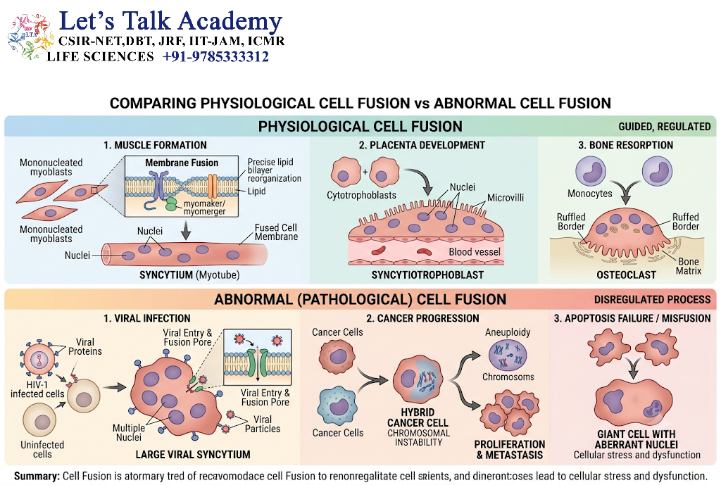
10.1 Characteristics of Physiological Fusion
Physiological fusion is generally:
- developmentally regulated,
- cell-type specific,
- spatially controlled,
- temporally regulated,
- molecularly coordinated,
- beneficial or necessary for tissue function.
Examples include:
- fertilization,
- skeletal muscle formation,
- placental syncytiotrophoblast formation,
- osteoclast development.
10.2 Characteristics of Pathological Fusion
Pathological fusion may be associated with:
- chronic inflammation,
- infection,
- tissue damage,
- cancer,
- foreign-body responses.
It may generate cells with abnormal chromosome numbers, altered gene expression, or unusual functional properties.
10.3 Comparative Overview
| Feature | Physiological Cell Fusion | Pathological Cell Fusion |
|---|---|---|
| Regulation | Highly controlled | May be dysregulated |
| Main purpose | Development, reproduction, tissue function | Often associated with disease or cellular stress |
| Cell specificity | Usually highly selective | May involve inappropriate cell types |
| Genetic outcome | Usually compatible with tissue function | May produce genomic instability |
| Examples | Fertilization, myoblast fusion, osteoclast formation | Viral syncytia, tumor-cell fusion, foreign-body giant cells |
| Biological consequence | Normal tissue development or maintenance | May contribute to pathological changes |
11. Mechanism of Membrane Fusion
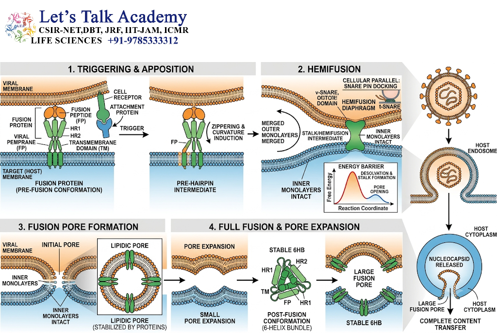
11.1 Initial Membrane Contact
The first physical requirement is close apposition of the two plasma membranes.
Because lipid bilayers naturally resist merging, specialized proteins and cellular forces are required.
11.2 Hemifusion
During hemifusion, the outer lipid layers of the two membranes merge while the inner layers remain separate.
This is an intermediate stage in many membrane-fusion processes.
11.3 Fusion Pore Formation
A small opening, known as a fusion pore, forms between the two cells.
The pore allows limited cytoplasmic continuity.
11.4 Expansion of the Fusion Pore
The fusion pore expands until the two cells become fully connected.
At this stage, molecules and organelles may move between the previously separate cytoplasmic compartments, depending on the size and organization of the resulting fused cell.
12. Role of the Cytoskeleton in Cell Fusion
12.1 Actin Filaments
Actin is one of the most important cytoskeletal components associated with cell fusion.
It can regulate:
- cell shape,
- membrane protrusion,
- cell adhesion,
- force generation,
- membrane remodeling.
12.2 Myosin
Myosin proteins interact with actin filaments and generate mechanical forces.
These forces can help organize the cell cortex and influence membrane behavior during fusion.
12.3 Microtubules
Microtubules contribute to intracellular transport, organelle positioning, and cell organization.
Because cell fusion involves major changes in cellular architecture, microtubule-dependent transport can also influence fusion-related processes.
13. Cell Fusion and Cell Signaling
Cell fusion requires communication between participating cells.
Signaling can occur through:
- receptor-ligand interactions,
- intracellular kinases,
- small GTPases,
- calcium signaling,
- transcriptional regulation,
- cytoskeletal signaling.
These pathways coordinate the transition from cell recognition to membrane fusion.
A key concept is that fusion is not simply a passive consequence of two membranes touching. It is an active, regulated biological event.
14. Cell Fusion in Development
Cell fusion contributes to the formation of specialized tissues during development.
14.1 Muscle Development
Fusion of myoblasts produces multinucleated muscle fibers.
14.2 Placental Development
Fusion of trophoblast cells generates the syncytiotrophoblast.
14.3 Bone Remodeling
Fusion of precursor cells generates multinucleated osteoclasts.
14.4 Reproductive Development
Fusion of gametes produces the zygote and initiates embryonic development.
These examples demonstrate that cell fusion is an important mechanism for increasing cellular size, generating multinucleated structures, and establishing specialized tissues.
15. Cell Fusion in Tissue Regeneration
Cell fusion has also been investigated in tissue repair and regeneration.
Cells involved in regeneration may interact with damaged cells and, under certain circumstances, undergo fusion.
Such events have been reported in experimental systems involving tissues such as:
- liver,
- skeletal muscle,
- heart,
- nervous system.
However, the functional importance of fusion during regeneration varies between tissues and experimental conditions.
Cell fusion should therefore be distinguished from other mechanisms of tissue repair, such as stem-cell differentiation, paracrine signaling, and direct cell replacement.
16. Cell Fusion in Cancer Biology
16.1 Tumor Cell Fusion
Tumor cells exist within a complex microenvironment containing immune cells, fibroblasts, endothelial cells, extracellular matrix, and other stromal components.
This environment can create opportunities for cellular interactions.
When tumor cells fuse with other cells, the resulting hybrid cells may have altered properties.
16.2 Hybrid Tumor Cells
Hybrid cells may acquire combinations of characteristics from both parental cells.
Potential consequences include changes in:
- migration,
- proliferation,
- metabolism,
- differentiation,
- drug response,
- interaction with immune cells.
However, these outcomes are highly context-dependent.
16.3 Fusion and Metastasis
Cell fusion has been investigated as one possible mechanism that could contribute to phenotypic changes associated with tumor progression and metastasis.
A fused cell may undergo genomic and epigenetic alterations that affect its behavior.
Nevertheless, fusion is only one of several mechanisms proposed to explain tumor heterogeneity and metastatic progression.
17. Cell Fusion in Immunology
Cell fusion can occur within the immune system, particularly among macrophage-lineage cells.
17.1 Macrophage Fusion
Macrophages may fuse when exposed to persistent inflammatory stimuli or large particles.
17.2 Multinucleated Giant Cells
Fusion can generate multinucleated giant cells that participate in chronic inflammatory reactions.
17.3 Granulomatous Inflammation
Multinucleated giant cells may occur within granulomatous inflammatory lesions.
Their presence reflects coordinated activity among macrophage-lineage cells in response to persistent stimuli.
18. Viral Cell Fusion
Viruses provide a particularly important example of membrane fusion.
18.1 Viral Fusion Proteins
Certain enveloped viruses use specialized viral proteins to mediate membrane fusion.
These proteins can facilitate:
Virus membrane + host-cell membrane → Fusion → Viral entry
18.2 Formation of Syncytia
When viral fusion proteins are expressed on the surface of infected cells, they may cause infected cells to fuse with neighboring cells.
This produces multinucleated syncytia.
18.3 Biological Consequences
Viral-induced cell fusion can:
- alter cellular architecture,
- disrupt normal cellular functions,
- facilitate viral spread,
- contribute to tissue damage,
- trigger immune responses.
19. Experimental Study of Cell Fusion
Cell fusion can be investigated using several experimental approaches.
19.1 Microscopy
Light microscopy can identify large multinucleated cells and morphological changes associated with fusion.
19.2 Fluorescence Microscopy
Different fluorescent labels can be used to mark two parental cell populations.
If the labeled cells combine into a single cellular structure, fluorescence can provide evidence of fusion.
19.3 Live-Cell Imaging
Time-lapse microscopy allows researchers to observe:
- cell migration,
- cell-cell contact,
- adhesion,
- membrane fusion,
- formation of multinucleated cells.
19.4 Molecular Markers
Specific proteins associated with fusion can be detected using:
- immunofluorescence,
- immunoblotting,
- flow cytometry,
- molecular assays.
19.5 Genetic Approaches
Gene knockout, gene silencing, overexpression, and genome-editing approaches can be used to investigate the function of fusion-associated genes.
These methods help establish whether particular proteins are required for recognition, adhesion, membrane fusion, or downstream responses.
20. Factors Affecting Cell Fusion
Several factors can influence the efficiency and outcome of cell fusion.
20.1 Cell Type
Different cell types possess different fusion capacities.
20.2 Cell Differentiation State
Some cells become fusion-competent only after differentiation.
20.3 Cell Density
Cell density influences the frequency with which cells encounter one another.
20.4 Membrane Composition
The lipid composition and organization of the plasma membrane can affect membrane fusion.
20.5 Cytoskeletal Organization
Changes in actin and other cytoskeletal components can alter cell shape and membrane dynamics.
20.6 Signaling Environment
Growth factors, cytokines, inflammatory mediators, and other extracellular signals can influence fusion.
20.7 Cellular Stress
Hypoxia, oxidative stress, infection, and tissue injury may modify cellular interactions and fusion behavior.
21. Biological Significance of Cell-Cell Fusion
Cell-cell fusion has several major biological functions.
21.1 Generation of Multinucleated Cells
Fusion allows several cells to contribute their nuclei to one large cytoplasmic compartment.
21.2 Tissue Formation
Fusion contributes directly to the formation of skeletal muscle and placenta.
21.3 Reproduction
Gamete fusion initiates the formation of a new organism.
21.4 Tissue Homeostasis
Fusion contributes to bone remodeling and certain immune responses.
21.5 Disease Processes
Abnormal fusion may contribute to infection, inflammation, genomic instability, and cancer-associated cellular changes.
22. Important Concepts to Understand
22.1 Syncytium
A syncytium is a multinucleated cellular structure in which nuclei share a common cytoplasmic compartment.
It can arise through cell fusion or, in some organisms and tissues, through other mechanisms such as incomplete cytokinesis.
22.2 Multinucleated Cell
A multinucleated cell contains multiple nuclei within a common cytoplasm.
Examples include skeletal muscle fibers and osteoclasts.
22.3 Fusogen
A fusogen is a protein or molecular machinery capable of promoting membrane fusion.
22.4 Karyogamy
Karyogamy refers specifically to the fusion of nuclei.
It should not be considered synonymous with plasma membrane fusion.
22.5 Heterokaryon
A heterokaryon is a cell containing genetically distinct nuclei within a common cytoplasm, often produced experimentally through cell fusion.
23. Cell Fusion and Heterokaryon Formation
Experimental fusion of different cell types can generate heterokaryons.
For example, two genetically distinct cells can be experimentally induced to fuse, resulting in a common cytoplasm containing two different nuclei.
Heterokaryons have historically been useful for studying:
- gene expression,
- nuclear-cytoplasmic interactions,
- chromosome behavior,
- cellular differentiation,
- nuclear reprogramming.
These studies helped demonstrate that cellular phenotype is influenced not only by the genome itself but also by regulatory factors present within the cellular environment.
24. Cell Fusion and Nuclear Reprogramming
One important consequence of cell fusion is that factors from one cell can influence the nucleus of another cell.
The cytoplasm contains:
- transcription factors,
- regulatory RNAs,
- enzymes,
- signaling proteins,
- epigenetic regulators.
After fusion, these molecules may interact with the nuclei of the parental cells.
This provides an experimental framework for studying cellular plasticity and nuclear reprogramming.
25. Distinction Between Cell Fusion and Cell Aggregation
Cell fusion must be distinguished from simple cell aggregation.
In aggregation:
Cells come together but remain separate.
In fusion:
Cells come together and their plasma membranes merge.
Therefore, physical proximity alone does not demonstrate cell fusion.
Evidence of membrane continuity or cytoplasmic mixing is generally needed to establish that genuine fusion has occurred.
26. Regulation and Prevention of Inappropriate Fusion
Because uncontrolled fusion can be harmful, cells possess mechanisms that limit inappropriate fusion.
These mechanisms can involve:
- restricted expression of fusogens,
- cell-type-specific recognition,
- inhibitory signaling,
- membrane organization,
- immune surveillance,
- regulation of cell-cell adhesion.
Such regulatory mechanisms ensure that fusion occurs primarily when biologically appropriate.
27. Integrated View of Normal and Abnormal Cell Fusion
Cell-cell fusion should be understood as a spectrum rather than as an inherently beneficial or harmful event.
Under normal conditions, fusion is essential for:
- reproduction,
- muscle formation,
- placental development,
- bone remodeling,
- selected immune responses.
Under pathological conditions, fusion may contribute to:
- viral syncytium formation,
- chronic inflammatory giant-cell formation,
- tumor-cell hybridization,
- genomic instability,
- altered cellular behavior.
Therefore, the biological outcome depends on the cell types involved, molecular mechanism, timing, tissue environment, and genetic consequences of fusion.


