
1. Introduction
The immune system must continuously distinguish between normal cellular components and molecules that indicate infection, cellular damage, or abnormal growth. T lymphocytes play a central role in this process, but they generally do not recognize intact protein antigens directly. Instead, proteins are broken down into smaller peptide fragments, and selected peptides are displayed on the cell surface in association with major histocompatibility complex (MHC) molecules. T-cell receptors then recognize these peptide–MHC complexes and initiate an appropriate immune response.
The complete sequence through which proteins are acquired, degraded into peptides, associated with MHC molecules, transported, and displayed at the cell surface is known as antigen processing and presentation. This process forms a critical bridge between innate sensing of foreign or abnormal material and antigen-specific adaptive immunity.
Two major antigen-presentation pathways are recognized:
- MHC class I antigen processing and presentation, which primarily displays peptides derived from proteins present within the cell and is generally recognized by CD8⁺ T cells.
- MHC class II antigen processing and presentation, which primarily displays peptides derived from extracellular proteins taken up by specialized antigen-presenting cells and is generally recognized by CD4⁺ T cells.
A third important mechanism, called cross-presentation, allows certain antigen-presenting cells, particularly dendritic cells, to present extracellular antigens on MHC class I molecules. This mechanism is particularly important for initiating CD8⁺ T-cell responses against antigens that may not be produced directly by the antigen-presenting cell.
Understanding these pathways requires connecting several concepts: antigen uptake, intracellular protein degradation, MHC biosynthesis, peptide transport, peptide loading, intracellular trafficking, cell-surface expression, and T-cell recognition. Each step is carefully regulated so that the immune system can monitor both intracellular and extracellular environments.
1.1 Meaning of Antigen Processing
Antigen processing refers to the intracellular conversion of protein antigens into smaller peptide fragments that can associate with MHC molecules.
Most proteins are too large to fit directly into the peptide-binding groove of MHC molecules. Therefore, they must first undergo controlled proteolytic degradation. Depending on where the source protein originates and which MHC molecule is involved, different intracellular compartments and proteolytic enzymes participate in this process.
For MHC class I presentation, intracellular proteins are commonly degraded by the proteasome, producing peptides that can enter the endoplasmic reticulum through the transporter associated with antigen processing, commonly called TAP.
For MHC class II presentation, extracellular proteins are internalized into endosomal and lysosomal compartments, where proteases generate peptides suitable for binding to MHC class II molecules.
Thus, antigen processing can be understood as the preparation of protein-derived peptides for recognition by T lymphocytes.
1.2 Meaning of Antigen Presentation
Antigen presentation is the process by which processed antigenic peptides are displayed on the cell surface in association with MHC molecules.
The MHC molecule acts like a molecular display platform. It holds a peptide fragment in a specific binding groove and presents that peptide to the extracellular portion of a T-cell receptor (TCR).
The TCR does not normally recognize the peptide alone. Instead, it recognizes the combined molecular structure formed by:
Peptide + MHC molecule + T-cell receptor
This interaction provides antigen-specific information to the T cell.
MHC class I molecules primarily interact with CD8⁺ T cells, whereas MHC class II molecules primarily interact with CD4⁺ T cells.
1.3 Why Antigen Processing and Presentation Are Important
Antigen processing and presentation are essential because the adaptive immune system needs a mechanism to inspect proteins produced within cells as well as proteins obtained from the extracellular environment.
The process contributes to:
- Detection of virus-infected cells
- Recognition of intracellular microbial proteins
- Elimination of abnormal or transformed cells
- Activation of helper T cells
- Activation of cytotoxic T cells
- Development of immune memory
- Regulation of immune tolerance
- Initiation of responses against extracellular pathogens
- Presentation of antigens acquired from dying or damaged cells
- Coordination between antigen-presenting cells and lymphocytes
The same general machinery also participates in immune tolerance by displaying peptides derived from self proteins. Consequently, antigen presentation is not simply a mechanism for recognizing foreign material; it is also important for maintaining immune balance and preventing inappropriate immune activation.
2. Major Histocompatibility Complex
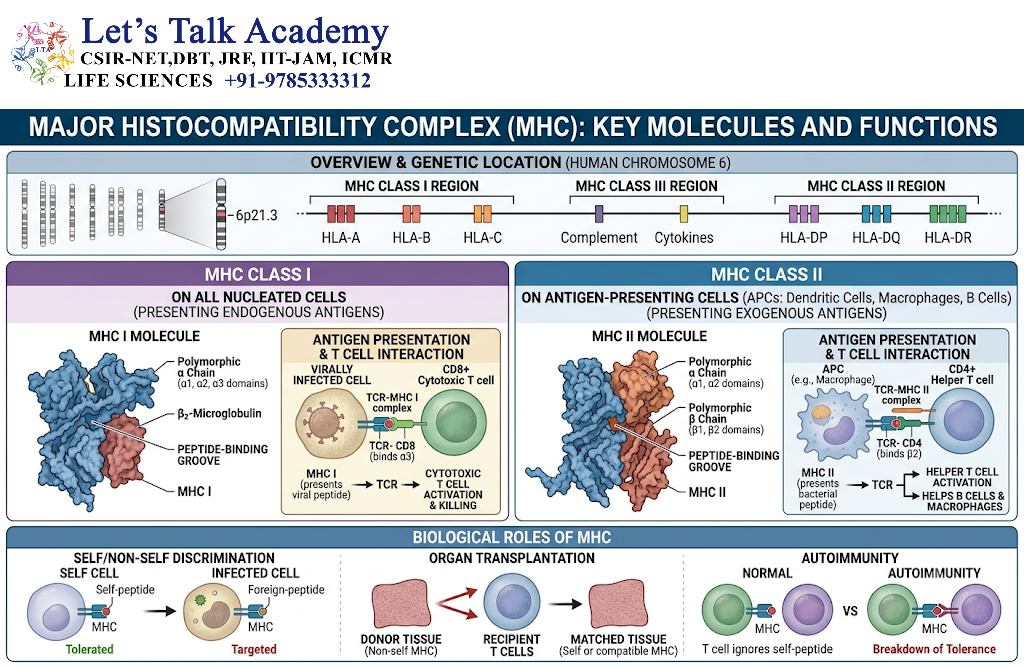
The major histocompatibility complex (MHC) is a genomic region containing genes that encode molecules involved in antigen presentation and other immune functions.
In humans, MHC molecules are commonly referred to as human leukocyte antigens (HLA).
MHC molecules are highly polymorphic. This means that many different allelic forms occur within populations. Such genetic variation influences which peptides can bind to particular MHC molecules and therefore affects antigen recognition and immune responses.
MHC molecules involved directly in conventional peptide presentation are divided into:
- MHC class I
- MHC class II
MHC class III genes are also present within the broader MHC genomic region, but their products are not conventional peptide-presenting molecules.
2.1 MHC Class I Molecules
MHC class I molecules are expressed on nearly all nucleated cells, although the level of expression can vary among cell types and physiological conditions.
A classical MHC class I molecule consists of:
- One polymorphic heavy α chain
- β2-microglobulin
- A peptide-binding groove
The α chain contains three extracellular domains:
- α1
- α2
- α3
The peptide-binding groove is formed primarily by the α1 and α2 domains.
The α3 domain contributes to interaction with the CD8 co-receptor.
MHC class I molecules generally bind relatively short peptides, commonly around 8–11 amino acids in length, although the precise peptide length can vary.
The major function of MHC class I is to display peptides generated from intracellular proteins to CD8⁺ T cells.
2.2 MHC Class II Molecules
MHC class II molecules are primarily expressed by professional antigen-presenting cells.
Important professional antigen-presenting cells include:
- Dendritic cells
- Macrophages
- B lymphocytes
MHC class II consists of two transmembrane chains:
- α chain
- β chain
Both chains contribute to the peptide-binding groove.
Unlike the closed-ended peptide-binding groove of classical MHC class I molecules, the MHC class II groove is open at both ends. Therefore, MHC class II can accommodate longer peptides, often approximately 13–18 amino acids or longer, with the exact length depending on the peptide and MHC molecule.
MHC class II molecules primarily present peptides generated from extracellular proteins that have been internalized by antigen-presenting cells.
3. Antigen-Presenting Cells
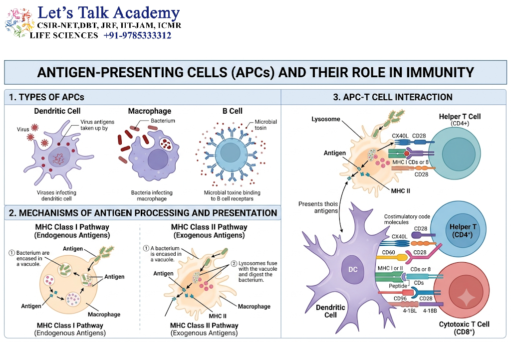
Cells that process and display antigen-derived peptides through MHC molecules are called antigen-presenting cells (APCs).
Although many nucleated cells can express MHC class I, specialized antigen-presenting cells have particularly important roles in initiating adaptive immune responses.
3.1 Dendritic Cells
Dendritic cells are highly specialized antigen-presenting cells that efficiently capture antigen in peripheral tissues and present processed peptides to T cells.
They are particularly important for the activation of naïve T cells.
Dendritic cells can present:
- Extracellular antigens through MHC class II
- Endogenous antigens through MHC class I
- Certain extracellular antigens through MHC class I by cross-presentation
Their ability to cross-present extracellular antigen is especially important for initiating CD8⁺ T-cell responses against antigens originating from cells that may not directly infect the dendritic cell.
3.2 Macrophages
Macrophages are professional phagocytic cells. They ingest microorganisms, cellular debris, and other particulate material.
After uptake, antigenic proteins can be degraded in intracellular vesicular compartments and presented through MHC class II.
Macrophages therefore have an important role in connecting phagocytosis with antigen-specific T-cell responses.
3.3 B Lymphocytes
B cells can internalize antigen through their B-cell receptor (BCR).
After antigen binding, the BCR–antigen complex can be internalized and delivered into intracellular processing compartments. Peptides generated from the antigen can then be loaded onto MHC class II and presented to CD4⁺ T cells.
This interaction is particularly important during T-cell-dependent B-cell responses.
4. MHC Class I Antigen Processing and Presentation
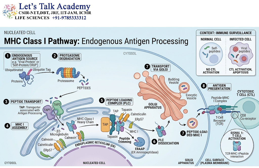
The term endogenous means that the source protein is generated within the cell.
Examples include proteins derived from:
- Viral proteins produced during infection
- Abnormal proteins produced by transformed cells
- Normal cellular proteins undergoing turnover
- Mutated or altered intracellular proteins
The overall pathway can be represented as:
Intracellular protein → Proteasomal degradation → Peptide transport by TAP → ER peptide loading → MHC class I–peptide complex → Golgi/secretory pathway → Cell surface → CD8⁺ T cell recognition
4.1 Generation of Endogenous Antigens
Proteins within cells undergo continuous synthesis and degradation.
Some proteins become substrates for the ubiquitin–proteasome system. Ubiquitination can mark proteins for degradation, after which the proteasome cleaves them into smaller peptide fragments.
The proteasome is therefore an important source of peptides used for MHC class I presentation.
Not every peptide generated by the proteasome becomes an antigenic peptide. Many peptides are further degraded into smaller components and eventually amino acids. Only a fraction enter the antigen-presentation pathway.
4.2 Role of the Proteasome
The proteasome is a large intracellular proteolytic complex located mainly in the cytosol and nucleus.
Its major functions include:
- Removal of damaged proteins
- Regulation of protein turnover
- Degradation of short-lived regulatory proteins
- Generation of peptide fragments for MHC class I presentation
During certain inflammatory conditions, cells can express alternative proteasomal components that form the immunoproteasome.
The immunoproteasome can alter the pattern of peptide cleavage and thereby influence the repertoire of peptides available for MHC class I presentation.
4.3 Transport of Peptides by TAP
Peptides generated in the cytosol need access to the compartment where MHC class I molecules are loaded.
This is facilitated by TAP, the transporter associated with antigen processing.
TAP consists primarily of two subunits:
- TAP1
- TAP2
TAP is an ATP-dependent transporter located in the membrane of the endoplasmic reticulum.
It transports suitable cytosolic peptides into the ER lumen.
This step is particularly important because newly synthesized MHC class I molecules undergo peptide loading within the ER in the classical pathway.
4.4 Peptide Trimming
Proteasomal products may not always possess the precise length or sequence required for optimal MHC class I binding.
Additional peptide trimming can therefore occur.
In the cytosol, various aminopeptidases may modify peptide fragments. In the ER, enzymes such as ERAP1 and, where expressed, ERAP2 can trim peptide N-termini.
This processing helps generate peptides with appropriate characteristics for MHC class I binding.
5. Assembly of MHC Class I Molecules
MHC class I assembly is a carefully coordinated process involving several molecular chaperones.
5.1 Synthesis of the MHC Class I Heavy Chain
The MHC class I α chain is synthesized on ribosomes associated with the rough endoplasmic reticulum.
The newly synthesized heavy chain initially requires assistance from molecular chaperones to achieve an appropriate conformation.
One important early chaperone is calnexin.
5.2 Association with β2-Microglobulin
After appropriate folding of the heavy chain, it associates with β2-microglobulin.
This interaction stabilizes the MHC class I molecule and permits further progression through the peptide-loading pathway.
5.3 Peptide-Loading Complex
The peptide-loading complex contains several proteins that coordinate the loading of peptides onto MHC class I.
Important components include:
- TAP
- Tapasin
- Calreticulin
- ERp57
- MHC class I
- β2-microglobulin
Tapasin connects MHC class I molecules functionally with TAP and contributes to peptide editing.
The peptide-loading machinery helps favor stable MHC class I molecules carrying peptides with suitable binding characteristics.
5.4 Tapasin and Peptide Editing
Tapasin is an important component of the MHC class I peptide-loading complex.
It promotes the selection of peptides that form stable MHC class I–peptide complexes.
This process is sometimes described as peptide editing.
The importance of peptide editing is that not every peptide entering the ER is equally suitable for long-term surface expression. Efficient peptide selection contributes to the quality of the peptide repertoire displayed to CD8⁺ T cells.
6. Transport of MHC Class I–Peptide Complexes to the Cell Surface
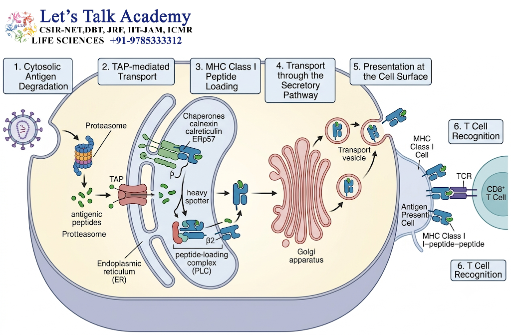
Once a suitable peptide is loaded, the stable MHC class I–peptide complex leaves the ER.
It proceeds through the secretory pathway, including the Golgi apparatus, and ultimately reaches the plasma membrane.
At the cell surface, the peptide is exposed within the MHC class I binding groove.
CD8⁺ T cells can then inspect the displayed peptide through their T-cell receptor.
This creates a continuous surveillance system in which cells effectively display a molecular sample of their intracellular protein environment.
6.1 Recognition by CD8⁺ T Cells
CD8⁺ T cells recognize antigenic peptides presented by MHC class I.
The interaction involves:
Peptide–MHC class I complex + TCR + CD8 co-receptor
When the appropriate antigen is recognized in the correct cellular context, signaling pathways are activated within the T cell.
Activated cytotoxic T lymphocytes can then develop effector functions that contribute to the elimination of infected or abnormal target cells.
7. MHC Class II Antigen Processing and Presentation
The MHC class II pathway primarily processes exogenous antigens.
Exogenous antigens originate outside the cell and enter antigen-presenting cells through mechanisms such as:
- Endocytosis
- Receptor-mediated uptake
- Phagocytosis
- Macropinocytosis
The overall pathway can be represented as:
Extracellular antigen → Endocytosis/phagocytosis → Endosomal processing → MHC class II peptide loading → Cell-surface expression → CD4⁺ T-cell recognition
8. Uptake of Extracellular Antigens
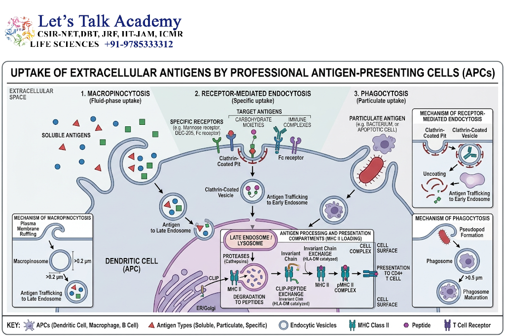
Antigen-presenting cells use several mechanisms to acquire extracellular proteins.
8.1 Endocytosis
Endocytosis allows cells to internalize soluble molecules and membrane-associated material.
The internalized antigen enters vesicular compartments that progressively mature and become more acidic.
8.2 Phagocytosis
Phagocytosis is particularly important for large particles such as:
- Bacteria
- Cellular debris
- Dead cells
- Large particulate material
Phagosomes can interact with endosomal and lysosomal compartments, allowing antigen degradation.
8.3 Macropinocytosis
Macropinocytosis allows cells, especially dendritic cells, to take up relatively large volumes of extracellular fluid.
This mechanism can contribute to antigen acquisition from the surrounding environment.
9. MHC Class II Biosynthesis and Invariant Chain
MHC class II molecules are synthesized in the endoplasmic reticulum.
However, the peptide-binding groove must initially be protected from inappropriate peptides present in the ER.
This protection is provided by the invariant chain, also called Ii.
9.1 Role of the Invariant Chain
The invariant chain associates with newly synthesized MHC class II molecules.
Its functions include:
- Stabilizing newly synthesized MHC class II molecules
- Preventing premature binding of ER-derived peptides
- Directing MHC class II molecules toward endosomal compartments
As the MHC class II–invariant chain complex moves through the endosomal system, the invariant chain undergoes proteolytic degradation.
9.2 Formation of CLIP
After progressive degradation of invariant chain, a small peptide fragment called CLIP, or class II-associated invariant chain peptide, remains within the MHC class II peptide-binding groove.
CLIP temporarily occupies the groove and prevents premature binding of unrelated peptides.
The replacement of CLIP by antigen-derived peptide is an essential stage of MHC class II antigen presentation.
10. Antigen Degradation in Endosomal and Lysosomal Compartments
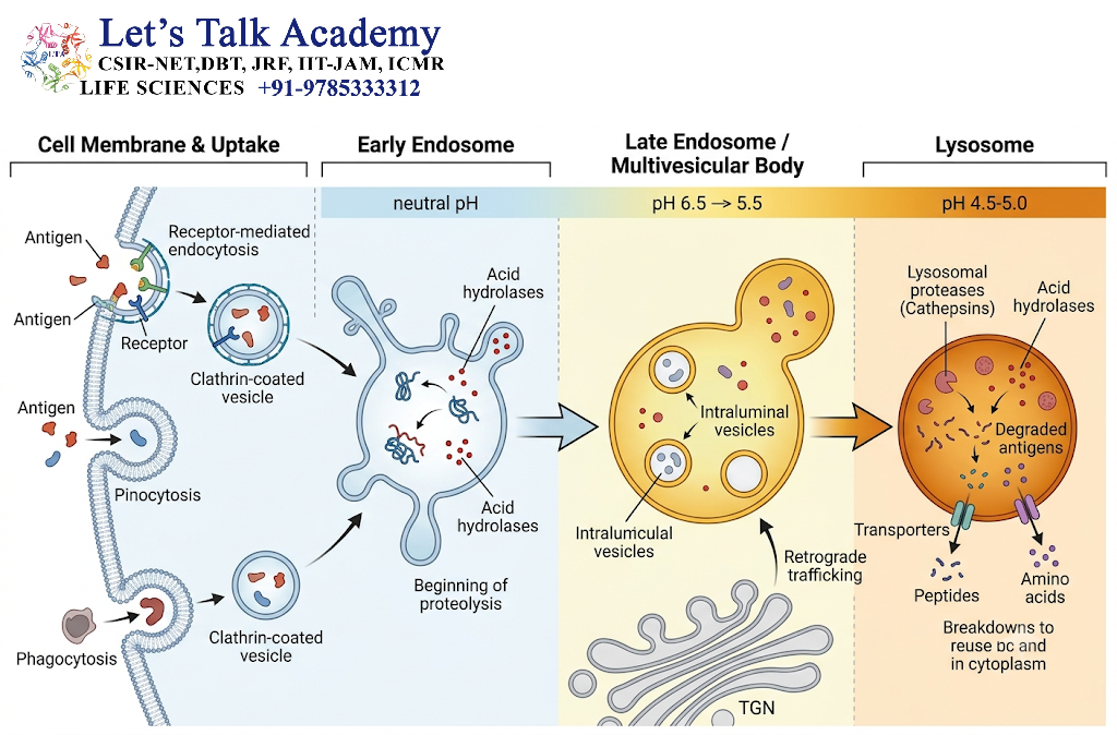
Extracellular proteins taken into antigen-presenting cells encounter progressively acidic vesicular compartments.
These compartments contain proteolytic enzymes, particularly cathepsins.
The acidic environment promotes protein degradation and contributes to the generation of peptides suitable for MHC class II binding.
The resulting peptides are generally longer than the peptides associated with classical MHC class I molecules because the MHC class II binding groove is open at both ends.
11. HLA-DM and Peptide Loading onto MHC Class II
The removal of CLIP and replacement with antigen-derived peptides is facilitated by HLA-DM.
HLA-DM functions as an important peptide-exchange and peptide-editing molecule.
It helps remove CLIP and promotes the loading of suitable antigen-derived peptides onto MHC class II.
This process is important because many different peptides may be present within endosomal compartments, but only some will form sufficiently stable MHC class II–peptide complexes.
11.1 Peptide Editing
HLA-DM contributes to the selection of peptides that form stable complexes with MHC class II.
Therefore, peptide editing is not simply about generating peptides; it also involves selecting which peptides are efficiently displayed.
This increases the functional quality of the peptide repertoire presented to CD4⁺ T cells.
12. MHC Class II–Peptide Complexes at the Cell Surface
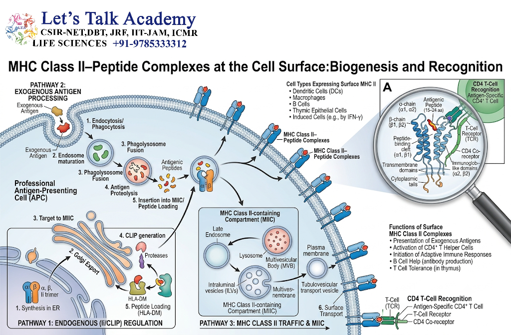
After peptide loading, MHC class II–peptide complexes are transported to the plasma membrane.
At the cell surface, the peptide is exposed to T cells.
CD4⁺ T cells recognize the antigenic peptide in association with MHC class II.
The interaction can be represented as:
Peptide–MHC class II + TCR + CD4 co-receptor
This interaction can contribute to activation and differentiation of CD4⁺ T cells, depending on the antigen-presenting cell, co-stimulatory signals, cytokine environment, and other regulatory factors.
13. Comparison of MHC Class I and MHC Class II Pathways
| Feature | MHC Class I | MHC Class II |
|---|---|---|
| Main antigen source | Primarily endogenous/intracellular | Primarily exogenous/extracellular |
| Major processing compartment | Cytosol and ER | Endosomal/lysosomal system |
| Major proteolytic machinery | Proteasome and other peptidases | Cathepsins and other endosomal proteases |
| Peptide loading | Mainly ER in classical pathway | Endosomal MHC class II compartments |
| Major transporter | TAP | No equivalent TAP-dependent classical loading step |
| Typical peptide length | Approximately 8–11 amino acids | Often approximately 13–18 amino acids or longer |
| Main T-cell population | CD8⁺ T cells | CD4⁺ T cells |
| Major immune function | Cellular immune surveillance | Helper T-cell responses |
| Typical expressing cells | Most nucleated cells | Mainly professional APCs |
| Important accessory proteins | Tapasin, calreticulin, ERp57 | Invariant chain, CLIP, HLA-DM |
These are general rules rather than absolute restrictions. Antigen processing is flexible, and exceptions such as cross-presentation demonstrate that extracellular antigens can sometimes enter the MHC class I pathway.
14. Cross-Presentation
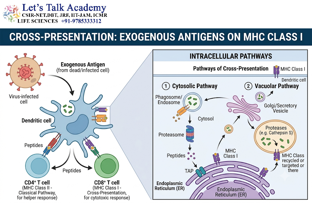
Cross-presentation is the process in which an extracellular antigen is presented on MHC class I molecules.
This is an important exception to the conventional division between:
Endogenous antigen → MHC class I
and
Exogenous antigen → MHC class II
During cross-presentation:
Exogenous antigen → antigen-presenting cell → MHC class I → CD8⁺ T cell
Dendritic cells are particularly important in this process.
15. Importance of Cross-Presentation
Cross-presentation is especially important when a dendritic cell acquires antigen from another cell.
For example, a dendritic cell can acquire proteins from:
- Virus-infected cells
- Tumor cells
- Dead or dying cells
- Extracellular pathogens
The acquired proteins can subsequently enter an MHC class I presentation pathway.
This enables dendritic cells to activate naïve CD8⁺ T cells even when the dendritic cell itself is not the original source of the antigen.
This process is closely associated with cross-priming, in which cross-presented antigen contributes to the activation of naïve CD8⁺ T cells.
16. Cytosolic Pathway of Cross-Presentation
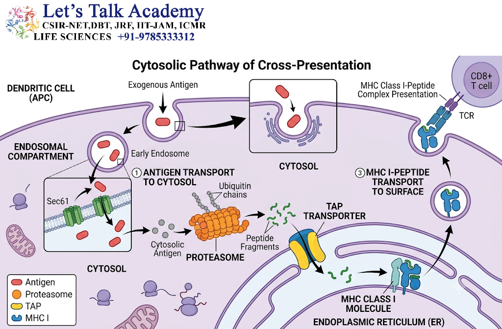
In one major form of cross-presentation, extracellular proteins are internalized into vesicular compartments and subsequently gain access to the cytosol.
Once in the cytosol, the antigen can be degraded by the proteasome.
The resulting peptides can then enter the conventional MHC class I processing pathway.
A simplified sequence is:
Extracellular protein → Endocytosis/phagocytosis → Cytosolic transfer → Proteasomal degradation → TAP-dependent peptide transport → MHC class I loading → Cell surface
This pathway uses many components associated with conventional MHC class I processing.
17. Vacuolar Pathway of Cross-Presentation
Another mechanism involves processing within intracellular vesicular compartments.
In this pathway, antigen degradation and peptide generation can occur within vesicles, and peptide loading onto MHC class I can occur without requiring all of the conventional cytosolic processing steps.
The precise molecular mechanisms of cross-presentation are complex and can vary according to the antigen, cell type, intracellular compartment, and experimental conditions. Current evidence supports more than one pathway rather than a single universal mechanism.
18. Antigen Presentation and T-Cell Activation
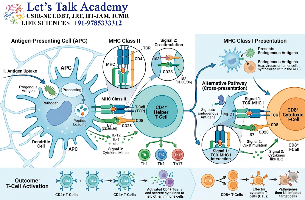
Presentation of a peptide by an MHC molecule is essential but is generally not sufficient by itself to produce a complete response from a naïve T cell.
Effective T-cell activation involves multiple signals.
18.1 Signal 1: Antigen Recognition
The first signal is generated when the T-cell receptor recognizes the peptide–MHC complex.
For CD8⁺ T cells, the relevant complex is generally:
Peptide–MHC class I
For CD4⁺ T cells:
Peptide–MHC class II
18.2 Signal 2: Co-Stimulation
T-cell activation also depends on co-stimulatory interactions.
Important examples include interactions involving:
- CD28 on T cells
- B7-1/CD80 on APCs
- B7-2/CD86 on APCs
The balance between antigen recognition and co-stimulation is important for determining whether a T cell becomes activated, remains unresponsive, or develops another functional state.
18.3 Signal 3: Cytokine Environment
Cytokines produced by antigen-presenting cells and surrounding cells influence T-cell differentiation and functional specialization.
Therefore, antigen presentation should not be viewed as simply placing a peptide on the cell surface. It occurs within a larger cellular signaling environment that determines the nature and magnitude of the immune response.
19. Antigen Processing and Immune Tolerance
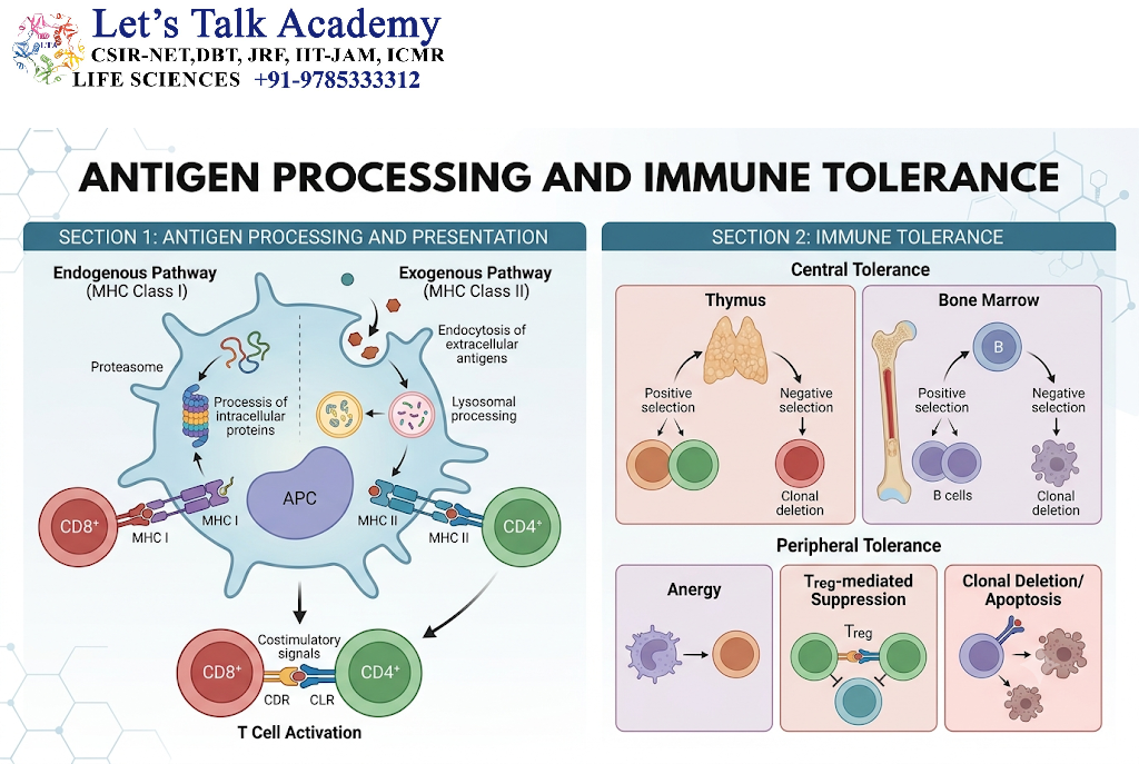
Antigen processing and presentation are also central to immune tolerance.
The immune system must distinguish harmful foreign or abnormal antigens from normal self components.
Self-derived peptides are continuously generated and presented through MHC molecules.
During T-cell development, recognition of self peptide–MHC complexes contributes to selection processes that shape the mature T-cell repertoire.
Peripheral presentation of self antigens can also contribute to mechanisms that prevent inappropriate immune activation.
Thus, antigen presentation has two complementary functions:
Immune defense against harmful antigens
and
Immune tolerance toward appropriate self components.
20. Antigen Presentation in Viral Infection
Viral infection provides a clear example of the importance of MHC class I presentation.
When a virus replicates inside a host cell, viral proteins can enter intracellular protein degradation pathways.
Peptides generated from viral proteins can be presented on MHC class I molecules.
CD8⁺ T cells can recognize these viral peptide–MHC class I complexes and develop cytotoxic functions against infected cells.
Viruses, however, have evolved multiple mechanisms that interfere with antigen processing and presentation. Such mechanisms may target peptide generation, TAP-mediated transport, MHC assembly, or intracellular trafficking.
This creates an ongoing molecular interaction between host immune surveillance and pathogen immune evasion.
21. Antigen Presentation in Cancer
Cancer cells can produce proteins that differ from normal cellular proteins because of genetic mutations, abnormal gene expression, viral oncogenic proteins, or other changes in cellular biology.
Peptides derived from such proteins may be presented by MHC class I molecules.
If recognized as abnormal, these peptide–MHC complexes can become targets of T-cell-mediated immune responses.
However, tumors can also alter antigen presentation and immune signaling, reducing the effectiveness of immune recognition.
The study of antigen processing and presentation is therefore closely connected with modern approaches to understanding antitumor immunity.
22. Antigen Processing and Vaccination
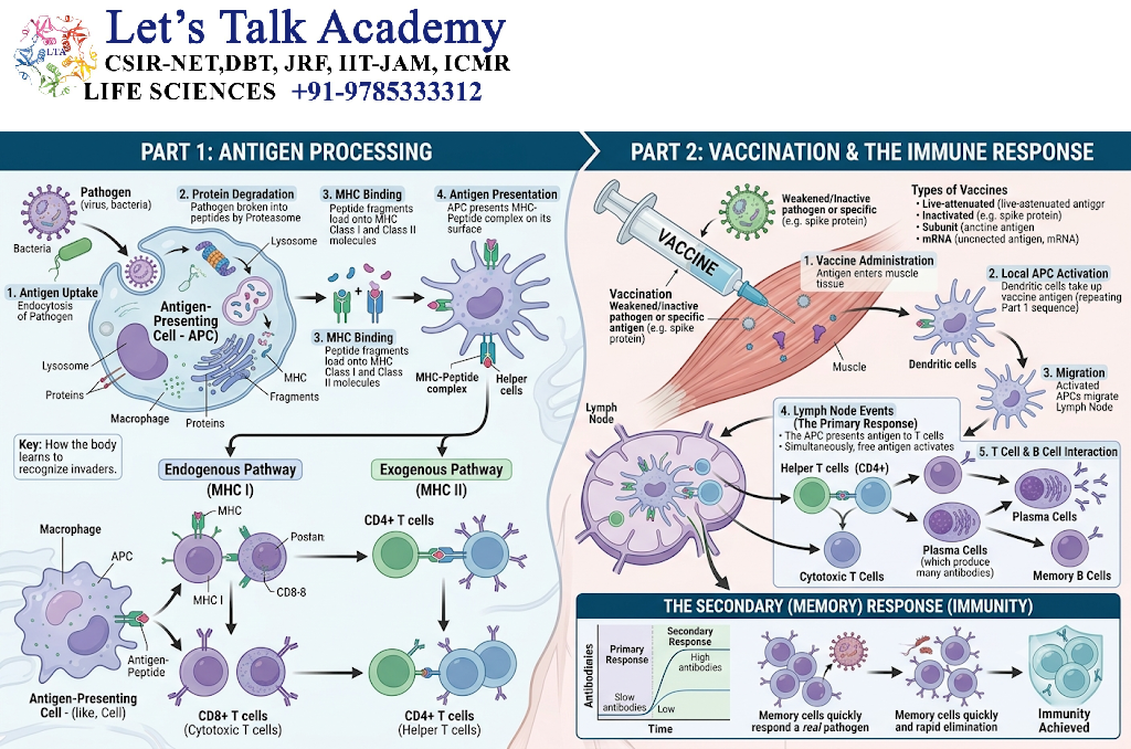
Vaccination depends on the immune system’s ability to recognize antigen and develop long-lasting immune memory.
Different vaccine platforms can deliver antigen to different cellular compartments and therefore influence the pathways through which antigen is processed and presented.
Protein antigens taken up by antigen-presenting cells commonly enter MHC class II pathways.
Some vaccine strategies can also generate or promote MHC class I presentation, including through mechanisms associated with cross-presentation.
The resulting activation of CD4⁺ and CD8⁺ T cells, together with B-cell responses, can contribute to immunological memory.
23. Role of Endoplasmic Reticulum in Antigen Presentation
The endoplasmic reticulum is a major site for the assembly and peptide loading of classical MHC class I molecules.
It provides:
- MHC class I heavy-chain synthesis
- Association with β2-microglobulin
- Access to TAP-transported peptides
- Peptide-loading machinery
- Quality-control mechanisms
The ER therefore acts as a critical checkpoint where MHC class I molecules are assembled and loaded before they are allowed to proceed toward the cell surface.
24. Role of Endosomes and Lysosomes
Endosomes and lysosomes are particularly important for MHC class II antigen processing.
They provide:
- Acidic environments
- Proteolytic enzymes
- Antigen degradation
- MHC class II trafficking
- Peptide loading and exchange
The progression of antigen through the endosomal system is highly regulated.
Early and late endosomal compartments differ in their acidity, protein composition, proteolytic activity, and trafficking characteristics.
These differences influence how efficiently antigen is degraded and presented.
25. Role of Proteases in Antigen Processing
Proteases are essential because intact proteins generally cannot bind directly to MHC peptide-binding grooves.
Important proteolytic systems include:
25.1 Proteasomes
Important for cytosolic protein degradation and classical MHC class I antigen processing.
25.2 Cathepsins
Important proteases within endosomal and lysosomal compartments, particularly in MHC class II processing.
25.3 ERAP Enzymes
Endoplasmic reticulum aminopeptidases can trim peptide N-termini and influence the repertoire of peptides available for MHC class I presentation.
The combined action of these enzymes determines which peptide fragments become available for MHC binding.
26. Peptide–MHC Stability
Not all peptide–MHC complexes are equally stable.
The stability of the interaction depends on:
- Peptide sequence
- Peptide length
- MHC allele
- Anchor residues
- Binding affinity
- Structural compatibility
Stable peptide–MHC complexes tend to persist longer at the cell surface, increasing the opportunity for recognition by T cells.
This explains why antigen presentation is not simply a matter of generating as many peptides as possible. The quality and stability of peptide binding are also important.
27. MHC Polymorphism and Peptide Binding
MHC molecules are among the most polymorphic proteins in the immune system.
Different MHC alleles can bind different sets of peptides.
This diversity has important consequences for immune recognition.
A peptide that binds efficiently to one MHC variant may bind poorly to another.
Consequently, genetic variation in MHC molecules contributes to differences among individuals in antigen presentation and immune responses.
At the population level, MHC polymorphism increases the range of microbial peptides that can potentially be presented across a population.
28. Antigen Processing and Immune Evasion
Successful pathogens often evolve mechanisms that interfere with antigen presentation.
Possible strategies include:
- Reducing MHC expression
- Interfering with proteasomal processing
- Blocking TAP-mediated transport
- Preventing MHC assembly
- Altering peptide loading
- Retaining MHC molecules within intracellular compartments
- Increasing degradation of MHC molecules
These mechanisms can reduce the visibility of infected cells to T lymphocytes.
The study of immune evasion therefore provides additional insight into why each stage of antigen processing is biologically important.
29. Antigen Presentation by Dendritic Cells
Dendritic cells are particularly efficient at linking antigen capture with T-cell activation.
Immature dendritic cells in peripheral tissues are adapted for antigen acquisition.
After appropriate activation, they undergo changes that promote migration toward lymphoid tissues and improve their ability to interact with T cells.
During this process, antigen processing and presentation are integrated with changes in:
- MHC expression
- Co-stimulatory molecule expression
- Cytokine production
- Cellular trafficking
- T-cell interaction
Thus, dendritic cells are not simply passive antigen display platforms. They actively regulate the environment in which antigen-specific T-cell responses develop.
30. Antigen Processing and Presentation: Integrated View
The entire process can be summarized as two major pathways.
30.1 Classical MHC Class I Pathway
Intracellular protein
↓
Ubiquitination and proteasomal degradation
↓
Generation of peptide fragments
↓
Cytosolic peptide trimming
↓
Transport into ER by TAP
↓
MHC class I assembly
↓
Peptide loading with assistance from the peptide-loading complex
↓
Peptide editing and stabilization
↓
Transport through Golgi and secretory pathway
↓
MHC class I–peptide complex on cell surface
↓
Recognition by CD8⁺ T cell
30.2 Classical MHC Class II Pathway
Extracellular protein
↓
Endocytosis or phagocytosis
↓
Entry into endosomal/phagosomal compartments
↓
Acidification and proteolysis
↓
MHC class II trafficking with invariant chain
↓
Invariant chain degradation
↓
CLIP formation
↓
HLA-DM-mediated peptide exchange
↓
Antigenic peptide loading
↓
MHC class II–peptide transport to cell surface
↓
Recognition by CD4⁺ T cell
31. Cross-Presentation: Integrated View
The major steps can be summarized as:
Extracellular antigen
↓
Uptake by dendritic cell or another capable APC
↓
Localization within intracellular vesicles
↓
Transfer of antigen or antigen-derived material into the cytosol, in one major pathway
↓
Proteasomal processing
↓
Generation of antigenic peptides
↓
Loading onto MHC class I
↓
Transport to cell surface
↓
Recognition by CD8⁺ T cells
Cross-presentation provides an important connection between extracellular antigen acquisition and MHC class I-restricted immunity.
32. Important Molecules Involved in Antigen Processing and Presentation
A useful way to understand the pathway is to remember the major molecules according to their functions.
| Molecule | Major role |
|---|---|
| Proteasome | Generates peptides from intracellular proteins |
| Immunoproteasome | Modified proteasomal machinery that influences peptide generation during immune activation |
| TAP1/TAP2 | Transports cytosolic peptides into the ER |
| β2-microglobulin | Stabilizes MHC class I |
| Calnexin | Assists MHC class I folding |
| Calreticulin | Participates in MHC class I peptide loading |
| ERp57 | Supports peptide-loading complex function |
| Tapasin | Connects MHC class I with TAP and contributes to peptide editing |
| ERAP1/ERAP2 | Trims peptides in the ER |
| Invariant chain | Protects MHC class II peptide-binding groove and directs trafficking |
| CLIP | Temporarily occupies the MHC class II groove |
| HLA-DM | Facilitates CLIP removal and peptide exchange |
| Cathepsins | Degrade proteins in endosomal/lysosomal compartments |
| MHC class I | Presents mainly endogenous peptides to CD8⁺ T cells |
| MHC class II | Presents mainly exogenous peptides to CD4⁺ T cells |
33. Key Differences Between Antigen Processing and Antigen Presentation
Although the two terms are often used together, they describe different stages.
Antigen processing means converting proteins into suitable peptide fragments.
Antigen presentation means displaying those peptide fragments in association with MHC molecules at the cell surface.
In simple terms:
Processing prepares the antigen.
Presentation displays the processed peptide.
Both steps are required for efficient T-cell recognition.


