
1. Introduction
The immune system protects the body by recognizing foreign or abnormal molecules and generating specific responses against them. One of the most important mechanisms of adaptive immunity is the interaction between an antigen and an antibody.
Antibodies, also called immunoglobulins, are produced by B lymphocytes and plasma cells. They recognize specific molecular regions called epitopes on antigens. The antibody-binding region that interacts with an epitope is called the paratope.
Antigen–antibody interaction is highly specific but is generally based on non-covalent molecular forces rather than permanent covalent bonds. The strength, specificity, and biological consequences of these interactions depend on factors such as antigen structure, antibody affinity, avidity, concentration, temperature, pH, ionic strength, and molecular accessibility.
These interactions are important not only for natural immune defense but also for laboratory diagnosis, research, biotechnology, vaccine development, and therapeutic antibody applications.
2. Definition of Antigen–Antibody Interaction
An antigen–antibody interaction is the specific and reversible binding of an antibody to a particular epitope present on an antigen.
The basic interaction can be represented as:
Antigen + Antibody ⇌ Antigen–Antibody Complex
The interaction is reversible because the molecules are held together primarily by non-covalent forces.
2.1 Antigen
An antigen is a molecule or molecular structure that can be specifically recognized by components of the adaptive immune system, including antibodies, B-cell receptors, or T-cell receptors.
Examples include:
- Proteins
- Polysaccharides
- Glycoproteins
- Lipids associated with proteins or carbohydrates
- Toxins
- Viral surface proteins
- Bacterial surface molecules
2.2 Antibody
An antibody is an immunoglobulin molecule produced by B cells and plasma cells in response to antigenic stimulation.
Its antigen-binding sites are located in the Fab regions, particularly within the variable domains.
2.3 Epitope and Paratope
An epitope is the specific region of an antigen recognized by an antibody.
A paratope is the complementary antigen-binding region of an antibody.
The interaction can therefore be simplified as:
Epitope ↔ Paratope
The molecular shapes, chemical properties, and spatial arrangement of the two regions determine how effectively they interact.
3. Molecular Basis of Antigen–Antibody Interaction
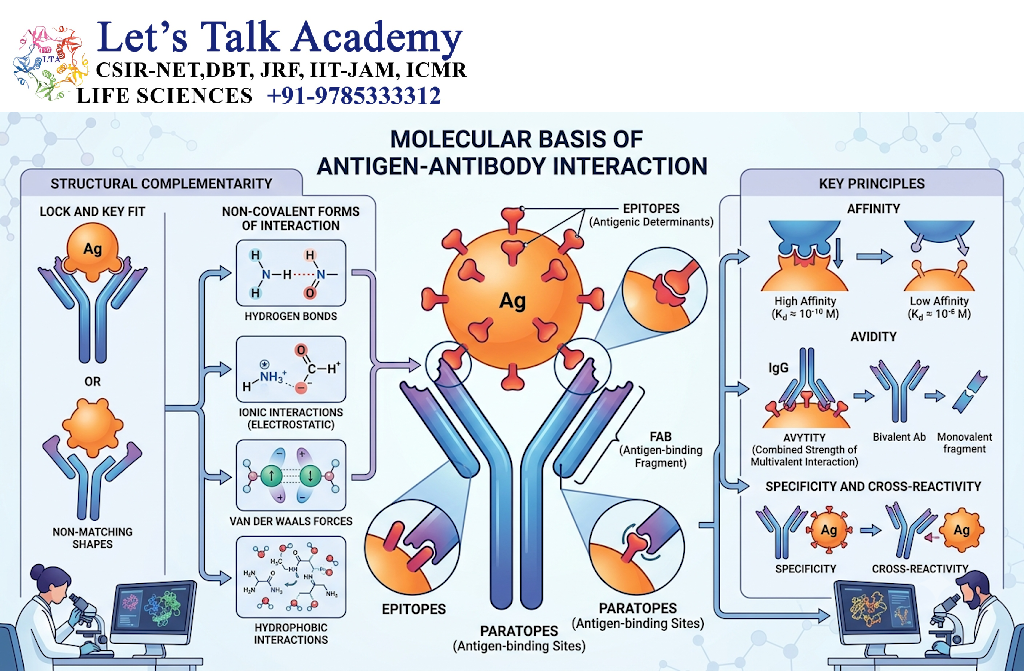
Antigen–antibody binding depends on complementary molecular characteristics.
The antibody-binding site contains amino acid residues that can interact with residues or chemical groups on the antigen.
Important characteristics include:
- Shape complementarity
- Electrostatic complementarity
- Hydrogen-bonding compatibility
- Hydrophobic interactions
- Van der Waals interactions
- Molecular accessibility
The interaction does not usually require the formation of a covalent bond.
4. Forces Involved in Antigen–Antibody Interactions
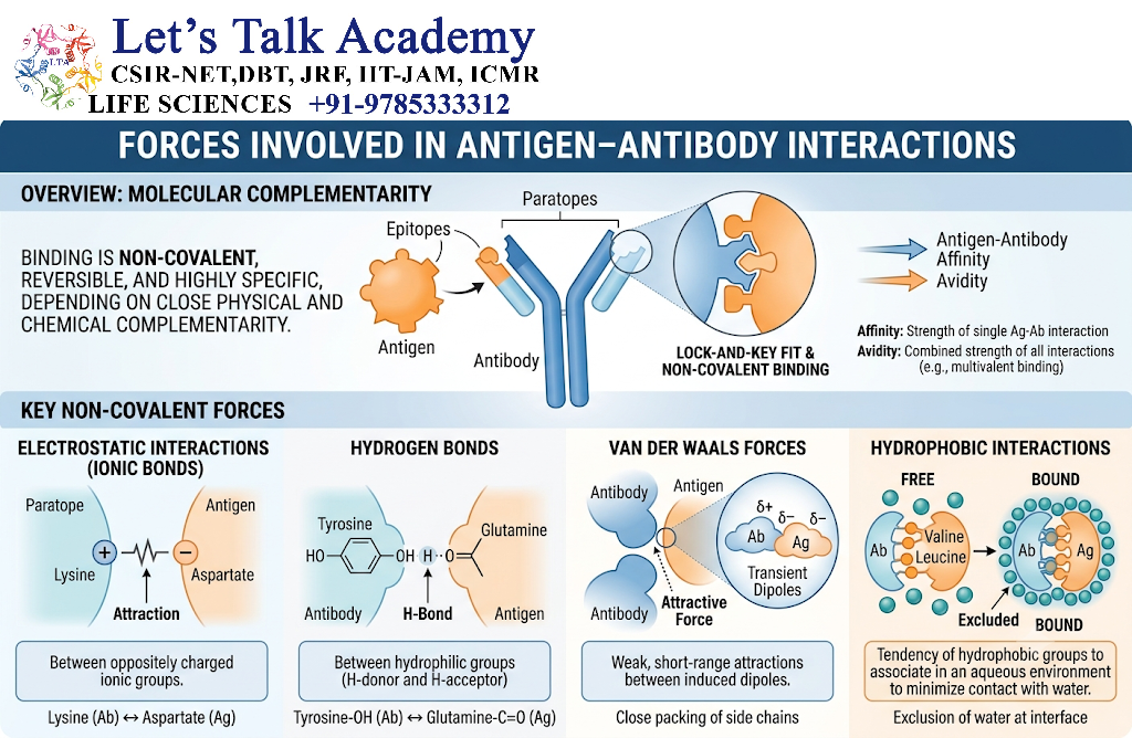
Several weak non-covalent forces collectively stabilize the antigen–antibody complex.
4.1 Electrostatic Interactions
Electrostatic interactions occur between oppositely charged groups.
For example:
Positive group ↔ Negative group
These interactions depend strongly on the distance between charged groups and the surrounding ionic environment.
4.2 Hydrogen Bonds
Hydrogen bonds can form between suitable hydrogen-bond donor and acceptor groups.
They contribute significantly to molecular recognition and specificity.
4.3 Hydrophobic Interactions
Hydrophobic regions tend to associate away from water.
When hydrophobic surfaces on an antigen and antibody come together, water molecules may be displaced, contributing to stabilization of the complex.
4.4 Van der Waals Forces
Van der Waals forces are weak attractive forces generated by temporary fluctuations in electron distribution.
Although individually weak, many such interactions can collectively contribute to antibody–antigen binding.
4.5 Combined Effect of Forces
No single interaction usually determines the entire binding process.
Instead:
Multiple weak forces → Complementary interactions → Stable antigen–antibody complex
5. Complementarity Between Antigen and Antibody
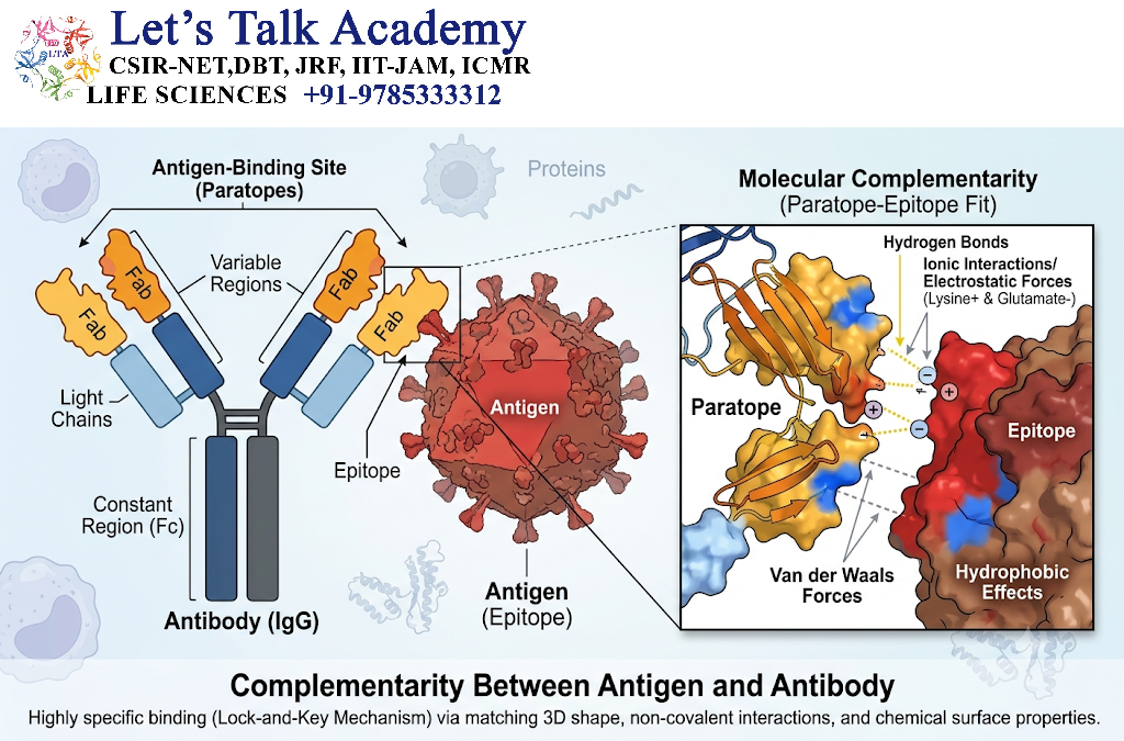
Antigen–antibody recognition depends on structural and chemical complementarity.
The older concept of a simple lock-and-key model illustrates that the antibody-binding site has a shape complementary to the antigen.
A more accurate concept is the induced-fit model, in which binding can involve limited conformational changes in the antibody, antigen, or both.
Thus:
Antigen approaches antibody → Molecular recognition → Conformational adjustment → Stable complex
6. Specificity of Antigen–Antibody Interaction
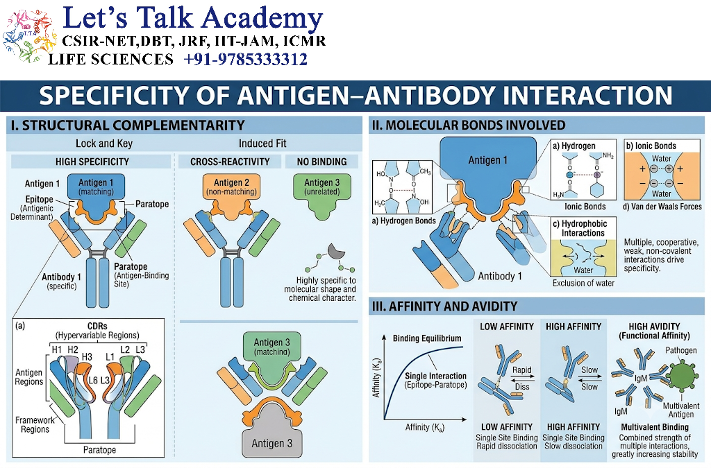
Specificity refers to the ability of an antibody to preferentially recognize a particular antigenic structure.
Specificity depends on:
- Shape complementarity
- Chemical compatibility
- Epitope accessibility
- Amino acid composition
- Spatial arrangement of interacting groups
An antibody generated against one epitope may not bind another antigen if the structural and chemical properties are sufficiently different.
However, specificity is not always absolute.
7. Cross-Reactivity
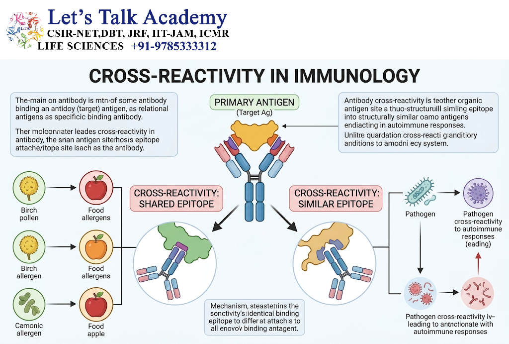
Cross-reactivity occurs when an antibody generated against one antigen also recognizes a structurally related epitope on another antigen.
This can occur when different antigens contain similar molecular structures.
For example:
Antigen A → Antibody A
If Antigen B contains a sufficiently similar epitope:
Antibody A → Antigen B
Cross-reactivity can be biologically useful, but it can also produce unwanted reactions in diagnostic tests.
8. Affinity
Affinity is the strength of interaction between a single antigenic epitope and a single antibody-binding site.
It reflects the equilibrium between antibody-bound and antibody-free states.
The general relationship is:
Antibody + Antigen ⇌ Antibody–Antigen complex
A higher affinity generally means that the antibody forms a more stable complex with the particular epitope.
8.1 Association and Dissociation
Two important processes are:
Association: Antigen and antibody come together.
Dissociation: The antigen–antibody complex separates.
The equilibrium can be described using association and dissociation constants.
The dissociation constant is commonly represented as:
Kd = [Antibody][Antigen] / [Antibody–Antigen]
A lower Kd generally indicates stronger binding affinity.
9. Avidity
Avidity is the overall strength of binding between a multivalent antibody and a multivalent antigen.
It depends on:
- Individual binding-site affinities
- Number of available binding sites
- Spatial arrangement of epitopes
- Simultaneous interactions
Avidity can therefore be much greater than the strength of any single antigen-binding interaction.
9.1 Affinity vs Avidity
| Feature | Affinity | Avidity |
|---|---|---|
| Meaning | Strength of one binding interaction | Overall strength of multiple interactions |
| Depends on | One epitope–paratope interaction | Multiple binding interactions |
| Molecular level | Individual site | Entire multivalent complex |
| Example | One antibody site binding one epitope | IgM binding multiple epitopes |
10. Valency of Antibodies
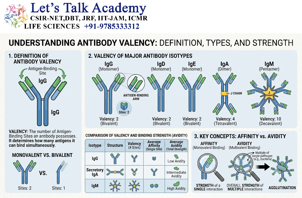
Valency refers to the number of antigen-binding sites available on an antibody.
Examples:
- IgG: generally bivalent
- Secretory IgA: commonly dimeric and functionally multivalent
- IgM: pentameric in its common secreted form and highly multivalent
Multivalency can increase overall binding strength through avidity.
11. Antigen–Antibody Equilibrium
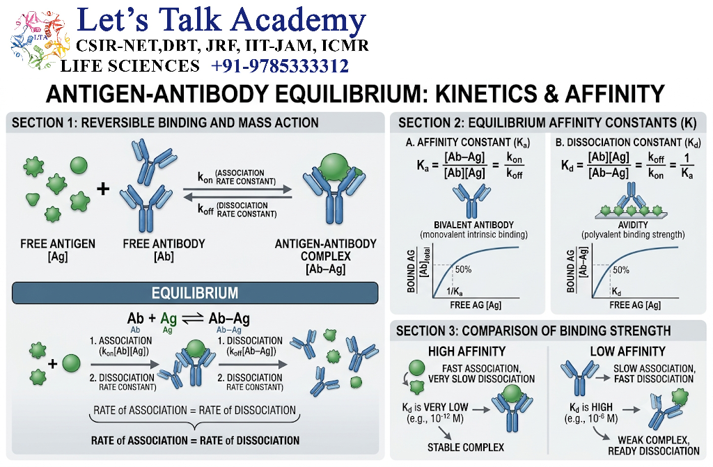
Antigen–antibody binding is dynamic and reversible.
The equilibrium can be represented as:
Ag + Ab ⇌ Ag–Ab
The equilibrium depends on:
- Association rate
- Dissociation rate
- Antigen concentration
- Antibody concentration
- Temperature
- pH
- Ionic strength
- Molecular structure
The binding kinetics can therefore influence the duration and stability of antigen recognition.
12. Factors Affecting Antigen–Antibody Interactions
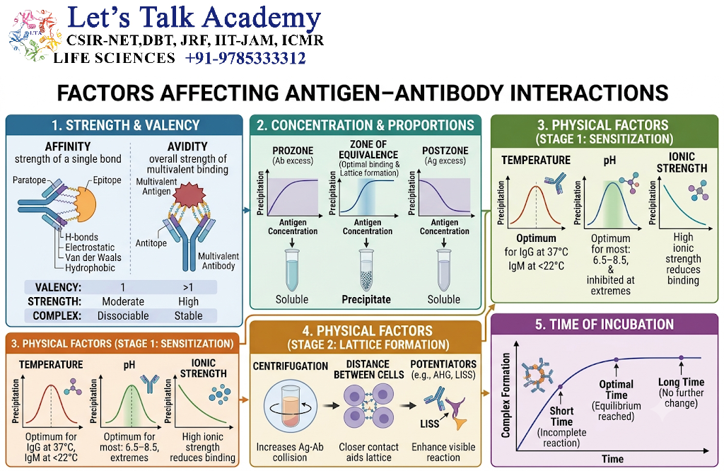
12.1 Antigen Concentration
The concentration of antigen can influence the amount of antigen–antibody complex formed.
12.2 Antibody Concentration
An adequate concentration of antibody is necessary for efficient binding and detection.
12.3 Temperature
Temperature can affect molecular motion, reaction kinetics, and stability of antigen–antibody complexes.
12.4 pH
Changes in pH can alter the ionization state of amino acid side chains and therefore influence electrostatic interactions.
12.5 Ionic Strength
The concentration of ions in the surrounding solution can affect electrostatic interactions and binding behavior.
12.6 Epitope Accessibility
An antibody cannot efficiently bind an epitope that is buried or physically inaccessible.
12.7 Structural Integrity
Changes in antigen conformation can expose, hide, or alter epitopes.
13. Antigen–Antibody Reaction Zones
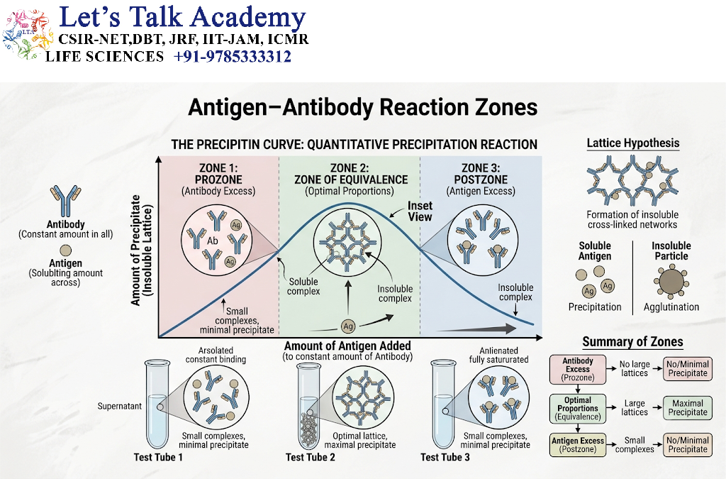
When soluble antigens and antibodies interact, the relative proportions of the two molecules can influence the formation of visible complexes.
Three important zones are:
- Prozone
- Equivalence zone
- Postzone
13.1 Prozone
The prozone occurs when antibody concentration is excessively high relative to antigen.
Although antigen–antibody binding occurs, large lattice formation may be reduced.
13.2 Equivalence Zone
At the equivalence zone, antigen and antibody are present in approximately suitable proportions for efficient lattice formation.
This region can produce strong visible reactions.
13.3 Postzone
The postzone occurs when antigen is present in excess relative to antibody.
Large immune-complex formation may again decrease.
13.4 Reaction Pattern
Antibody excess → Prozone
Optimal proportion → Equivalence
Antigen excess → Postzone
These effects are important in immunological assays because an unexpectedly weak reaction does not always mean that antigen or antibody is absent.
14. Precipitation Reactions
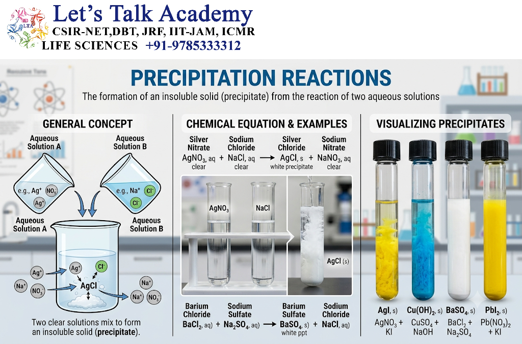
Precipitation occurs when a soluble antigen interacts with a soluble antibody and forms sufficiently large immune complexes that become insoluble or visible.
General mechanism:
Soluble antigen + Soluble antibody → Immune complex → Lattice formation → Precipitate
14.1 Lattice Formation
Multivalent antigens and antibodies can cross-link with one another.
This produces a network or lattice.
When the complexes become sufficiently large, they may precipitate from solution.
15. Types of Precipitation Reactions
Important precipitation methods include:
- Ring precipitation
- Immunodiffusion
- Single radial immunodiffusion
- Double immunodiffusion
- Immunoelectrophoresis
These techniques have been widely used for studying and detecting soluble antigens.
16. Agglutination Reactions
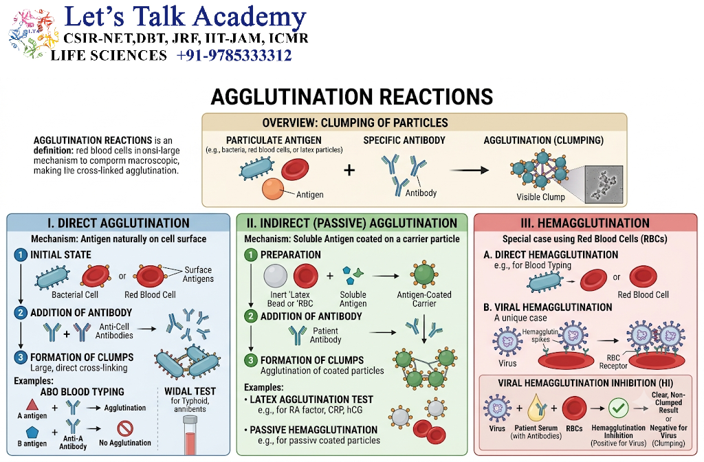
Agglutination occurs when antibodies cross-link particulate antigens, producing visible clumping.
Particulate antigens may include:
- Bacterial cells
- Red blood cells
- Latex particles
- Other cellular particles
General mechanism:
Particulate antigen + Antibody → Cross-linking → Clumping
17. Types of Agglutination
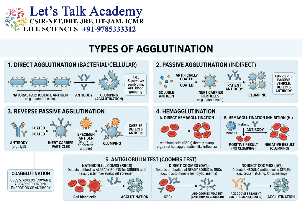
17.1 Direct Agglutination
Antibodies directly bind naturally occurring antigens on the surface of cells or particles.
17.2 Passive Agglutination
Soluble antigens are artificially attached to particles such as latex beads.
Antibodies then cause visible agglutination.
17.3 Reverse Passive Agglutination
Antibodies are attached to particles, and the particles react with the target antigen.
17.4 Hemagglutination
Agglutination involving red blood cells is called hemagglutination.
18. Precipitation vs Agglutination
| Feature | Precipitation | Agglutination |
|---|---|---|
| Antigen | Soluble | Particulate |
| Visible reaction | Precipitate | Clumps |
| Main process | Lattice formation | Particle cross-linking |
| Example | Immunodiffusion | Hemagglutination |
19. Neutralization
Neutralization occurs when antibodies bind to biologically active molecules or pathogens and prevent them from interacting effectively with their target cells.
Examples include antibodies against:
- Viral surface proteins
- Bacterial toxins
- Other pathogenic molecules
19.1 Viral Neutralization
A neutralizing antibody may bind a viral surface protein required for attachment or entry.
The simplified process is:
Virus → Antibody binding → Blocked attachment/entry → Reduced infection
19.2 Toxin Neutralization
Antibodies can bind toxins and prevent their interaction with cellular receptors.
20. Opsonization
Antibodies can enhance the uptake of pathogens by phagocytic cells.
In antibody-mediated opsonization:
Pathogen → Antibody coating → Fc receptor recognition → Phagocyte attachment → Phagocytosis
IgG antibodies are particularly important in this process.
21. Complement Activation
Certain antigen–antibody complexes can activate the complement system.
For example, antibodies such as IgM and some IgG subclasses can participate in classical complement activation when appropriately bound to antigen.
General pathway:
Antigen–antibody complex → Complement activation → Complement cascade → Effector functions
Consequences can include:
- Opsonization
- Inflammation
- Enhanced phagocytosis
- Membrane attack complex formation in susceptible targets
22. Antibody-Dependent Cellular Cytotoxicity
In antibody-dependent cellular cytotoxicity (ADCC), antibodies bind antigens on a target cell.
Immune cells recognize the Fc region of the bound antibodies.
For example:
Target cell → IgG binding → Fc receptor-bearing immune cell recognition → Cytotoxic response
Natural killer cells are important mediators of IgG-dependent ADCC.
23. Immune Complex Formation
When antibodies bind soluble antigens, they can form immune complexes.
These complexes may:
- Remain soluble
- Become deposited in tissues
- Be cleared by phagocytic systems
- Activate complement
The biological effect depends on their size, composition, location, and clearance.
24. Antibody Binding to Conformational Epitopes
Some antibodies recognize conformational epitopes.
These epitopes are formed by amino acid residues that may be separated in the primary sequence but brought together by protein folding.
Therefore:
Protein folding → Three-dimensional epitope formation → Antibody recognition
Denaturation may destroy such epitopes.
25. Antibody Binding to Linear Epitopes
A linear epitope consists of a continuous sequence of residues.
Some antibodies can recognize such epitopes even when the antigen has been partially or completely denatured.
This principle is particularly important in certain laboratory techniques such as immunoblotting.
26. Specificity and Cross-Reactivity
Specificity and cross-reactivity represent related but distinct properties.
| Property | Specificity | Cross-reactivity |
|---|---|---|
| Meaning | Preferential recognition of a target | Recognition of related structures |
| Basis | Molecular complementarity | Structural similarity |
| Biological effect | Target-specific recognition | Recognition beyond original antigen |
| Diagnostic importance | Improves selectivity | Can produce false-positive reactions |
27. Antigen–Antibody Interaction in Immunodiagnostics
Antigen–antibody interactions form the basis of many laboratory diagnostic techniques.
Important methods include:
- ELISA
- Western blotting
- Immunofluorescence
- Immunohistochemistry
- Flow cytometry
- Immunoprecipitation
- Lateral-flow immunoassays
- Immunodiffusion
- Agglutination assays
28. ELISA
Enzyme-linked immunosorbent assay (ELISA) uses antigen–antibody binding together with an enzyme-linked detection system.
A simplified format is:
Antigen/antibody immobilization → Specific binding → Enzyme-linked detection reagent → Substrate reaction → Detectable signal
The intensity of the signal can be related to the amount of target under appropriately validated assay conditions.
29. Western Blotting
Western blotting is used to detect specific proteins.
General workflow:
Protein separation → Transfer to membrane → Primary antibody binding → Secondary antibody binding → Signal detection
The primary antibody recognizes the target protein.
30. Immunofluorescence
In immunofluorescence, antibodies are linked directly or indirectly to fluorescent labels.
After antibody binding, the target can be visualized using fluorescence microscopy.
It is useful for studying:
- Protein localization
- Cellular structures
- Tissue antigens
- Microbial components
31. Immunohistochemistry
Immunohistochemistry uses antibodies to detect specific molecules in tissue sections.
It is widely used in biological research and diagnostic pathology.
General process:
Tissue preparation → Antigen accessibility → Primary antibody binding → Detection system → Microscopic visualization
32. Flow Cytometry
Flow cytometry uses fluorescently labeled antibodies to identify and characterize cells based on surface or intracellular markers.
For example:
Cell → Fluorescent antibody binding → Laser excitation → Fluorescence detection → Cell population analysis
33. Lateral-Flow Immunoassays
Lateral-flow assays use antibody-based molecular recognition on a membrane-based test strip.
They can provide rapid detection of specific antigens or antibodies.
Their general principle involves:
Sample → Migration → Specific binding → Capture → Visible signal
34. Immunoprecipitation
Immunoprecipitation uses an antibody to selectively capture a target antigen, commonly a protein, from a complex biological mixture.
The antibody–target complex can then be isolated and analyzed.
35. Factors Determining Diagnostic Accuracy
The performance of an antigen–antibody assay depends on:
- Antibody specificity
- Antibody affinity
- Antigen concentration
- Epitope accessibility
- Cross-reactivity
- Sample quality
- Assay design
- Detection sensitivity
- Background binding
- Proper controls
Therefore, a strong antigen–antibody interaction alone does not guarantee a perfect diagnostic test.
36. Primary and Secondary Antibodies
In many immunoassays, two antibody types are used.
36.1 Primary Antibody
The primary antibody directly recognizes the target antigen.
36.2 Secondary Antibody
The secondary antibody recognizes the primary antibody and often carries a detectable label.
Advantages of secondary detection include:
- Signal amplification
- Flexibility
- Reduced need to label every primary antibody
37. Monoclonal and Polyclonal Antibodies in Antigen Detection
Monoclonal Antibodies
Monoclonal antibodies recognize a defined epitope or closely related epitope.
Advantages include:
- High specificity
- Consistent properties
- Reproducible production
Polyclonal Antibodies
Polyclonal antibodies contain a mixture of antibodies recognizing multiple epitopes on the same antigen.
Advantages can include:
- Stronger recognition of complex antigens
- Recognition of multiple epitopes
- Potentially greater tolerance of some structural variation
38. Antigen–Antibody Interactions in Biological Defense
These interactions contribute to immune defense through several mechanisms.
Antigen recognition → Antibody binding → Functional consequence
Possible consequences include:
- Neutralization
- Opsonization
- Complement activation
- Agglutination
- Immune complex formation
- ADCC
- Enhanced antigen clearance
39. Antigen–Antibody Interaction Flowchart
Antigen enters or is encountered by the immune system
                    ↓
Antigenic epitope becomes recognized
                    ↓
Specific B-cell response
                    ↓
Antibody production
                    ↓
Antibody binds epitope
                    ↓
Antigen–antibody complex
                    ↓
 ┌──────────┬────────────┬──────────────┐
 ↓          ↓            ↓              ↓
Neutral.  Opsonization  Complement   Agglutination
 ↓          ↓            ↓              ↓
Reduced    Phagocytosis  Immune       Particle
activity                 effector      removal
                         functions
40. Biological Significance
Antigen–antibody interactions are essential because they provide molecular specificity to humoral immunity.
They allow the immune system to:
- Recognize foreign structures
- Block pathogen attachment
- Neutralize toxins
- Mark pathogens for phagocytosis
- Activate complement
- Remove particulate antigens
- Participate in immune regulation
- Generate measurable laboratory signals
41. Applications of Antigen–Antibody Interactions
41.1 Clinical Diagnostics
Used in detection of:
- Pathogen antigens
- Patient antibodies
- Hormones
- Proteins
- Biomarkers
- Cellular markers
41.2 Research
Used for:
- Protein localization
- Protein purification
- Molecular characterization
- Cell identification
- Protein interaction studies
41.3 Therapeutics
Engineered antibodies can be designed to:
- Neutralize disease-associated molecules
- Block receptors
- Activate or inhibit immune pathways
- Target specific cells
- Deliver therapeutic payloads
41.4 Biotechnology
Antibody-based technologies are widely used in:
- Protein purification
- Biosensors
- Diagnostic kits
- Targeted delivery systems
- Biomarker detection


