
1. Introduction to Gap Junctions
Cells in multicellular organisms do not function as isolated units. They constantly communicate with neighboring cells to coordinate growth, metabolism, movement, development, and responses to environmental changes. Although cells can communicate through secreted chemical signals, membrane receptors, and direct cell-to-cell contact, some tissues require a particularly rapid and direct mechanism of communication.
Gap junctions are specialized intercellular communication structures that allow neighboring cells to exchange ions and small molecules directly through channels connecting their cytoplasm. These channels create a pathway for electrical signals and selected chemical messengers to move from one cell to another without first entering the extracellular environment.
Gap junctions are especially important in tissues where groups of cells must behave in a coordinated manner. For example, cardiac muscle cells use gap junctions to spread electrical excitation efficiently, helping the heart contract in a synchronized manner. Similarly, gap junctions in smooth muscle, epithelial tissues, and developing embryos help coordinate cellular activities.
Unlike many forms of receptor-mediated signaling, gap junctional communication does not necessarily require a signal to bind to a receptor on the receiving cell. Instead, the two neighboring cells are physically connected through intercellular channels.
1.1 Definition of Gap Junctions
Gap junctions are specialized regions of direct intercellular contact in which membrane-spanning protein channels connect the cytoplasm of adjacent cells, permitting the exchange of ions and small, water-soluble molecules between them.
These channels are formed by proteins belonging primarily to the connexin family in vertebrates. In invertebrates, related proteins called innexins perform comparable functions, although connexins and innexins are structurally distinct protein families.
Gap junction channels are selective rather than completely open pores. Their permeability depends on the size, charge, and chemical properties of the transported substances, as well as the molecular composition and regulatory state of the channel.
1.2 General Characteristics of Gap Junctions
The major characteristics of gap junctions include:
-
Direct cytoplasmic communication: Gap junctions provide a continuous pathway between the cytoplasm of neighboring cells.
-
Rapid signal transmission: Small ions and signaling molecules can move directly between connected cells.
-
Selective permeability: Only molecules of suitable size and physicochemical properties can pass efficiently.
-
Bidirectional communication: Molecules can generally move in either direction, depending on concentration and electrical gradients.
-
Electrical coupling: Gap junctions can allow ionic currents to pass between cells.
-
Regulated channel activity: Gap junction channels can open, close, or alter their conductance in response to cellular conditions.
-
Tissue coordination: They help neighboring cells coordinate their physiological and metabolic activities.
-
Dynamic organization: Gap junctions can be assembled, internalized, degraded, and remodeled in response to developmental and environmental signals.
1.3 Historical Background
The concept of direct intercellular communication developed through ultrastructural studies of closely apposed cell membranes. Electron microscopy revealed specialized regions where adjacent plasma membranes were separated by a very narrow, regular intercellular space.
The term gap junction refers to the distinctive appearance of these membrane contacts in electron micrographs. Subsequent structural and electrophysiological studies demonstrated that these junctions contain channels capable of transferring ions and small molecules between cells.
Molecular studies later identified connexin proteins as the principal structural components of vertebrate gap junction channels. Advances in cryo-electron microscopy and molecular biology have since provided increasingly detailed insights into channel architecture, gating, assembly, and regulation.
2. Molecular Structure of Gap Junctions
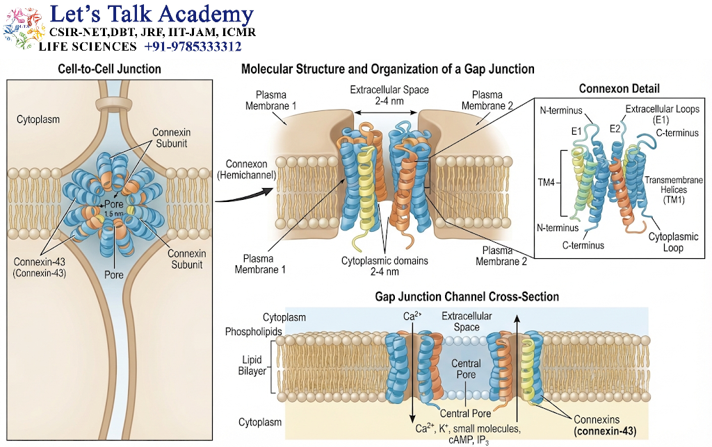
Understanding the structure of gap junctions is essential for understanding how they facilitate intercellular communication. Their function depends on the precise organization of membrane proteins in two neighboring cells.
2.1 Overall Organization of a Gap Junction
A typical gap junction is formed when two adjacent cells bring specialized regions of their plasma membranes into close proximity. Each cell contributes a membrane protein assembly called a hemichannel, also known as a connexon.
One hemichannel is present in the membrane of each participating cell. When the two hemichannels align across the narrow extracellular space and dock with one another, they form a continuous intercellular channel.
The resulting channel connects the cytoplasm of one cell with the cytoplasm of its neighbor.
The main structural components are:
-
Connexin proteins
-
Hemichannels or connexons
-
Intercellular channel pore
-
Cytoplasmic regulatory regions
-
Closely apposed plasma membranes of neighboring cells
2.2 Connexins: The Building Blocks of Gap Junctions
Connexins are integral membrane proteins that assemble to form gap junction channels in vertebrates. Different connexin proteins are encoded by distinct genes and have different tissue distributions and functional properties.
Connexins are commonly designated using the abbreviation Cx, followed by a number corresponding to their approximate molecular mass in kilodaltons. For example, connexin 43 is commonly written as Cx43, and connexin 32 as Cx32.
The molecular mass-based naming system is useful for identifying different connexin proteins, although the number does not directly indicate the size of the channel pore or its permeability.
2.2.1 General Structure of a Connexin Protein
A typical connexin protein contains:
-
Four transmembrane α-helical domains.
-
Two extracellular loops.
-
One intracellular loop.
-
An intracellular N-terminal region.
-
An intracellular C-terminal region.
The four transmembrane segments anchor the protein within the plasma membrane. The extracellular loops participate in the recognition and docking of hemichannels from neighboring cells.
The intracellular loop and C-terminal tail contribute to channel regulation, protein interactions, trafficking, and cellular signaling. Their properties vary among connexin types.
2.2.2 Transmembrane Domains
The four transmembrane domains span the lipid bilayer. They form much of the structural framework of the connexin and help shape the channel pore.
The arrangement of these helices is important for:
-
Maintaining membrane insertion.
-
Creating the appropriate channel architecture.
-
Influencing ion and solute permeability.
-
Supporting channel opening and closing.
-
Mediating interactions between connexin subunits.
Changes in connexin sequence can influence channel conductance, selectivity, stability, and regulation.
2.2.3 Extracellular Loops
Each connexin contains two extracellular loops. These loops are essential for the correct recognition and docking of hemichannels.
When two compatible hemichannels from adjacent cells interact, their extracellular regions align to form a continuous passageway. The extracellular loops also contribute to the specificity of interactions between connexin-containing hemichannels.
Mutations affecting these regions can interfere with gap junction formation or channel function.
2.2.4 Intracellular Loop and C-Terminal Tail
The intracellular loop and C-terminal tail are exposed to the cytoplasm and participate in regulatory processes.
The C-terminal region of some connexins, particularly Cx43, contains sites that can undergo phosphorylation. These modifications influence channel assembly, turnover, localization, and gating.
The intracellular regions can also interact with signaling proteins, scaffolding proteins, and cytoskeletal components. Consequently, gap junctions are not merely passive channels; they can participate in broader signaling networks within the cell.
2.3 Connexons or Hemichannels
A connexon is a hemichannel composed of six connexin subunits in a typical vertebrate gap junction.
Six connexin molecules assemble around a central pore to create a hemichannel within the plasma membrane of a single cell.
When two compatible connexons from neighboring cells dock together, they form a complete gap junction channel.
2.3.1 Homomeric and Heteromeric Connexons
Depending on the connexin composition, a hemichannel may be classified as:
Homomeric connexon: A hemichannel composed of one type of connexin.
Heteromeric connexon: A hemichannel composed of more than one type of connexin.
The combination of connexin isoforms can influence the channel’s permeability, conductance, voltage sensitivity, and regulatory properties.
2.3.2 Homotypic and Heterotypic Gap Junction Channels
Gap junction channels can also be classified according to the connexins present in the two interacting cells.
Homotypic channel: Both hemichannels contain the same connexin composition.
Heterotypic channel: The two hemichannels contain different connexin compositions.
These structural variations can produce differences in channel behavior and compatibility. Not every connexin-containing hemichannel can dock efficiently with every other type.
2.4 Gap Junction Plaques
Gap junction channels generally occur in clusters called gap junction plaques. A plaque consists of multiple gap junction channels concentrated in a specialized region of contact between two cells.
The number of channels within a plaque can vary considerably. Some plaques are small, whereas others contain large assemblies of interconnected channels.
Plaque organization is influenced by:
-
Connexin synthesis and delivery to the plasma membrane.
-
Interactions with scaffolding proteins.
-
Cell adhesion and membrane organization.
-
Phosphorylation and other regulatory processes.
-
Endocytosis and degradation.
Gap junction plaques are dynamic structures. Channels can be added or removed depending on the physiological requirements of the tissue.
2.5 Intercellular Channel and Pore
The complete gap junction channel creates a hydrophilic pathway between the cytoplasm of two adjacent cells. It permits the movement of selected ions and small molecules.
The pore is not an unrestricted opening. Its permeability depends on the channel’s molecular structure and the properties of the transported substance.
Many gap junction channels permit the passage of:
-
Inorganic ions such as potassium, sodium, and calcium.
-
Small second messengers, including cyclic AMP in suitable contexts.
-
Inositol trisphosphate (IP₃), depending on connexin composition.
-
Small metabolites such as some sugars and other low-molecular-weight compounds.
The permeability of a particular channel depends on connexin identity, molecular size, charge, concentration gradients, and channel state.
2.6 Intercellular Gap and Membrane Arrangement
In a conventional gap junction, the plasma membranes of the two adjacent cells are separated by a narrow extracellular gap. The two hemichannels bridge this region and dock to form a continuous channel.
The extracellular spacing and highly organized arrangement of the membrane proteins distinguish gap junctions from other types of cell–cell contact.
This close membrane arrangement is important because it permits direct transfer between cells while keeping the exchanged molecules largely within the connected cytoplasmic compartments.
3. Formation and Assembly of Gap Junctions
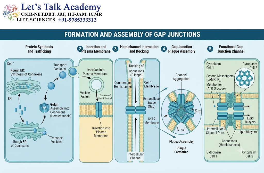
Gap junctions are not formed simply by placing two cells next to each other. Their formation involves a coordinated sequence of molecular events, including connexin synthesis, membrane insertion, oligomerization, transport, docking, and plaque organization.
The entire process is carefully regulated because the number and distribution of gap junction channels determine how effectively neighboring cells can communicate.
3.1 Synthesis of Connexin Proteins
Connexin proteins are synthesized by ribosomes associated with the endoplasmic reticulum (ER). Because connexins are integral membrane proteins, their transmembrane domains become embedded in the ER membrane during synthesis.
After translation, connexins undergo folding and quality-control processes that help ensure that functional proteins are produced.
The major steps involved in connexin synthesis include:
-
Transcription of the connexin gene.
-
Translation of connexin messenger RNA.
-
Insertion of the nascent protein into the ER membrane.
-
Folding and quality control.
-
Oligomerization into hemichannels.
-
Transport toward the plasma membrane.
-
Incorporation into gap junction plaques.
The efficiency of these processes can vary between connexin isoforms.
3.2 Oligomerization of Connexins
Oligomerization is the process by which individual connexin molecules assemble into larger protein complexes.
In many connexins, six protein subunits form a hemichannel. The assembly process is influenced by the molecular characteristics of the connexin and the cellular machinery responsible for protein folding and trafficking.
Connexin oligomerization may occur in the endoplasmic reticulum, Golgi apparatus, or other intracellular compartments, depending on the connexin type.
The correct assembly of hemichannels is important because improperly assembled connexins may fail to reach the cell surface or may be targeted for degradation.
3.3 Transport of Connexons to the Plasma Membrane
Once connexons have formed, they must be transported to the plasma membrane.
Transport involves intracellular trafficking pathways that deliver connexin-containing structures to appropriate regions of the cell surface. Interactions with cytoskeletal components and regulatory proteins can influence this process.
At the plasma membrane, connexons may accumulate at specialized cell–cell contact sites. These sites provide the physical environment required for gap junction formation.
Connexin trafficking is a dynamic process. Newly synthesized channels can be added to existing plaques, while older channels are removed and degraded.
3.4 Docking of Hemichannels
Gap junction channel formation requires the docking of two hemichannels located in the membranes of adjacent cells.
The extracellular loops of the connexins participate in the recognition and alignment of compatible hemichannels. Molecular interactions between the two hemichannels help create a continuous aqueous pathway.
The docking process produces a complete channel with a pore extending from the cytoplasm of one cell to the cytoplasm of the neighboring cell.
An important point is that a hemichannel in one cell must not be confused with a complete gap junction channel. A hemichannel occupies one membrane, whereas a gap junction channel spans the membranes of two cells.
3.5 Formation of Gap Junction Plaques
Following the formation of functional channels, additional channels may accumulate in the same region of cell–cell contact.
This produces a gap junction plaque, which contains many channels organized within a specialized membrane domain.
Plaque size and composition are influenced by:
-
Connexin expression.
-
Protein delivery to the cell surface.
-
Channel assembly and docking.
-
Cell adhesion.
-
Endocytosis and degradation.
-
Signaling pathways that control junctional turnover.
A plaque may contain channels with different properties if more than one connexin isoform is expressed in the participating cells.
3.6 Turnover and Degradation of Gap Junctions
Gap junctions are continuously remodeled. Their channels have a limited lifespan, and the cell must replace them to maintain effective communication.
Old gap junction channels can be internalized through endocytic mechanisms. In some cases, portions of a junctional plaque are removed from one cell while the corresponding membrane from the neighboring cell remains associated with the internalized structure.
These internalized structures are often called annular gap junctions or connexosomes. They can subsequently be degraded through lysosomal or other intracellular pathways.
Regulated turnover helps cells adjust the amount of intercellular communication according to changing physiological conditions.
4. Mechanism of Intercellular Communication Through Gap Junctions
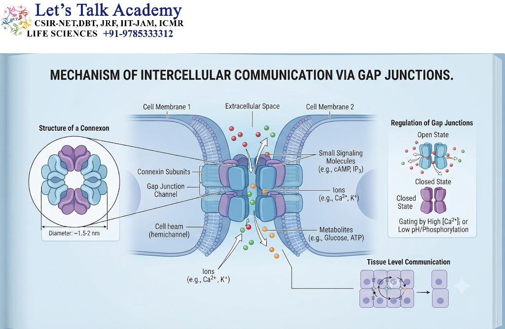
Gap junctions allow neighboring cells to communicate by providing a direct route for the movement of selected substances between their cytoplasms.
This mechanism differs from communication through secreted hormones or neurotransmitters, where a signal is released into the extracellular space and detected by a receptor on another cell.
4.1 Direct Cytoplasmic Exchange
A functional gap junction channel connects the cytoplasm of two adjacent cells.
When a suitable ion or small molecule is present at a higher effective electrochemical potential in one cell than in the other, it can move through the channel toward the lower-potential side.
The direction and magnitude of transport depend on the relevant concentration gradients, electrical gradients, and channel properties.
For example, ions can move between electrically coupled cells, allowing changes in membrane potential to influence neighboring cells.
4.2 Movement of Ions
Ions are among the most important substances transferred through gap junction channels.
Commonly transported ions include:
-
Potassium ions (K⁺).
-
Sodium ions (Na⁺).
-
Calcium ions (Ca²⁺).
-
Chloride ions (Cl⁻), depending on channel properties.
The movement of ions permits electrical coupling between cells. This is especially important in cardiac and smooth muscle tissues, where the coordinated movement of ions supports synchronized contraction.
The precise conductance and ionic selectivity of a gap junction channel depend on its connexin composition.
4.3 Movement of Small Metabolites
Gap junctions also allow the exchange of certain small metabolites and signaling molecules.
Examples include some:
-
Amino acids.
-
Nucleotides.
-
Metabolic intermediates.
-
Second messengers.
-
Small water-soluble nutrients.
The transfer of these substances helps neighboring cells share metabolic resources and coordinate biochemical activities.
For example, metabolic coupling can help groups of cells respond collectively to local changes in nutrient availability.
However, not every small molecule can pass through every gap junction channel. Permeability varies with molecular size, charge, shape, and connexin composition.
4.4 Electrical Coupling
Electrical coupling occurs when ions pass directly between cells through gap junction channels, allowing changes in membrane potential to spread from one cell to another.
If one electrically excitable cell becomes depolarized, current can flow through gap junctions into adjacent cells. This can bring the neighboring cells closer to the threshold required for electrical activation.
Electrical coupling is particularly important in:
-
Cardiac muscle.
-
Some smooth muscle tissues.
-
Certain neuronal circuits.
-
Specialized sensory systems.
In the heart, electrical coupling through gap junctions contributes to the coordinated propagation of excitation across cardiac tissue.
4.5 Chemical and Metabolic Coupling
Gap junctions contribute to chemical coupling by allowing selected intracellular signaling molecules to move between connected cells.
For instance, the movement of inositol trisphosphate (IP₃) or other small messengers can help coordinate intracellular signaling across neighboring cells, depending on the channel’s permeability.
Metabolic coupling may also help cells distribute small molecules and buffer local differences in metabolic conditions.
The consequences of this coupling depend on the tissue and the direction of the concentration gradients.
4.6 Gap Junctions and Calcium Signaling
Calcium ions are important intracellular messengers that regulate processes such as contraction, secretion, metabolism, and gene expression.
Gap junction channels can contribute to intercellular calcium signaling by permitting the transfer of calcium ions or certain calcium-related signaling molecules.
However, calcium signals can also spread through extracellular pathways involving ATP release, purinergic receptors, and other mechanisms. Therefore, a calcium wave observed across a tissue does not necessarily indicate direct calcium transfer through gap junctions.
The relative contribution of each mechanism depends on the tissue, experimental conditions, and connexin composition.
5. Major Functions of Gap Junctions
Gap junctions perform diverse functions in multicellular organisms. Their importance extends beyond simple molecular transport because they help coordinate the behavior of groups of cells.
5.1 Coordination of Electrical Activity
One of the best-known functions of gap junctions is the coordination of electrical activity.
In electrically excitable tissues, cells must often respond in a synchronized manner. Gap junction channels provide low-resistance pathways for electrical current to pass between neighboring cells.
This enables the rapid spread of electrical signals without requiring the release of chemical neurotransmitters.
5.2 Synchronization of Cardiac Muscle Contraction
Cardiac muscle cells must contract in a coordinated sequence to pump blood efficiently.
Gap junctions located in specialized regions of cardiac cell contact, particularly within intercalated discs, allow electrical signals to spread between adjacent cardiomyocytes.
Connexin 43 is a major gap junction protein in ventricular myocardium, while other connexins contribute to communication in different regions of the heart.
Through electrical coupling, groups of cardiac cells can depolarize in a coordinated manner.
5.2.1 Importance in Cardiac Physiology
The coordinated transmission of excitation contributes to:
-
Synchronization of cardiac muscle contraction.
-
Efficient propagation of electrical impulses.
-
Maintenance of organized cardiac rhythm.
Disruption of gap junction distribution or function can interfere with electrical propagation and may contribute to cardiac arrhythmias.
However, cardiac rhythm is also controlled by ion channels, conduction-system anatomy, autonomic regulation, and other factors. Gap junctions are one component of this complex system.
5.3 Communication in Smooth Muscle
Smooth muscle is present in the walls of blood vessels, the digestive tract, the urinary bladder, and several other organs.
In many smooth muscle tissues, gap junctions allow electrical signals and small intracellular messengers to spread between neighboring cells.
This coupling helps coordinate contraction across groups of cells.
For example, coordinated activity in gastrointestinal smooth muscle contributes to organized patterns of movement that support the transport of food through the digestive tract.
The extent of gap junctional coupling differs among smooth muscle tissues and can change in response to physiological and hormonal signals.
5.4 Coordination of Metabolic Activities
Cells within a tissue may experience differences in nutrient supply, metabolic demand, and signaling conditions.
Gap junctions can help neighboring cells share selected small metabolites and signaling molecules. This process is called metabolic coupling.
Metabolic coupling may support:
-
Distribution of small metabolic intermediates.
-
Coordination of biosynthetic activity.
-
Maintenance of local cellular homeostasis.
-
Sharing of selected nutrients and signaling compounds.
In some tissues, this function is particularly important for maintaining the health of cells that are poorly supplied by direct access to certain metabolites.
5.5 Regulation of Cell Growth and Differentiation
Gap junctional communication can influence cellular growth and differentiation.
During development, cells respond to signals from their neighbors, and direct communication through gap junctions may contribute to the coordination of these responses.
Connexins can also influence cellular processes independently of their role as intercellular channels. For example, some connexin proteins interact with intracellular signaling proteins and structural components.
The relationship between gap junctional communication and cell proliferation is complex and depends on cell type, developmental stage, and the particular connexin involved.
5.6 Role in Embryonic Development
During embryonic development, cells undergo coordinated proliferation, migration, differentiation, and tissue organization.
Gap junctions may contribute to these processes by allowing developing cells to exchange signaling molecules and ions.
The expression of connexin proteins changes during development, and some connexins are associated with specific developmental stages or tissues.
In certain developmental contexts, gap junctional communication helps coordinate groups of cells that must respond to shared signals.
Nevertheless, embryonic development is controlled by many overlapping mechanisms, including morphogen gradients, cell adhesion, transcriptional regulation, and extracellular signaling.
5.7 Role in the Nervous System
Gap junctions have important functions in the nervous system, where they can form electrical synapses.
Electrical synapses allow current to pass directly between neurons through gap junction channels.
This type of communication can provide rapid transmission and synchronization of neuronal activity.
Gap junctions are also present between certain glial cells, including astrocytes. Glial gap junctions can contribute to metabolic and ionic homeostasis within nervous tissue.
5.7.1 Electrical Synapses
At an electrical synapse, the membranes of two neurons are connected through gap junction channels.
When electrical activity occurs in one neuron, current can flow through the junction and influence the membrane potential of the other neuron.
Electrical synapses can support:
-
Rapid signal transmission.
-
Synchronous firing.
-
Coordinated rhythmic activity.
-
Bidirectional electrical communication in many arrangements.
The physiological properties of electrical synapses depend on the connexin type, channel organization, and electrical characteristics of the participating neurons.
5.7.2 Connexin 36 in Neuronal Communication
Connexin 36, encoded by the GJD2 gene, is an important connexin in many neuronal electrical synapses.
It is expressed in several neuronal populations and contributes to electrical coupling between specific groups of neurons.
Its presence can support the synchronization of neuronal activity, although the exact function depends on the neural circuit involved.
5.8 Role in Epithelial Tissue
Epithelial cells form protective and functional barriers in tissues such as the skin, intestine, and kidney.
Gap junctions in epithelial tissues help neighboring cells exchange selected ions, metabolites, and signaling molecules.
This communication can support:
-
Coordination of cellular differentiation.
-
Regulation of tissue homeostasis.
-
Response to injury.
-
Coordination of cellular growth.
-
Maintenance of tissue function.
Gap junctions do not generally form the primary seal that prevents substances from passing between epithelial cells through the extracellular space. That barrier function is primarily associated with tight junctions.
5.9 Role in Wound Healing
When tissue is injured, neighboring cells must coordinate migration, proliferation, and repair.
Gap junctional communication can influence wound-healing responses by allowing cells to exchange small signaling molecules and by participating in regulatory signaling networks.
Changes in connexin expression and localization have been observed during tissue injury and repair.
The effects of a particular connexin can differ depending on the tissue, the stage of healing, and whether its channel-dependent or channel-independent functions are involved.
5.10 Role in Homeostasis
Homeostasis refers to the maintenance of relatively stable internal conditions within a biological system.
Gap junctions contribute to homeostasis by coordinating activities among groups of cells.
They can help regulate:
-
Ionic balance.
-
Metabolic cooperation.
-
Electrical activity.
-
Intercellular signaling.
-
Responses to local environmental changes.
By linking neighboring cells, gap junctions enable tissues to function as coordinated units rather than as isolated collections of cells.
6. Regulation of Gap Junctional Communication
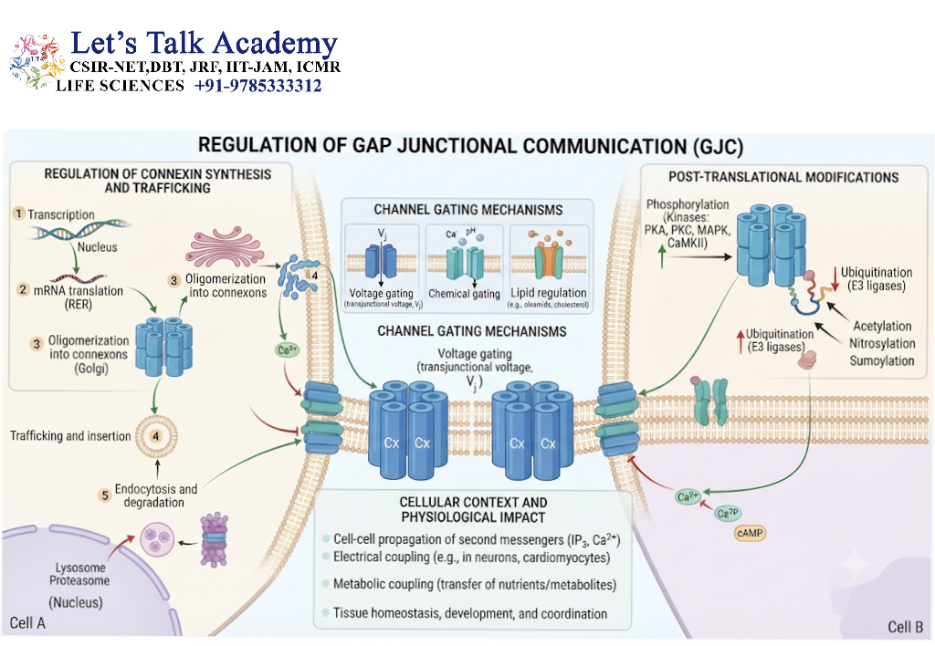
Gap junctions are dynamic communication systems whose activity can change according to the physiological state of the cell. The number of channels, their molecular composition, their cellular location, and their opening and closing behavior all influence intercellular communication.
Gap junctional communication is regulated at multiple levels, from gene expression to the activity of individual channels.
6.1 Regulation at the Gene Expression Level
The first level of regulation involves controlling the production of connexin proteins.
Connexin genes are transcribed in response to developmental signals, hormones, growth factors, tissue-specific transcription factors, and environmental conditions.
Changes in connexin gene expression can alter the number of functional gap junction channels in a tissue.
For example, a cell may increase the expression of a particular connexin during differentiation, thereby changing its ability to communicate with neighboring cells.
Gene expression is regulated through mechanisms such as:
-
Activation or repression of transcription factors.
-
Epigenetic modifications.
-
Changes in messenger RNA stability.
-
Regulation of protein translation.
-
Tissue-specific signaling pathways.
Different tissues express different combinations of connexin isoforms, producing tissue-specific communication properties.
6.2 Regulation Through Protein Trafficking
After synthesis, connexins must be transported to the appropriate cellular location.
Changes in intracellular trafficking can affect gap junctional communication even when connexin gene expression remains unchanged.
Trafficking regulation may influence:
-
Transport of connexins from the ER.
-
Assembly of connexons.
-
Delivery of connexons to the plasma membrane.
-
Incorporation of channels into plaques.
-
Removal of channels from the cell surface.
Defects in trafficking may prevent functional gap junctions from forming.
6.3 Regulation by Phosphorylation
Phosphorylation is a reversible post-translational modification in which a phosphate group is added to a protein by a kinase.
Connexins can be phosphorylated at specific amino acid residues. Phosphorylation may influence channel assembly, trafficking, conductance, gating, and degradation.
Connexin 43 is particularly well studied because its C-terminal tail contains multiple regulatory phosphorylation sites.
The consequences of phosphorylation depend on:
-
The connexin isoform.
-
The particular amino acid residue modified.
-
The kinase or phosphatase involved.
-
The physiological condition of the cell.
-
Whether the modification affects assembly, channel gating, or turnover.
Phosphorylation should not be considered universally activating or inhibitory. Its effect is context-dependent.
6.4 Voltage-Dependent Gating
Gap junction channels can respond to differences in electrical potential between the cytoplasms of the two connected cells.
The voltage difference across a gap junction is called the transjunctional voltage.
Here, V1V_1 and V2V_2 represent the electrical potentials of the two cells.
Changes in transjunctional voltage can influence channel conductance. Depending on the connexin type and the direction of the voltage difference, channels may reduce their conductance or adopt different gating states.
Voltage-dependent gating helps regulate the transfer of current and ions between coupled cells.
6.5 Chemical Gating
Gap junction channels are sensitive to several chemical and intracellular conditions.
Important regulatory factors include:
-
Intracellular pH.
-
Calcium concentration.
-
Redox state.
-
Phosphorylation status.
-
Certain metabolites and signaling molecules.
6.5.1 Regulation by Intracellular pH
A decrease in intracellular pH, known as intracellular acidification, can reduce gap junctional communication in several connexin systems.
This response may be physiologically relevant during metabolic stress, ischemia, or other conditions that alter cellular metabolism.
Acidification can influence the conformation of regulatory regions within the channel, reducing its conductance.
However, the exact mechanism and sensitivity vary among connexin isoforms.
6.5.2 Regulation by Calcium Ions
Calcium ions play important roles in cellular signaling and can influence gap junction channels and unapposed hemichannels.
A rise in intracellular calcium can alter gap junctional conductance under certain conditions. Calcium-dependent signaling pathways may also modify connexins through phosphorylation or other regulatory mechanisms.
It is important to distinguish between calcium regulation of complete gap junction channels and calcium-related effects on hemichannels.
6.5.3 Regulation by Redox Conditions
The redox state of a cell reflects the balance between oxidizing and reducing chemical processes.
Changes in cellular redox conditions can affect connexin proteins and gap junctional communication.
Oxidative stress may influence:
-
Connexin phosphorylation.
-
Channel gating.
-
Protein stability.
-
Channel trafficking.
-
Junctional turnover.
The effects depend on the connexin type and the intensity and duration of the stress.
6.6 Regulation Through Connexin Degradation
Gap junctional communication depends partly on how long connexin proteins remain functional at the cell surface.
Cells regulate communication by removing older channels and replacing them with newly synthesized ones.
Connexin degradation may occur through lysosomal pathways, proteasomal mechanisms, and other protein-quality-control processes.
Increased degradation can reduce the number of functional channels, whereas enhanced delivery and assembly can increase intercellular coupling.
6.7 Regulation by Cellular Stress
Various stress conditions can affect gap junctional communication.
Examples include:
-
Hypoxia.
-
Ischemia.
-
Oxidative stress.
-
Inflammation.
-
Mechanical injury.
-
Metabolic disturbances.
-
Changes in extracellular signaling.
Stress can alter connexin phosphorylation, localization, degradation, and channel activity.
In some tissues, reduced coupling may protect neighboring cells by limiting the spread of damaging signals. In other circumstances, reduced coupling may interfere with normal tissue function.
Therefore, the consequences of gap junctional regulation depend on the biological context.
7. Gap Junctions and Hemichannels: A Functional Distinction
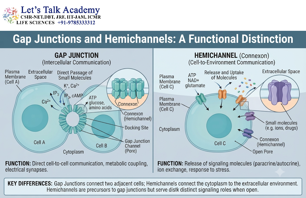
A clear understanding of the difference between complete gap junction channels and hemichannels is essential for studying intercellular communication.
7.1 Complete Gap Junction Channels
A complete gap junction channel is formed by the docking of two connexons contributed by two neighboring cells.
It creates a direct cytoplasmic connection between the cells.
Its principal functions include:
-
Electrical coupling.
-
Transfer of selected small molecules.
-
Metabolic communication.
-
Coordination of intracellular signaling.
7.2 Unapposed Hemichannels
A hemichannel is a connexon that has not docked with a hemichannel from an adjacent cell.
Under some circumstances, unapposed hemichannels can provide a pathway between the cytoplasm and the extracellular environment.
However, hemichannel activity must be tightly regulated because excessive opening can disturb ionic balance and cellular homeostasis.
Hemichannels may participate in the release of ATP and other signaling substances under particular physiological or pathological conditions.
The behavior of hemichannels is influenced by extracellular calcium, voltage, membrane conditions, and other regulatory factors.
7.3 Comparison Between Gap Junction Channels and Hemichannels
|
Feature |
Complete gap junction channel |
Unapposed hemichannel |
|---|---|---|
|
Structural organization |
Two docked connexons |
One connexon |
|
Location |
Between adjacent cells |
In a single cell membrane |
|
Main pathway |
Cytoplasm to cytoplasm |
Cytoplasm to extracellular space |
|
Typical function |
Direct intercellular communication |
Regulated exchange with the extracellular environment |
|
Regulation |
Voltage, chemical factors, phosphorylation, and other mechanisms |
Voltage, extracellular conditions, intracellular signaling, and other mechanisms |
|
Potential risk of excessive activity |
Abnormal ionic or metabolic coupling |
Loss of homeostasis and uncontrolled solute exchange |
Not all connexin hemichannels are equally likely to open under physiological conditions. Their behavior depends on the particular connexin and cellular environment.
8. Types of Gap Junction Proteins in Different Organisms
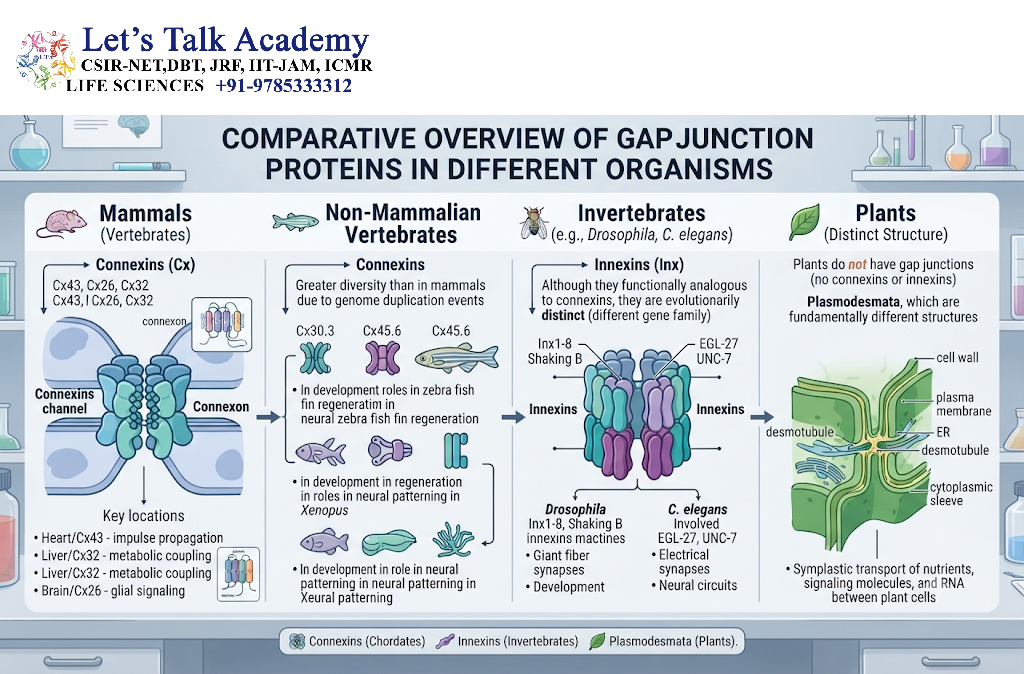
Gap junction-like communication systems occur in many multicellular organisms. However, the proteins responsible for forming these channels are not identical across all animal groups.
8.1 Connexins in Vertebrates
In vertebrates, gap junction channels are primarily composed of connexin proteins.
Humans possess a diverse connexin gene family, with approximately 21 connexin genes commonly recognized in the human genome.
Different connexins have characteristic patterns of tissue expression and functional properties.
Examples include:
|
Connexin |
Common designation |
Important sites or roles |
|---|---|---|
|
Cx26 |
GJB2 |
Inner ear, skin, and other tissues |
|
Cx32 |
GJB1 |
Peripheral nervous system and liver |
|
Cx36 |
GJD2 |
Neuronal electrical coupling |
|
Cx43 |
GJA1 |
Heart, connective tissues, and other organs |
|
Cx46 |
GJA3 |
Lens |
|
Cx50 |
GJA8 |
Lens development and physiology |
The expression of a connexin in a tissue does not necessarily mean that it is the only connexin present there. Multiple connexins may coexist and form channels with distinct properties.
8.2 Innexins in Invertebrates
In many invertebrate animals, gap junction-like intercellular channels are formed by proteins called innexins.
Innexins are structurally and evolutionarily distinct from connexins, although they perform related functions in direct intercellular communication.
They are found in organisms such as:
-
Nematodes.
-
Insects.
-
Other invertebrate groups.
In the fruit fly Drosophila melanogaster, innexin proteins contribute to electrical and metabolic communication between cells.
8.3 Pannexins in Vertebrates
Pannexins are a family of proteins related in broad structural organization to innexins.
Vertebrates possess pannexin proteins, including pannexin 1, pannexin 2, and pannexin 3.
Pannexins are particularly associated with membrane channels involved in communication between the cell and its extracellular environment.
Unlike connexins, native pannexins are generally not regarded as the primary structural components of conventional vertebrate cell-to-cell gap junctions.
Their functions include roles in extracellular ATP release and signaling in certain physiological and pathological contexts.
9. Physiological Significance of Gap Junctions in Different Tissues
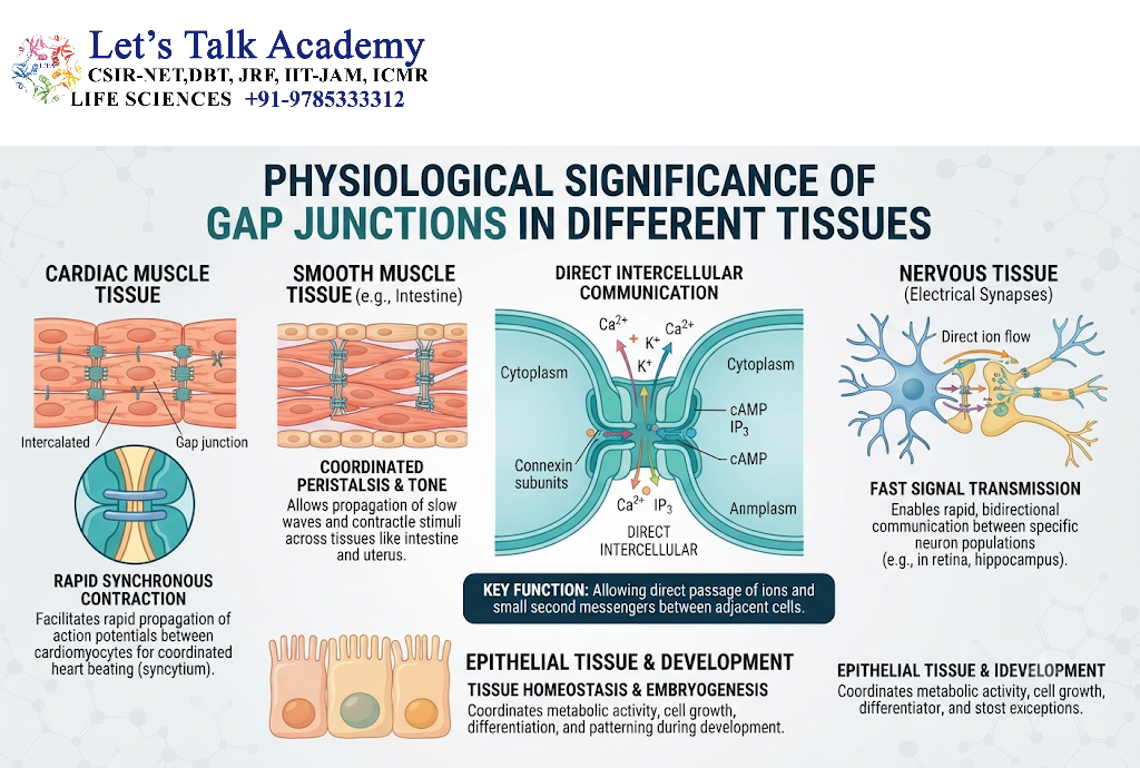
Gap junctions are distributed across numerous tissues, where they support functions adapted to the requirements of each organ.
9.1 Gap Junctions in the Heart
The heart requires coordinated electrical activity to produce effective contraction.
Gap junctions between cardiomyocytes allow electrical current to spread from one cell to another.
Connexin 43 is an important gap junction protein in ventricular cardiac muscle, while other connexins are involved in specialized regions of the heart.
The arrangement and density of gap junctions influence the direction and efficiency of electrical impulse propagation.
Disorganization of gap junctions can contribute to conduction abnormalities.
9.2 Gap Junctions in the Brain
Gap junctions occur between specific populations of neurons and glial cells.
In neurons, they form electrical synapses that can synchronize neuronal activity.
In astrocytes and other glial populations, gap junctions can support the exchange of ions and metabolites.
Astrocytic coupling can help maintain the chemical environment around neurons and facilitate the distribution of selected metabolic substances.
9.3 Gap Junctions in the Retina
The retina contains several types of electrically coupled neuronal networks.
Gap junctions contribute to communication between particular retinal neurons and help coordinate responses to visual stimuli.
Connexin 36, for example, participates in electrical coupling within several neuronal circuits.
The precise function of coupling depends on the retinal cell type and the organization of the neural circuit.
9.4 Gap Junctions in the Liver
Hepatocytes, the principal cells of the liver, express connexins that support intercellular communication.
Gap junctional communication in the liver can contribute to the coordination of metabolic activities and the transmission of intracellular signaling molecules.
Connexin 32 is an important hepatic connexin, although other connexins may also be expressed.
The activity of gap junctions can be influenced by hormones, metabolic changes, inflammation, and liver injury.
9.5 Gap Junctions in the Lens
The lens of the eye is a specialized tissue that must maintain transparency and support the movement of nutrients and waste products.
As lens fiber cells mature, they lose many of the organelles required for conventional metabolism. Intercellular communication becomes important for maintaining the internal environment of the lens.
Connexins such as Cx46 and Cx50 contribute to communication among lens cells.
Alterations in these connexins can interfere with lens physiology and are associated with certain inherited cataracts.
9.6 Gap Junctions in the Skin
The skin contains several types of cells that express connexins.
Gap junctional communication can influence:
-
Epidermal differentiation.
-
Cellular growth.
-
Tissue repair.
-
Intercellular signaling.
-
Maintenance of tissue homeostasis.
Connexin 26 and connexin 43 are among the connexins studied in skin biology.
The relationship between connexins and skin health is complex because connexins can have both channel-dependent and channel-independent effects.
9.7 Gap Junctions in Reproductive Tissues
Gap junctions are important in several reproductive tissues.
In ovarian follicles, communication between granulosa cells and other follicular cells helps coordinate metabolic and signaling activities.
Connexin 43 is widely studied in relation to follicular development and cellular communication within the ovary.
Gap junctions also contribute to communication in other reproductive tissues, although the specific connexins and their roles vary.
10. Gap Junctions and Cell Signaling Networks
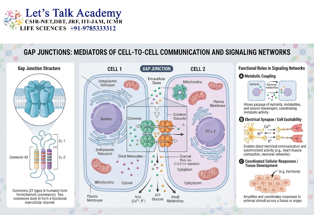
Gap junctions are integrated into broader cellular communication systems. They do not work independently of other signaling pathways.
10.1 Interaction With Intracellular Signaling Pathways
Connexins can interact with proteins involved in intracellular signaling.
These interactions may influence:
-
Protein kinase activity.
-
Cytoskeletal organization.
-
Cell adhesion.
-
Cell proliferation.
-
Cell survival.
-
Junctional assembly.
For this reason, connexins are increasingly understood as components of signaling complexes rather than simply as structural channel proteins.
10.2 Relationship With Tight Junctions
Tight junctions and gap junctions are both involved in cell–cell organization, but their primary functions differ.
Tight junctions help regulate the movement of substances through the space between epithelial cells and contribute to cell polarity.
Gap junctions provide direct communication between the cytoplasms of neighboring cells.
These junctional systems can influence one another through shared cellular signaling and structural networks.
10.3 Relationship With Adherens Junctions
Adherens junctions are cell–cell adhesion structures that help maintain tissue architecture.
They connect neighboring cells through cadherin-based adhesion systems and are linked to the actin cytoskeleton.
Gap junctions and adherens junctions may be organized within related regions of cell–cell contact. Their interactions can influence junctional stability, cellular organization, and tissue behavior.
10.4 Connexin Functions Independent of Channel Formation
Some connexins have biological functions that do not depend directly on the passage of substances through a gap junction channel.
For example, connexin proteins may interact with intracellular proteins, affect cell signaling, and influence cell growth or differentiation.
These channel-independent functions are an important area of research because they show that connexins can act as multifunctional regulatory proteins.
11. Clinical Significance of Gap Junctions
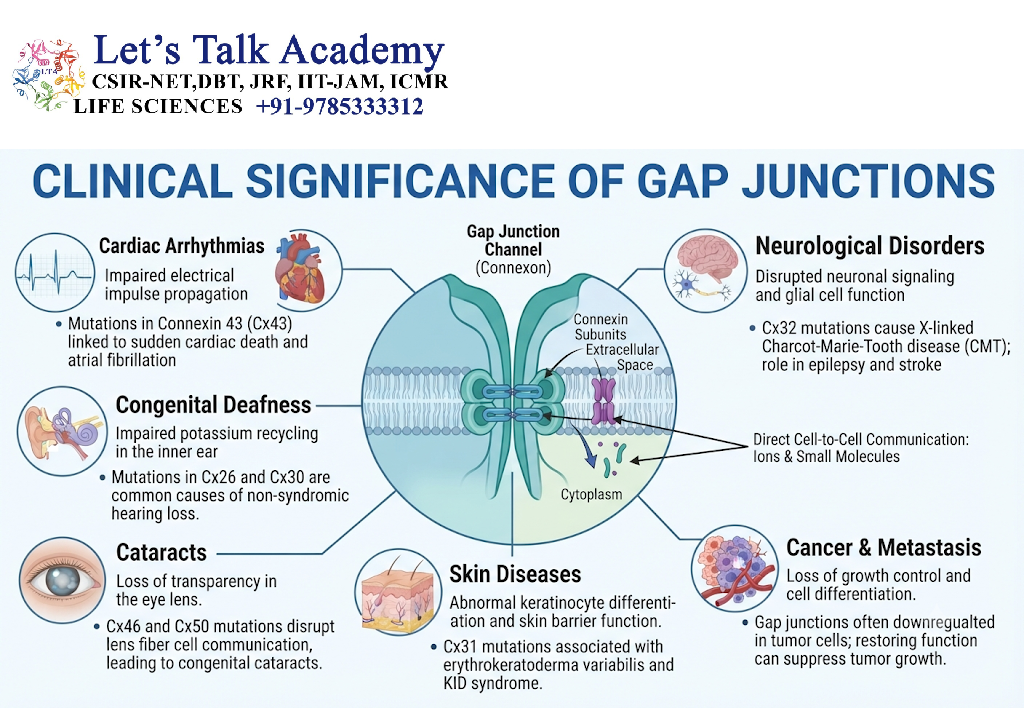
Changes in connexin structure, expression, localization, or channel function can be associated with human disease.
Some disorders arise from inherited mutations, whereas others involve acquired changes in connexin regulation.
11.1 Connexin Mutations and Disease
Mutations in connexin genes can affect the formation or function of gap junction channels.
A mutation may cause:
-
Incorrect protein folding.
-
Defective trafficking.
-
Impaired oligomerization.
-
Abnormal channel gating.
-
Altered molecular permeability.
-
Reduced intercellular communication.
The consequences depend on the gene involved and the tissues in which it is expressed.
11.2 Connexin 26 and Hearing Loss
Mutations in the GJB2 gene, which encodes connexin 26, are an important genetic cause of certain forms of hereditary hearing loss.
Connexin 26 is expressed in tissues of the inner ear and contributes to the specialized cellular environment required for normal auditory function.
Disruption of connexin 26 can interfere with the ionic and metabolic conditions necessary for hearing.
Some GJB2 variants are also associated with skin disorders.
11.3 Connexin 32 and Peripheral Neuropathy
Connexin 32 is encoded by the GJB1 gene.
Certain GJB1 mutations cause X-linked Charcot–Marie–Tooth disease, a hereditary peripheral neuropathy.
Connexin 32 is expressed in Schwann cells, where it contributes to communication within myelinated peripheral nerves.
Mutations can disrupt normal Schwann-cell function and impair peripheral nerve physiology.
11.4 Connexins and Cataracts
Connexin 46 and connexin 50 contribute to communication within the eye lens.
Mutations in the genes encoding these proteins, GJA3 and GJA8, respectively, have been associated with inherited forms of cataract.
Altered connexin function can interfere with lens homeostasis and transparency.
11.5 Gap Junctions and Cardiac Arrhythmias
Cardiac gap junctions contribute to the propagation of electrical excitation through heart muscle.
Changes in connexin expression, localization, or regulation can affect electrical conduction.
Such changes may be associated with an increased risk of abnormal cardiac rhythms in certain disease settings.
However, arrhythmias have many possible causes, and gap junction dysfunction is only one contributing factor.
11.6 Gap Junctions and Cancer Biology
Gap junctions have been studied extensively in relation to cancer development and progression.
Changes in connexin expression or gap junctional communication have been observed in various tumor types.
Some connexins and their associated signaling pathways may influence cell proliferation, migration, differentiation, or interactions with the tumor microenvironment.
However, connexins do not have a universally tumor-suppressive or tumor-promoting role. Their effects depend on the tissue, the connexin involved, and the cellular context.
11.7 Gap Junctions During Inflammation and Tissue Injury
Inflammation and tissue injury can alter connexin expression and channel activity.
These changes may affect communication between immune cells, epithelial cells, stromal cells, and other tissue components.
Gap junctional signaling can influence wound responses and the spread of intracellular signals during injury.
The outcome may be beneficial or harmful depending on the type and duration of the response.
12. Experimental Methods Used to Study Gap Junctions
Researchers use several experimental techniques to investigate the structure, function, and regulation of gap junctions.
12.1 Electron Microscopy
Electron microscopy is used to examine the ultrastructure of gap junctions.
It can reveal:
-
Closely apposed plasma membranes.
-
Gap junction plaques.
-
The organization of membrane-associated channels.
-
Changes in junctional architecture.
Transmission electron microscopy and freeze-fracture techniques have historically played important roles in studying gap junction structure.
12.2 Immunofluorescence Microscopy
Immunofluorescence microscopy uses antibodies against specific connexin proteins.
This technique allows researchers to determine the location and distribution of connexins within cells and tissues.
It can be used to study:
-
Connexin expression.
-
Gap junction plaque formation.
-
Changes in connexin localization.
-
Differences between healthy and diseased tissues.
12.3 Dye-Coupling Assays
Dye-coupling experiments are used to investigate the transfer of small fluorescent molecules between neighboring cells.
A fluorescent tracer is introduced into one cell, and researchers observe whether it spreads to adjacent cells.
The transfer of a suitable tracer provides evidence of functional intercellular coupling.
However, the absence of dye transfer does not necessarily prove that all electrical or molecular communication is absent.
12.4 Electrophysiological Techniques
Electrophysiological methods measure electrical currents and voltage changes associated with gap junctional communication.
The dual whole-cell voltage-clamp technique is frequently used to study electrical coupling between paired cells.
Researchers can measure:
-
Junctional conductance.
-
Current–voltage relationships.
-
Voltage-dependent gating.
-
Single-channel activity.
-
Changes in channel properties.
These techniques are particularly useful for studying electrical synapses and cardiac gap junctions.
12.5 Molecular Biology Techniques
Molecular biology methods help researchers investigate the genes and proteins involved in gap junction formation.
Common techniques include:
-
Polymerase chain reaction (PCR).
-
DNA sequencing.
-
Quantitative reverse-transcription PCR.
-
Western blotting.
-
Gene cloning.
-
RNA interference.
-
Gene knockout and gene-editing approaches.
These methods help determine how changes in connexin expression affect cellular communication.
12.6 Cryo-Electron Microscopy
Cryo-electron microscopy has helped researchers investigate connexin channel architecture at high resolution.
It provides structural information about:
-
Transmembrane helices.
-
Channel pores.
-
Extracellular docking regions.
-
Connexin subunit arrangement.
-
Structural features associated with gating.
High-resolution structural studies have expanded our understanding of how connexin sequence differences can influence channel behavior.


