
1. Introduction
Cells in multicellular organisms do not exist as isolated units. They interact continuously with neighboring cells, the extracellular matrix (ECM), and surrounding tissues. These interactions are essential for maintaining tissue architecture, coordinating cellular behavior, and allowing cells to communicate with their physical environment.
One of the most important mechanisms responsible for these interactions is cell adhesion.
Cell adhesion is the process by which cells attach to:
- Other cells
- Extracellular matrix components
- Basement membranes
- Tissue surfaces
Cell adhesion is mediated primarily by specialized proteins called cell adhesion molecules (CAMs).
Adhesion molecules do more than simply “hold cells together.” They also transmit mechanical and biochemical signals that influence:
- Cell shape
- Cell polarity
- Migration
- Proliferation
- Differentiation
- Survival
- Tissue development
- Immune-cell movement
- Wound healing
Therefore, cell adhesion is both a structural and signaling process.
2. Definition of Cell Adhesion
Cell adhesion is the process through which cells attach to neighboring cells or to components of the extracellular matrix through specialized adhesion molecules.
These interactions may be:
- Cell-to-cell
- Cell-to-extracellular-matrix
- Transient
- Stable
- Homophilic
- Heterophilic
2.1 Basic Principle
The general principle can be represented as:
Adhesion molecule on cell
↓
Binding partner on another cell or ECM
↓
Physical attachment
↓
Cytoskeletal connection
↓
Signal transmission
↓
Cellular response
3. Cell Adhesion Molecules
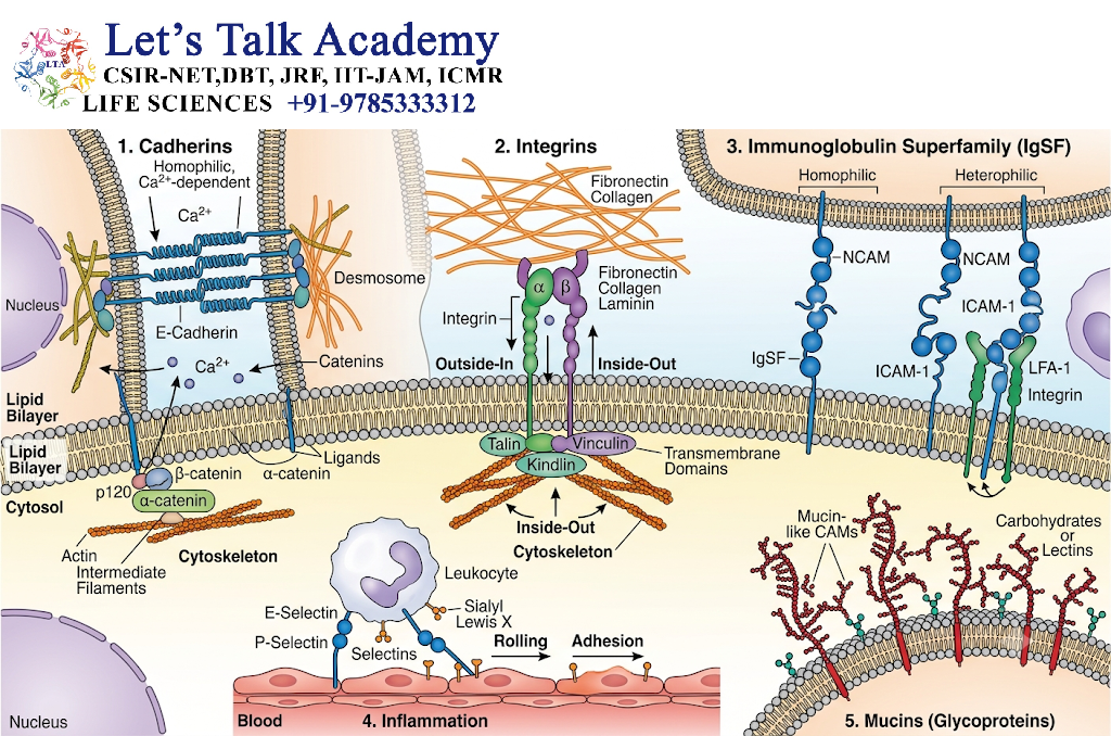
Cell adhesion molecules (CAMs) are proteins located on the cell surface that mediate interactions between cells or between cells and the extracellular matrix.
Major families include:
- Cadherins
- Integrins
- Selectins
- Immunoglobulin superfamily CAMs
- Certain other specialized adhesion proteins
Each family has different structural characteristics, binding properties, and biological functions.
4. Major Types of Cell Adhesion
Cell adhesion can broadly be divided into two major categories.
4.1 Cell-Cell Adhesion
In cell-cell adhesion, one cell attaches directly to another cell.
Examples include:
- Cadherin-mediated adhesion
- Selectin-mediated adhesion
- Immunoglobulin superfamily-mediated adhesion
4.2 Cell-Matrix Adhesion
In cell-matrix adhesion, cells attach to extracellular matrix components.
Integrins are the major molecules responsible for this type of adhesion.
Examples of ECM ligands include:
- Fibronectin
- Laminin
- Collagen
- Vitronectin
5. Homophilic and Heterophilic Adhesion
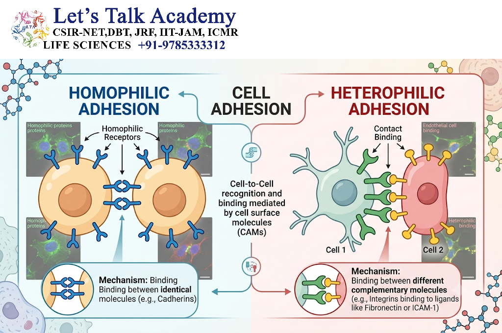
5.1 Homophilic Adhesion
In homophilic adhesion, the same type of adhesion molecule on two cells interacts.
For example:
Cadherin on Cell A ↔ Same cadherin on Cell B
This mechanism is particularly important in tissue organization.
5.2 Heterophilic Adhesion
In heterophilic adhesion, different molecules interact.
For example:
Integrin ↔ Fibronectin
or
Selectin ↔ Carbohydrate ligand
Thus, adhesion can occur through either similar or different molecular partners.
6. Structure of Cell Adhesion Molecules
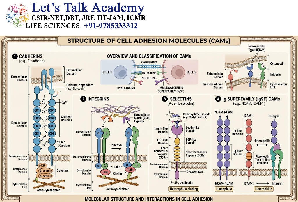
Although adhesion molecules differ considerably, many contain three functional regions:
6.1 Extracellular Domain
This region interacts with:
- Other adhesion molecules
- ECM proteins
- Carbohydrate ligands
6.2 Transmembrane Domain
This region anchors the adhesion molecule within the plasma membrane.
6.3 Cytoplasmic Domain
The intracellular region can interact with:
- Cytoskeletal proteins
- Adaptor proteins
- Signaling molecules
Therefore, many adhesion molecules form a molecular bridge between the external environment and the cytoskeleton.
7. Cadherins
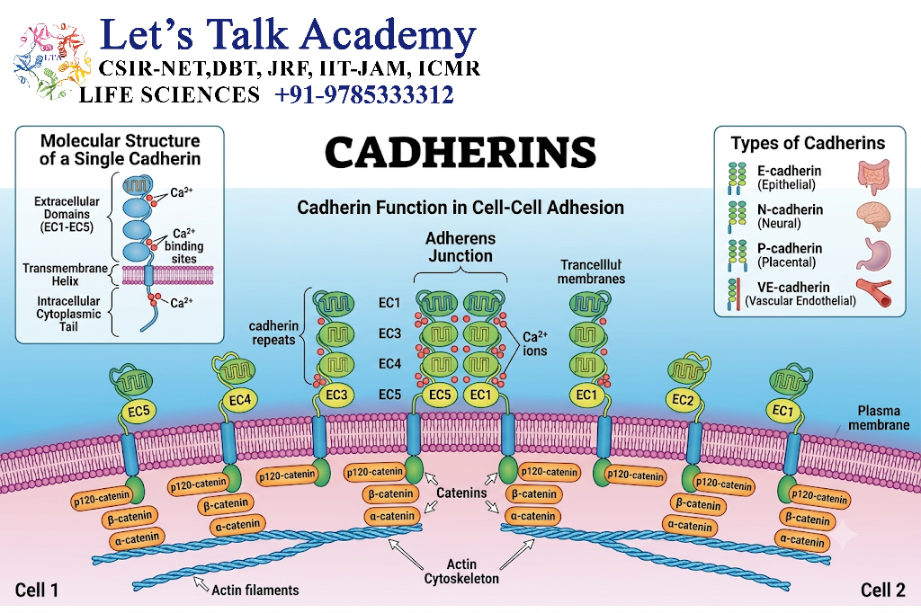
Cadherins are calcium-dependent cell-cell adhesion molecules that generally mediate strong and selective cell-cell interactions.
They are particularly important for maintaining tissue organization.
The name “cadherin” comes from calcium-dependent adhesion.
7.1 Characteristics of Cadherins
Cadherins generally:
- Mediate cell-cell adhesion
- Require Ca²⁺ for stable extracellular interactions
- Often participate in homophilic binding
- Connect to the actin cytoskeleton through catenins
- Contribute to tissue architecture
7.2 Classical Cadherins
Examples include:
- E-cadherin – commonly associated with epithelial tissues
- N-cadherin – important in neural and other tissues
- P-cadherin – found in several epithelial and developmental contexts
8. Cadherin-Catenin Complex
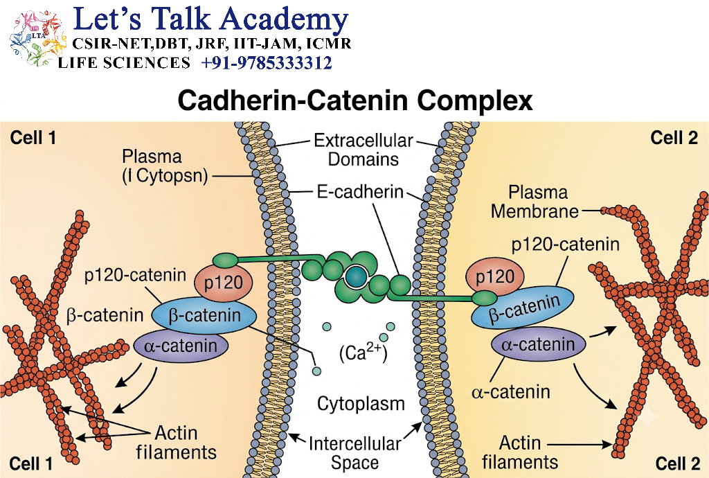
Cadherins are connected to the actin cytoskeleton through intracellular proteins called catenins.
A simplified organization is:
Cadherin
↓
β-catenin / p120-catenin
↓
α-catenin
↓
Actin cytoskeleton
This complex forms an important structural connection between neighboring cells and the actin cytoskeleton.
8.1 Role of Cadherins
Cadherins contribute to:
- Tissue integrity
- Cell sorting
- Morphogenesis
- Cell polarity
- Mechanical force transmission
- Development
- Maintenance of epithelial organization
9. E-Cadherin
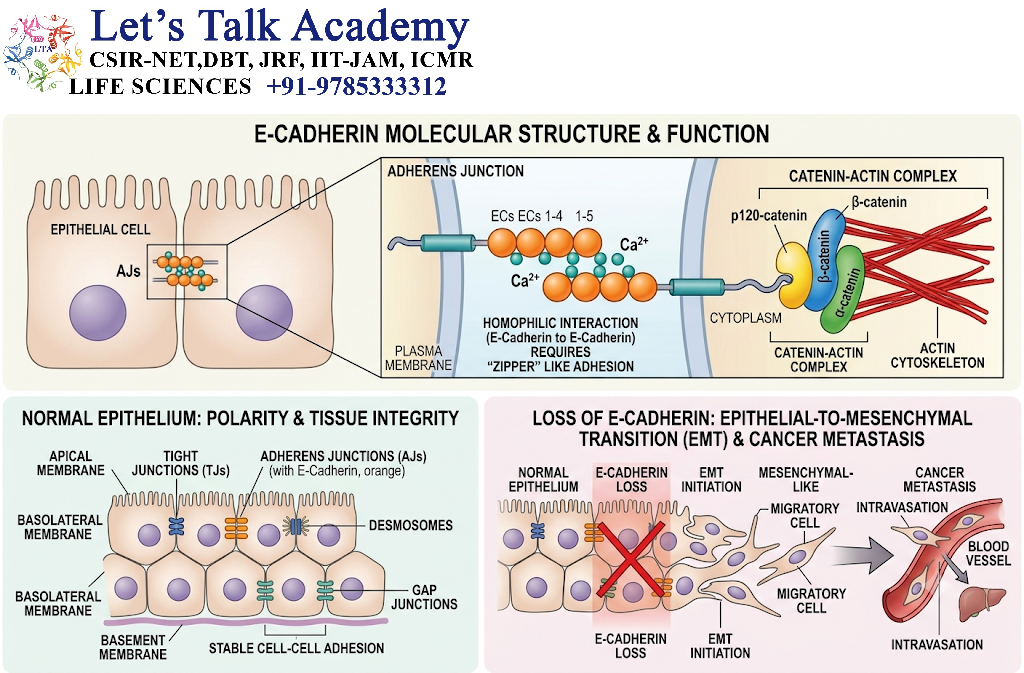
E-cadherin is an important adhesion molecule in epithelial tissues.
It contributes to the formation and maintenance of adherens junctions.
E-cadherin-mediated adhesion helps epithelial cells remain organized into continuous layers.
Reduced E-cadherin function can alter cell-cell adhesion and cellular organization and is associated with changes in tumor-cell behavior in several cancers.
10. Integrins
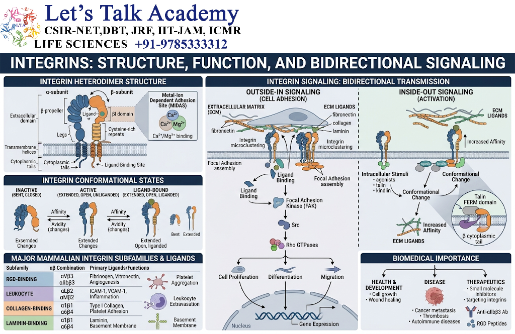
Integrins are transmembrane adhesion receptors that primarily mediate cell-extracellular matrix interactions and also participate in signaling.
Unlike cadherins, integrins are mainly involved in cell-matrix adhesion.
Integrins are heterodimeric proteins composed of:
- α subunit
- β subunit
Thus:
α + β → Integrin heterodimer
Different combinations of α and β subunits recognize different extracellular ligands.
11. Functions of Integrins
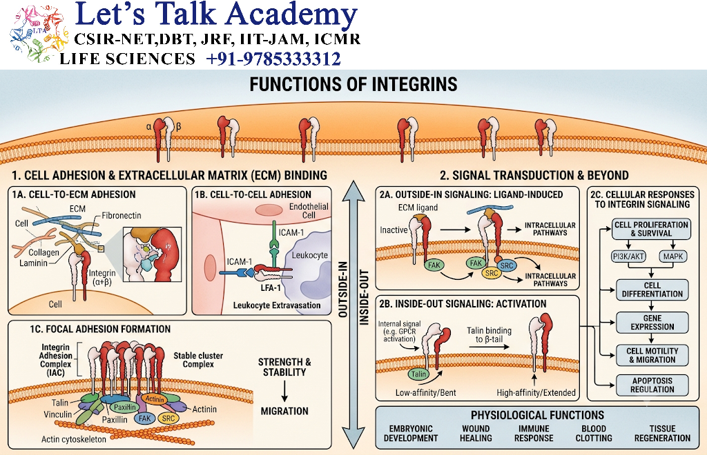
Integrins perform several important functions:
- Cell adhesion to ECM
- Cell migration
- Cell spreading
- Cell survival
- Mechanosensing
- Cell proliferation
- Differentiation
- Signal transduction
Integrins are therefore both adhesion receptors and signaling receptors.
12. Integrin Ligands
Different integrins can recognize different ECM molecules.
Important ECM ligands include:
| ECM molecule | Role |
|---|---|
| Fibronectin | Cell adhesion and migration |
| Laminin | Basement membrane adhesion |
| Collagen | Structural support and adhesion |
| Vitronectin | Cell-matrix interactions |
13. Integrin Activation
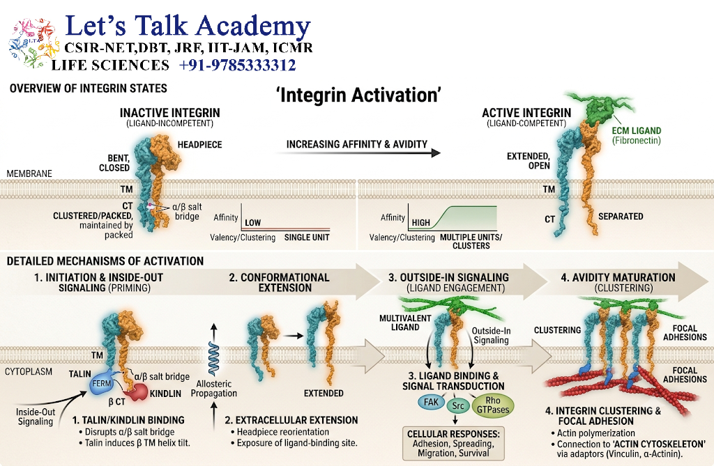
Integrins can exist in different conformational states.
13.1 Inside-Out Signaling
Intracellular signaling proteins can increase integrin affinity for extracellular ligands.
General pathway:
Intracellular signal
↓
Integrin conformational change
↓
Increased ligand-binding activity
↓
Cell adhesion
13.2 Outside-In Signaling
Binding of an extracellular matrix ligand to an integrin can activate intracellular signaling.
ECM binding
↓
Integrin clustering
↓
Adaptor and signaling-protein recruitment
↓
Kinase activation
↓
Cytoskeletal and cellular responses
14. Focal Adhesions
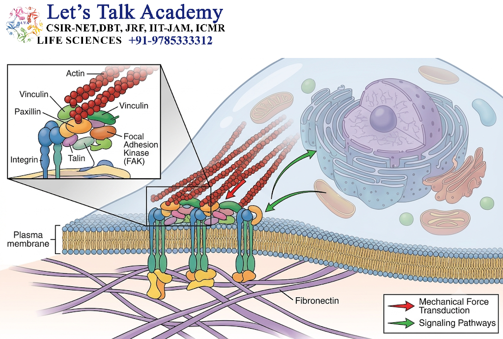
A major structure formed by integrins is the focal adhesion.
Focal adhesions connect the extracellular matrix to the actin cytoskeleton.
A simplified arrangement is:
ECM
↓
Integrin
↓
Talin / kindlin and other adaptor proteins
↓
Signaling proteins
↓
Actin cytoskeleton
Focal adhesions are important for:
- Cell spreading
- Migration
- Mechanical sensing
- Force transmission
- Survival signaling
15. Selectins
Selectins are cell adhesion molecules that recognize specific carbohydrate-containing ligands.
They are particularly important in the immune system.
Major selectins include:
- E-selectin
- P-selectin
- L-selectin
15.1 Main Function of Selectins
Selectins mediate relatively weak and transient interactions between cells.
They are especially important for the movement of leukocytes from the bloodstream into tissues.
16. Selectins in Leukocyte Recruitment
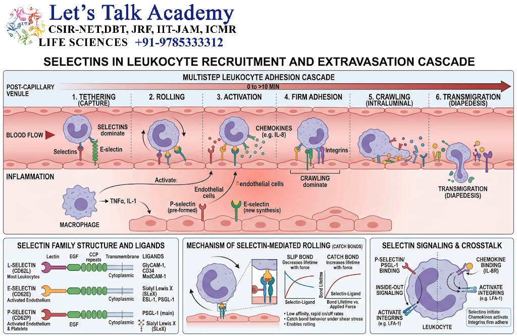
During inflammation, leukocytes must leave the bloodstream and enter affected tissues.
A simplified sequence is:
Blood flow
↓
Selectin-mediated rolling
↓
Chemokine signaling
↓
Integrin activation
↓
Firm adhesion
↓
Transmigration
↓
Movement into tissue
Selectins therefore help initiate the interaction between circulating leukocytes and vascular endothelial cells.
17. E-Selectin
E-selectin is expressed primarily by activated endothelial cells.
It contributes to:
Leukocyte rolling on activated endothelium
18. P-Selectin
P-selectin is found in:
- Activated endothelial cells
- Platelets
It participates in interactions between blood cells and the vascular surface.
19. L-Selectin
L-selectin is expressed on several leukocyte populations.
It contributes to leukocyte interactions with specialized endothelial cells and lymphoid tissues.
20. Immunoglobulin Superfamily CAMs
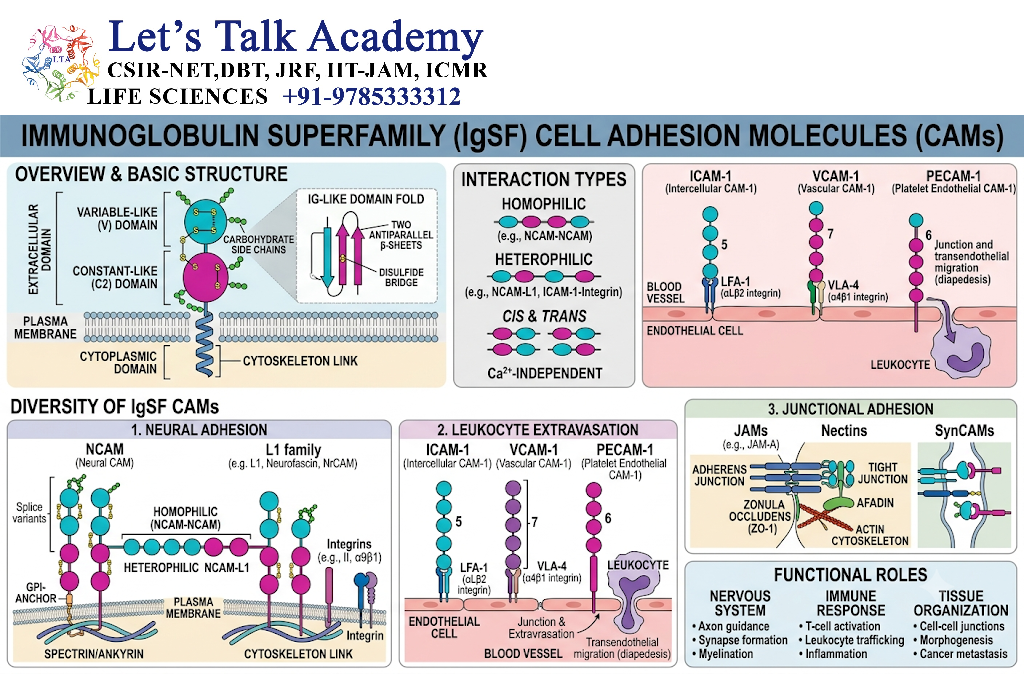
The immunoglobulin superfamily (IgSF) contains many cell-surface proteins involved in cell adhesion and recognition.
Examples include:
- ICAMs
- VCAMs
- NCAM
- PECAM-1
These molecules generally contain immunoglobulin-like domains in their extracellular regions.
21. ICAMs
Intercellular adhesion molecules (ICAMs) are important in cell-cell interactions, particularly during immune responses.
For example:
ICAM-1 on endothelial cells ↔ integrins on leukocytes
This interaction contributes to firm leukocyte adhesion.
22. VCAMs
Vascular cell adhesion molecule-1 (VCAM-1) is expressed on activated endothelial cells.
It interacts with specific leukocyte integrins and contributes to recruitment of immune cells to tissues.
23. NCAM
Neural cell adhesion molecule (NCAM) is particularly important in the nervous system.
It contributes to:
- Neuronal adhesion
- Neurite growth
- Neuronal development
- Synaptic organization
- Cell recognition
NCAM is an example of an Ig-superfamily adhesion molecule involved in neural development.
24. PECAM-1
Platelet endothelial cell adhesion molecule-1 (PECAM-1) is involved in cell-cell interactions within the vascular system.
It has an important role in:
- Leukocyte transmigration
- Endothelial-cell interactions
- Vascular organization
25. Major Cell Adhesion Junctions
Cell adhesion molecules organize into specialized junctions.
Major junctional structures include:
- Tight junctions
- Adherens junctions
- Desmosomes
- Hemidesmosomes
- Focal adhesions
- Gap junctions
Not all of these are primarily adhesion structures. For example, gap junctions are mainly specialized for intercellular communication, while tight junctions primarily regulate paracellular permeability but also contribute to cell-cell organization.
26. Adherens Junctions
Adherens junctions are cell-cell adhesion structures primarily based on cadherins.
Typical organization:
Cadherin
↓
Catenins
↓
Actin cytoskeleton
They help maintain:
- Cell shape
- Tissue integrity
- Epithelial organization
- Mechanical coupling
27. Desmosomes
Desmosomes are strong cell-cell adhesion structures that connect cells to intermediate filaments.
Important components include:
- Desmogleins
- Desmocollins
- Plakoglobin
- Plakophilins
- Desmoplakin
The basic arrangement is:
Cadherin-type desmosomal proteins
↓
Plaque proteins
↓
Intermediate filaments
Desmosomes provide mechanical strength to tissues that experience physical stress.
They are especially important in:
- Skin
- Cardiac muscle
28. Hemidesmosomes
Hemidesmosomes connect epithelial cells to the basement membrane.
They use integrins to connect cells with ECM components such as laminin.
Simplified organization:
Basement membrane
↓
Laminin
↓
Integrin
↓
Adaptor proteins
↓
Intermediate filaments
Hemidesmosomes help anchor epithelial cells firmly to the underlying extracellular matrix.
29. Tight Junctions
Tight junctions form seals between adjacent epithelial cells.
Important proteins include:
- Claudins
- Occludin
- Junctional adhesion molecules
Their major functions include:
- Restricting paracellular movement
- Maintaining epithelial barrier function
- Maintaining cell polarity
- Separating apical and basolateral membrane domains
Although their primary function is barrier formation rather than mechanical adhesion, tight junctions are closely integrated with cell adhesion systems.
30. Focal Adhesions vs Cell-Cell Junctions
| Feature | Focal adhesion | Adherens junction |
|---|---|---|
| Main interaction | Cell-ECM | Cell-cell |
| Major molecule | Integrin | Cadherin |
| Cytoskeletal connection | Actin | Actin |
| Major function | ECM adhesion and signaling | Cell-cell adhesion and tissue organization |
| Important feature | Mechanosensing | Mechanical coupling |
31. Role of Cell Adhesion in Cell Migration
Cell migration requires controlled formation and removal of adhesion contacts.
A simplified cycle is:
Adhesion formation
↓
Cell protrusion
↓
Actin organization
↓
Forward movement
↓
Adhesion release
↓
New adhesion formation
Integrins are especially important in cell migration through their interactions with ECM.
Selectins and integrins cooperate during immune-cell migration.
32. Cell Adhesion and Tissue Development
Cell adhesion plays a major role during embryonic development.
It regulates:
- Cell sorting
- Tissue formation
- Morphogenesis
- Cell migration
- Organ development
- Neuronal development
Differential expression of adhesion molecules can cause cells with similar adhesion properties to associate preferentially with one another.
33. Cell Adhesion and Cell Polarity
Adhesion molecules contribute to the establishment and maintenance of cell polarity.
In epithelial cells, coordinated adhesion and junction formation help distinguish:
- Apical surface
- Lateral surface
- Basal surface
This organization is essential for directional transport and epithelial function.
34. Cell Adhesion and Mechanotransduction
Cells experience physical forces from:
- Neighboring cells
- ECM
- Fluid flow
- Tissue stretching
- Mechanical compression
Adhesion complexes can detect these forces and convert them into biochemical signals.
This process is called mechanotransduction.
Integrins and cadherin-based junctions are major participants.
The general process is:
Mechanical force
↓
Adhesion complex deformation
↓
Conformational / biochemical changes
↓
Signaling pathway activation
↓
Changes in gene expression or cell behavior
35. Cell Adhesion and Cell Survival
Adhesion to the appropriate extracellular environment can promote cell survival.
Integrin-mediated signaling can activate pathways such as:
- FAK
- Src-family kinases
- PI3K-AKT
These pathways can influence survival and proliferation.
Loss of appropriate cell-ECM attachment can contribute to a specialized form of programmed cell death called anoikis in susceptible cells.
36. Cell Adhesion and Immune Responses
Cell adhesion molecules are essential for immune-cell communication and migration.
During inflammation:
Endothelial activation
↓
Selectin-mediated leukocyte rolling
↓
Chemokine-mediated integrin activation
↓
Integrin-mediated firm adhesion
↓
Transmigration
↓
Movement through tissue
Thus, multiple adhesion-molecule families cooperate rather than acting independently.
37. Cell Adhesion and Wound Healing
Cell adhesion is essential during tissue repair.
During wound healing:
- Cells detect tissue damage.
- Cells alter adhesion properties.
- Cells migrate toward the damaged area.
- ECM interactions guide migration.
- New cell-cell contacts form.
- Tissue architecture is gradually restored.
Integrins, cadherins and other adhesion molecules participate in these processes.
38. Cell Adhesion and Cancer
Changes in cell adhesion can contribute to abnormal tissue behavior.
Alterations may involve:
- Reduced cell-cell adhesion
- Altered integrin expression
- Abnormal ECM interactions
- Changes in adhesion signaling
- Increased cell migration
- Altered invasion
For example, changes in E-cadherin-mediated adhesion are associated with altered epithelial organization in several cancers.
However, cancer progression is a complex process involving many genetic, biochemical and microenvironmental changes.
39. Cell Adhesion and Metastasis
For metastatic spread, cancer cells must interact dynamically with:
- Neighboring cells
- Basement membranes
- ECM
- Blood-vessel surfaces
- Distant tissues
Adhesion molecules can therefore influence multiple stages of metastatic dissemination.
The process can involve:
Loss or alteration of cell-cell adhesion
↓
Cell detachment and migration
↓
ECM interaction
↓
Entry into circulation
↓
Endothelial interaction
↓
Exit from blood vessel
↓
Adhesion to new tissue environment
40. Comparison of Major Adhesion Molecules
| Adhesion molecule | Main binding type | Major partner | Cytoskeletal/signaling association | Major role |
|---|---|---|---|---|
| Cadherins | Cell-cell | Cadherins | Actin/intermediate filaments | Tissue organization |
| Integrins | Cell-ECM | ECM proteins | Actin/intermediate filaments + signaling | Adhesion, migration, mechanotransduction |
| Selectins | Cell-cell | Carbohydrate ligands | Signaling-associated | Leukocyte rolling |
| ICAMs | Cell-cell | Leukocyte integrins | Signaling-associated | Immune-cell adhesion |
| VCAMs | Cell-cell | Specific integrins | Signaling-associated | Leukocyte recruitment |
| NCAM | Cell-cell | Cell-surface ligands/NCAM and others | Cytoskeletal/signaling networks | Neural adhesion and development |
| PECAM-1 | Cell-cell | PECAM-1 and other partners | Cytoskeletal/signaling networks | Vascular interactions and transmigration |
41. Cadherins vs Integrins
| Feature | Cadherins | Integrins |
|---|---|---|
| Major function | Cell-cell adhesion | Cell-ECM adhesion |
| Main binding | Often homophilic | Usually heterophilic |
| Calcium dependence | Yes | Depends on integrin and cation environment |
| Structure | Single-pass adhesion proteins | α/β heterodimers |
| Cytoskeleton | Actin or intermediate filaments depending on junction | Actin or intermediate filaments depending on complex |
| Signaling | Yes | Strong signaling role |
| Major example | E-cadherin | Fibronectin-binding integrins |


