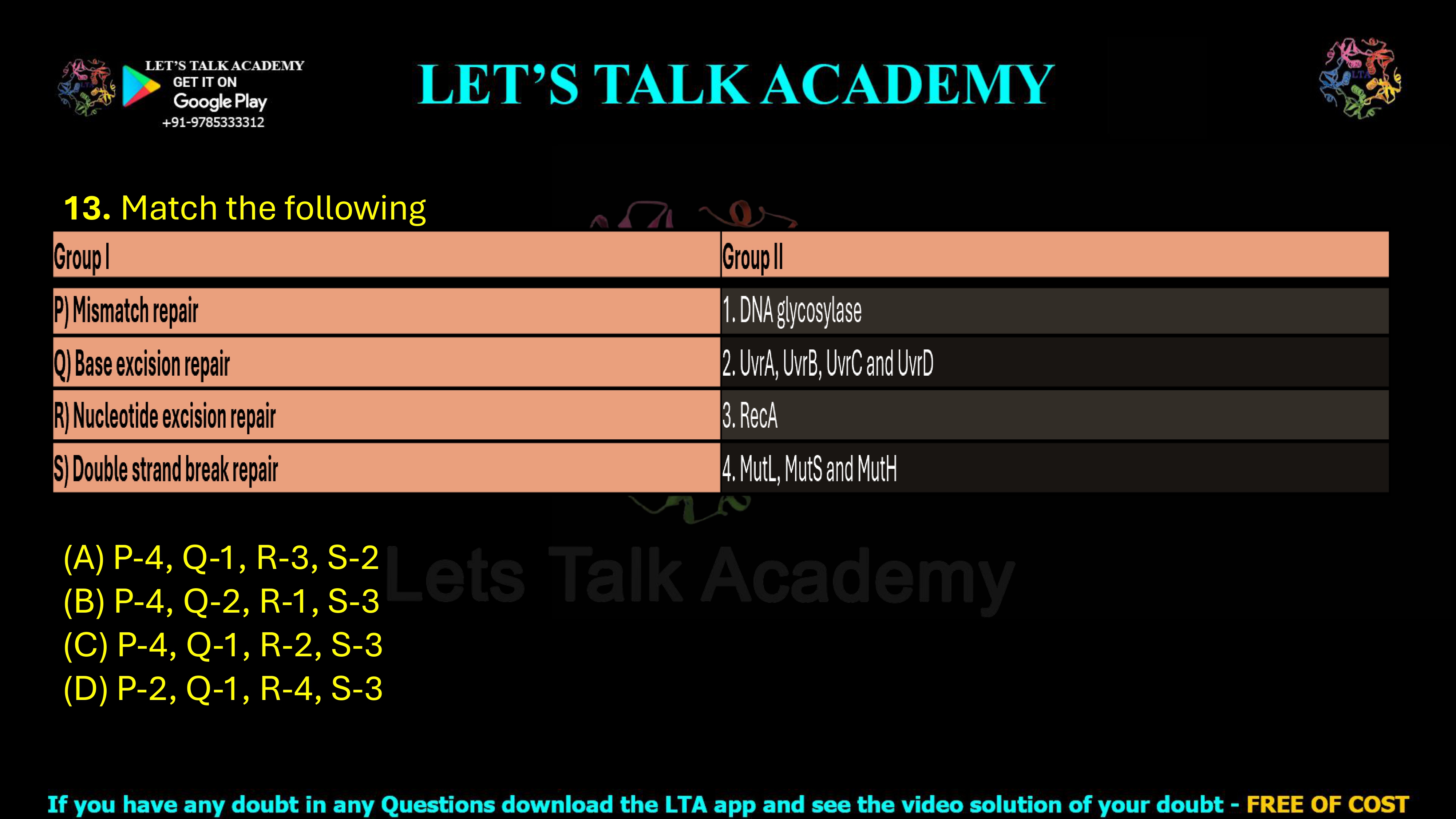
13. Match the following
| Group I | Group II |
| P) Mismatch repair | 1. DNA glycosylase |
| Q) Base excision repair | 2. UvrA, UvrB, UvrC and UvrD |
| R) Nucleotide excision repair | 3. RecA |
| S) Double strand break repair | 4. MutL, MutS and MutH |
(A) P-4, Q-1, R-3, S-2
(B) P-4, Q-2, R-1, S-3
(C) P-4, Q-1, R-2, S-3
(D) P-2, Q-1, R-4, S-3
DNA Repair Mechanisms: Match Mismatch Repair, Base Excision Repair, Nucleotide Excision Repair and Double-Strand Break Repair
Introduction
DNA is continuously exposed to different types of damage arising from spontaneous chemical reactions, errors during DNA replication, ultraviolet radiation, ionizing radiation, reactive oxygen species, and various chemical mutagens. If these alterations are not corrected, they can become permanent mutations and interfere with replication, transcription, chromosome stability, and cell survival. Cells therefore possess several specialized DNA repair pathways, with each pathway designed to recognize and repair a particular category of DNA damage.
Among the most important repair mechanisms are mismatch repair (MMR), base excision repair (BER), nucleotide excision repair (NER), and double-strand break repair. These pathways use different proteins and enzymes because the DNA lesions they recognize are fundamentally different. In bacteria, mismatch repair is associated with MutS, MutL, and MutH; base excision repair begins with DNA glycosylases; nucleotide excision repair involves UvrA, UvrB, UvrC, and UvrD; and homologous recombination-mediated repair of double-strand breaks depends strongly on RecA.
Correct Answer
Correct Answer: (C) P-4, Q-1, R-2, S-3
Detailed Explanation
The correct matching can be established by identifying the characteristic protein or enzyme associated with each DNA repair pathway. Mismatch repair in bacteria uses the MutS-MutL-MutH system, base excision repair begins with a DNA glycosylase, bacterial nucleotide excision repair depends on the Uvr proteins, and RecA is a central protein in homologous recombination-based repair of double-strand DNA breaks.
Therefore, the correct relationship is:
P → 4, Q → 1, R → 2, S → 3
Explanation of P: Mismatch Repair → MutL, MutS and MutH
Correct Match: P-4
Mismatch repair corrects errors that escape the proofreading activity of DNA polymerase during DNA replication. These errors include incorrectly paired bases, such as G-T or A-C mismatches, as well as small insertion-deletion loops. In bacteria such as Escherichia coli, the classical mismatch repair system depends on the proteins MutS, MutL, and MutH.
MutS is the primary mismatch-recognition protein. It scans newly replicated DNA and binds to an incorrectly paired region. MutL then interacts with MutS and coordinates the subsequent repair steps. MutH helps distinguish the newly synthesized strand from the parental strand and introduces a nick into the strand that contains the replication error. The damaged section is then removed, resynthesized by DNA polymerase, and sealed by DNA ligase.
Therefore:
Mismatch repair → MutL, MutS and MutH
Explanation of Q: Base Excision Repair → DNA Glycosylase
Correct Match: Q-1
Base excision repair removes relatively small, non-helix-distorting DNA lesions. These include uracil produced by cytosine deamination, oxidized bases, alkylated bases, and several other chemically altered nitrogenous bases. The pathway begins when a specific DNA glycosylase recognizes the damaged base.
DNA glycosylase cleaves the N-glycosidic bond connecting the damaged nitrogenous base to the deoxyribose sugar. This removes only the abnormal base and produces an apurinic or apyrimidinic site, commonly called an AP site. An AP endonuclease subsequently cuts the DNA backbone, the damaged region is processed, DNA polymerase fills the missing nucleotide or nucleotides, and DNA ligase seals the remaining nick.
Therefore:
Base excision repair → DNA glycosylase
Explanation of R: Nucleotide Excision Repair → UvrA, UvrB, UvrC and UvrD
Correct Match: R-2
Nucleotide excision repair removes bulky DNA lesions that distort the normal structure of the double helix. A classic example is the formation of pyrimidine dimers following exposure to ultraviolet radiation. Unlike base excision repair, which removes an individual damaged base, nucleotide excision repair removes a short stretch of nucleotides containing the lesion.
In bacteria, the major proteins involved are UvrA, UvrB, UvrC, and UvrD. UvrA and UvrB participate in damage recognition and verification. UvrC makes incisions on both sides of the damaged DNA segment, while UvrD helicase helps remove the excised oligonucleotide. DNA polymerase then fills the resulting gap, and DNA ligase restores the continuity of the DNA strand.
Therefore:
Nucleotide excision repair → UvrA, UvrB, UvrC and UvrD
Explanation of S: Double-Strand Break Repair → RecA
Correct Match: S-3
A double-strand break is one of the most dangerous forms of DNA damage because both strands of the DNA molecule are broken. If such damage is not repaired correctly, it can cause chromosome fragmentation, loss of genetic information, genome instability, or cell death.
In bacteria, the RecA protein plays a central role in homologous recombination and the repair of double-strand DNA breaks. RecA binds to single-stranded DNA generated during break processing and promotes the search for a homologous DNA sequence. It then facilitates strand invasion into the homologous DNA duplex, allowing the undamaged homologous sequence to serve as a template for accurate repair.
Therefore:
Double-strand break repair → RecA
Complete Correct Matching
| DNA Repair Pathway | Associated Protein or Enzyme | Correct Match |
|---|---|---|
| Mismatch repair | MutL, MutS and MutH | P-4 |
| Base excision repair | DNA glycosylase | Q-1 |
| Nucleotide excision repair | UvrA, UvrB, UvrC and UvrD | R-2 |
| Double-strand break repair | RecA | S-3 |
Explanation of Option (A)
Option (A): P-4, Q-1, R-3, S-2 is incorrect.
The first two matches are correct because mismatch repair is associated with MutL, MutS, and MutH, while base excision repair is initiated by DNA glycosylase. However, the last two matches are reversed. RecA is associated primarily with homologous recombination and double-strand break repair, whereas UvrA, UvrB, UvrC, and UvrD function in nucleotide excision repair. Therefore, option (A) is incorrect.
Explanation of Option (B)
Option (B): P-4, Q-2, R-1, S-3 is incorrect.
This option correctly matches mismatch repair with MutL, MutS, and MutH and correctly associates RecA with double-strand break repair. However, it incorrectly exchanges the proteins involved in base excision repair and nucleotide excision repair. DNA glycosylase belongs to base excision repair, while the Uvr proteins belong to bacterial nucleotide excision repair. Therefore, option (B) is incorrect.
Explanation of Option (C)
Option (C): P-4, Q-1, R-2, S-3 is correct.
Every pairing in this option is biologically accurate. Mismatch repair uses MutL, MutS, and MutH; base excision repair begins with DNA glycosylase; nucleotide excision repair uses UvrA, UvrB, UvrC, and UvrD; and double-strand break repair through homologous recombination involves RecA. Therefore, option (C) is the correct answer.
Explanation of Option (D)
Option (D): P-2, Q-1, R-4, S-3 is incorrect.
This option correctly associates base excision repair with DNA glycosylase and double-strand break repair with RecA. However, it incorrectly assigns the Uvr proteins to mismatch repair and the Mut proteins to nucleotide excision repair. In reality, these relationships are exactly the opposite: MutL, MutS, and MutH function in mismatch repair, while UvrA, UvrB, UvrC, and UvrD participate in nucleotide excision repair.
Comparison of Major DNA Repair Mechanisms
| Repair Mechanism | Major Type of Damage | Key Protein or Enzyme |
|---|---|---|
| Mismatch Repair | Replication errors and mismatched bases | MutS, MutL and MutH |
| Base Excision Repair | Small chemically altered bases | DNA glycosylase |
| Nucleotide Excision Repair | Bulky helix-distorting lesions | UvrA, UvrB, UvrC and UvrD |
| Double-Strand Break Repair | Breakage of both DNA strands | RecA |
How Mismatch Repair Works
Mismatch Recognition
MutS recognizes and binds to an incorrectly paired region of newly replicated DNA. This step provides the specificity required to distinguish a true mismatch from correctly paired DNA.
Repair Complex Formation
MutL binds to the MutS-DNA complex and acts as a coordinator between mismatch recognition and strand-directed repair.
Removal and Resynthesis
MutH participates in strand discrimination and incision in the classical bacterial system. The error-containing DNA region is removed, DNA polymerase resynthesizes the missing sequence, and DNA ligase seals the final nick.
How Base Excision Repair Works
Removal of the Damaged Base
A lesion-specific DNA glycosylase identifies the chemically damaged base and removes it by cleaving the N-glycosidic bond. This creates an AP site in the DNA.
DNA Backbone Incision and Repair
An AP endonuclease cuts the DNA backbone near the AP site. The remaining damaged sugar-phosphate residue is removed, DNA polymerase restores the correct nucleotide sequence, and DNA ligase completes the repair.
How Nucleotide Excision Repair Works
Recognition of DNA Distortion
The bacterial Uvr proteins detect structural abnormalities in DNA rather than recognizing only one specific damaged base. This makes nucleotide excision repair suitable for removing many different bulky lesions.
Dual Incision and Oligonucleotide Removal
UvrC cuts the damaged strand on both sides of the lesion. UvrD helps release the damaged oligonucleotide, after which DNA polymerase fills the gap and DNA ligase seals the repaired strand.
How RecA Supports Double-Strand Break Repair
RecA is essential for homologous recombination in bacteria. After a double-strand break is processed to expose single-stranded DNA, RecA forms a nucleoprotein filament on the single-stranded region. This filament searches for homologous DNA and promotes strand invasion. The homologous chromosome or DNA molecule then provides an intact template that allows accurate restoration of the damaged genetic information.
Biological Significance
DNA repair systems protect the genome by correcting different forms of DNA damage before they become permanent mutations. The presence of multiple specialized pathways is necessary because a mismatched base, an oxidized nucleotide, a UV-induced pyrimidine dimer, and a double-strand break cannot all be repaired by the same molecular mechanism.
Mismatch repair preserves replication fidelity, base excision repair removes subtle chemical modifications, nucleotide excision repair eliminates bulky DNA distortions, and recombination-mediated repair restores broken chromosomes. Together, these pathways maintain genome stability and support normal cell survival and reproduction.
Final Answer
P) Mismatch repair → 4) MutL, MutS and MutH
Q) Base excision repair → 1) DNA glycosylase
R) Nucleotide excision repair → 2) UvrA, UvrB, UvrC and UvrD
S) Double-strand break repair → 3) RecA
Correct Matching: P-4, Q-1, R-2, S-3
Correct Answer: (C)


