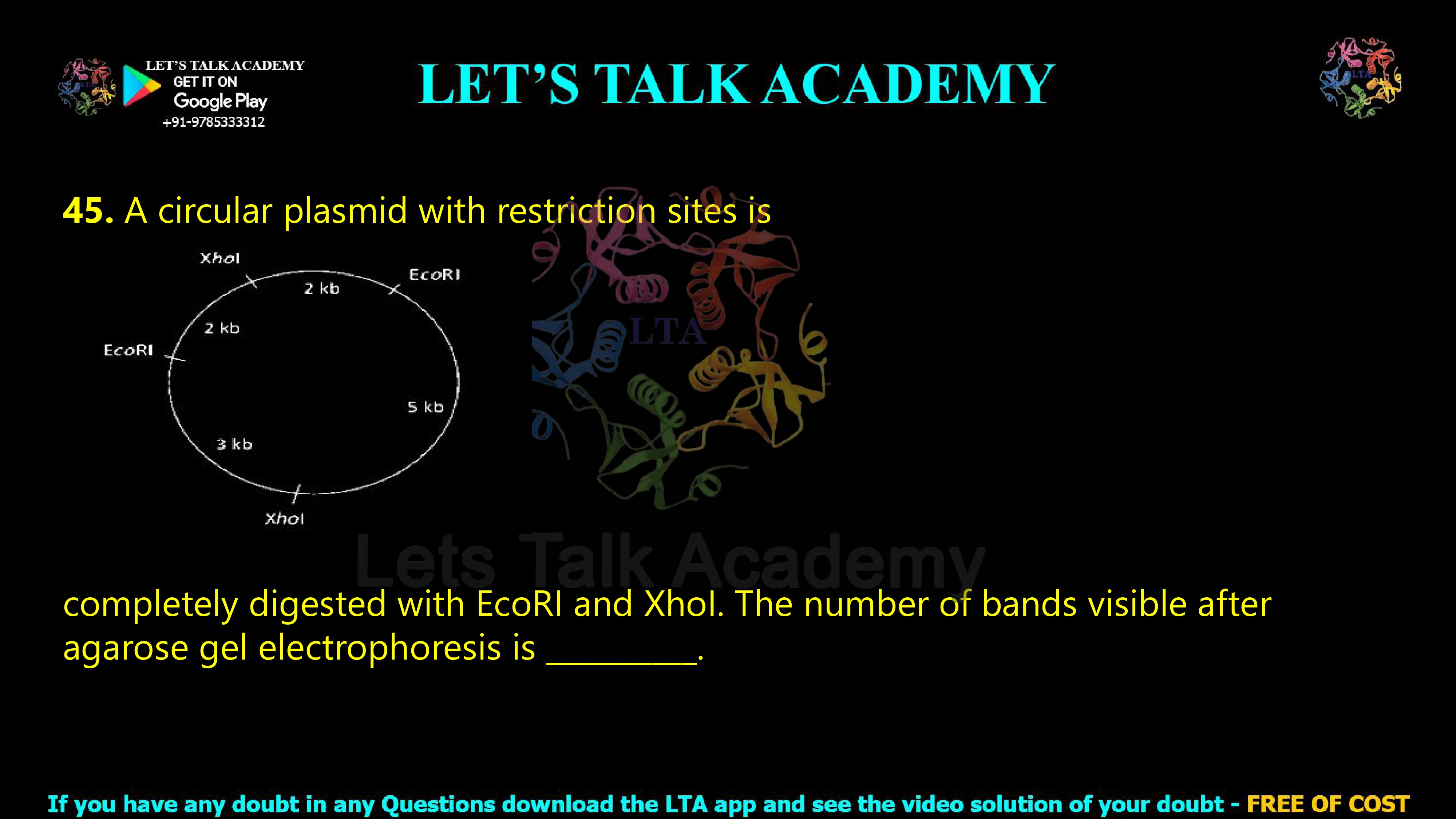
44. A circular plasmid with restriction sites is
completely digested with EcoRI and XhoI. The number of bands visible after agarose gel electrophoresis is __________.
Number of Bands After Complete EcoRI and XhoI Digestion of a Circular Plasmid
Correct Answer
Answer: 3 bands
The circular plasmid contains a total of four restriction sites: two EcoRI sites and two XhoI sites. Complete digestion with both enzymes therefore cuts the circular plasmid at four positions and generates four DNA fragments. According to the restriction map, the sizes of these fragments are 2 kb, 2 kb, 3 kb, and 5 kb.
However, the number of DNA fragments produced is not always equal to the number of bands visible on an agarose gel. The two fragments of 2 kb have exactly the same length and therefore migrate together during agarose gel electrophoresis. They appear as a single band at the 2 kb position.
Thus, the distinct DNA fragment sizes visible on the gel are:
5 kb, 3 kb, and 2 kb
Therefore, the total number of visible bands is:
3 bands
Understanding the Circular Plasmid Restriction Map
A restriction map shows the positions at which particular restriction enzymes cut a DNA molecule. In this question, the DNA molecule is a circular plasmid containing recognition sites for two restriction enzymes, EcoRI and XhoI. The positions of these restriction sites divide the plasmid into DNA segments of different lengths.
Moving around the circular plasmid from one restriction site to the next, the DNA segments shown in the diagram have lengths of 2 kb, 2 kb, 5 kb, and 3 kb. These four segments together make up the complete plasmid.
The total size of the plasmid can therefore be calculated as:
2 kb + 2 kb + 5 kb + 3 kb = 12 kb
Thus, the original circular plasmid is 12 kb in total length.
How Many Restriction Sites Are Present in the Plasmid?
EcoRI Restriction Sites
The diagram shows two EcoRI recognition sites. One EcoRI site is located on the left side of the circular plasmid, while the second EcoRI site is located toward the upper-right side.
Therefore:
Number of EcoRI sites = 2
XhoI Restriction Sites
The diagram also shows two XhoI recognition sites. One XhoI site is located near the top of the plasmid, while the other is located near the bottom.
Therefore:
Number of XhoI sites = 2
The total number of restriction sites recognized by the two enzymes is therefore:
2 EcoRI sites + 2 XhoI sites = 4 restriction sites
What Happens During Complete Digestion with EcoRI and XhoI?
A complete restriction digestion means that every recognition site for the restriction enzymes is cleaved. Since the plasmid contains two EcoRI sites and two XhoI sites, treatment with both enzymes cuts the plasmid at all four positions.
For a circular DNA molecule, cutting at four different positions produces four DNA fragments. Therefore, the complete double digestion generates the following fragments:
Fragment 1 = 2 kb
Fragment 2 = 2 kb
Fragment 3 = 3 kb
Fragment 4 = 5 kb
Thus, the total number of DNA fragments physically produced is four.
Why Does a Circular DNA Molecule Cut at Four Sites Produce Four Fragments?
The relationship between the number of cuts and the number of fragments depends on whether the original DNA molecule is circular or linear. This distinction is essential for solving restriction mapping questions correctly.
In a circular DNA molecule, there are no free ends before digestion. The first cut opens the circular molecule and converts it into a linear molecule. Each additional cut then produces one more fragment. Therefore, when a circular DNA molecule is completely cut at n distinct positions, it produces n DNA fragments.
In this question:
Number of cuts = 4
Therefore:
Number of DNA fragments produced = 4
This gives four physical fragments with sizes of 2 kb, 2 kb, 3 kb, and 5 kb.
Why Are Only Three Bands Visible on the Agarose Gel?
This is the most important concept in the question. Although four DNA fragments are produced after complete digestion, only three bands are visible after agarose gel electrophoresis.
Agarose gel electrophoresis separates DNA fragments mainly according to their size. Smaller DNA fragments move more rapidly through the pores of the agarose gel, whereas larger DNA fragments migrate more slowly.
DNA fragments having the same size migrate at essentially the same rate. Therefore, when two DNA fragments are both 2 kb long, they travel together through the agarose gel and occupy the same position.
The gel does not normally display these two equal-sized fragments as two separate bands. Instead, both 2 kb fragments co-migrate and appear as a single 2 kb band.
Therefore:
5 kb fragment → one band
3 kb fragment → one band
2 kb fragment + 2 kb fragment → one band
Total visible bands:
1 + 1 + 1 = 3 bands
Step-by-Step Solution of the Question
Step 1: Count the Total Restriction Sites
The circular plasmid contains two EcoRI sites and two XhoI sites. Therefore, complete digestion creates a total of four cuts.
Total cuts = 2 + 2 = 4
Step 2: Determine the Number of DNA Fragments
For circular DNA, the number of fragments produced after complete digestion is equal to the number of cuts.
4 cuts → 4 DNA fragments
Step 3: Identify the Fragment Sizes
The restriction map shows four segments between consecutive restriction sites:
2 kb, 2 kb, 3 kb, and 5 kb
Step 4: Identify the Number of Distinct Fragment Sizes
The four fragments do not have four different sizes. The 2 kb size occurs twice. Therefore, the distinct fragment sizes are only:
2 kb, 3 kb, and 5 kb
Step 5: Calculate the Number of Visible Bands
Fragments of identical size migrate together during agarose gel electrophoresis. The two 2 kb fragments therefore form one band.
Number of distinct DNA fragment sizes = Number of visible bands = 3
DNA Fragments Produced Versus Bands Visible
A major conceptual point in restriction digestion questions is the difference between the number of DNA fragments produced and the number of bands visible. These two values are equal only when every DNA fragment has a different size.
In the present question, complete digestion produces four fragments:
2 kb + 2 kb + 3 kb + 5 kb = 12 kb
However, the gel separates DNA according to size. Since two fragments have the same size of 2 kb, they migrate together and form a single band.
Therefore:
Number of DNA fragments produced = 4
Number of distinct fragment sizes = 3
Number of visible bands = 3
Expected Band Pattern on the Agarose Gel
The largest fragment, 5 kb, migrates the slowest and therefore remains closest to the well. The 3 kb fragment migrates farther than the 5 kb fragment. The 2 kb fragments migrate the farthest because they are the smallest DNA fragments in the digest.
The expected order of bands from the top of the gel toward the bottom is:
5 kb band
3 kb band
2 kb band
The 2 kb band represents two DNA fragments of identical length. Under suitable staining conditions, this band may contain approximately twice as much DNA as a band representing only one fragment of the same molar abundance. As a result, the 2 kb band may appear more intense than the 3 kb or 5 kb bands, although the question asks only for the number of visible bands.
Importance of Complete Digestion in This Question
The phrase “completely digested” is important because it indicates that every EcoRI and every XhoI recognition site has been cleaved. No intact plasmid or partially digested DNA molecules need to be included in the theoretical calculation.
If digestion were incomplete or partial, additional DNA molecules containing uncut restriction sites could be present. These molecules could generate extra bands of different sizes. However, complete digestion produces only the fragments defined by the distances between consecutive restriction sites.
Therefore, only the four expected fragments of 2 kb, 2 kb, 3 kb, and 5 kb need to be considered.
Difference Between Circular and Linear DNA Digestion
For a circular DNA molecule, the number of fragments produced after complete digestion is equal to the number of restriction cuts. Therefore, four cuts in a circular plasmid produce four fragments.
For a linear DNA molecule, the situation is different because the molecule already has two free ends. If a linear DNA molecule is cut at four internal positions, it produces five fragments.
Thus:
Circular DNA: n cuts → n fragments
Linear DNA: n cuts → n + 1 fragments
In this question, the DNA is specifically described as a circular plasmid. Therefore, four restriction cuts produce four DNA fragments.
Why the Answer Is Not Four Bands
It may initially appear that four restriction fragments should produce four bands. However, agarose gel electrophoresis cannot separate two DNA fragments if they have exactly the same length under ordinary conditions. The two 2 kb fragments migrate to the same position and overlap.
Thus, four fragments do not necessarily mean four visible bands. The number of visible bands depends on the number of different fragment sizes.
The four fragments are:
2 kb, 2 kb, 3 kb, and 5 kb
The three unique sizes are:
2 kb, 3 kb, and 5 kb
Therefore, only three bands are visible.
Restriction Mapping and Agarose Gel Electrophoresis Concept
Restriction mapping is a fundamental molecular biology technique used to determine the positions of restriction enzyme recognition sites within a DNA molecule. When DNA is digested with one or more restriction enzymes, the resulting fragment sizes provide information about the arrangement of restriction sites.
Agarose gel electrophoresis then separates these fragments according to size. By comparing the migration positions of unknown DNA fragments with a DNA size marker or ladder, researchers can estimate fragment lengths and reconstruct restriction maps.
In questions involving restriction maps, it is therefore necessary to consider both the physical number of fragments and the number of unique fragment sizes. Equal-sized fragments co-migrate and produce fewer visible bands than the total number of fragments.
Why This Question Is Important for Life Science and Biotechnology Exams
Restriction digestion and agarose gel electrophoresis are among the most frequently tested topics in molecular biology and biotechnology examinations. Questions may ask students to calculate the number of fragments, determine the number of visible bands, calculate the total size of a plasmid, identify single and double digestion patterns, or reconstruct a restriction map.
This concept is especially relevant for CSIR NET Life Science, DBT JRF, GATE Biotechnology, IIT JAM Biotechnology, ICMR JRF, and other life science examinations. The key to solving such questions is to carefully identify whether the DNA is circular or linear, count the restriction sites, determine all fragment sizes, and finally count only the distinct sizes when the question asks for visible bands.
Concept Summary
The circular plasmid contains four restriction sites in total: two EcoRI sites and two XhoI sites. Complete digestion with both enzymes cuts the circular plasmid at all four positions and generates four DNA fragments with sizes of 2 kb, 2 kb, 3 kb, and 5 kb.
During agarose gel electrophoresis, the two 2 kb DNA fragments migrate together because they have identical lengths. They therefore appear as a single band. The three distinct fragment sizes visible on the gel are 5 kb, 3 kb, and 2 kb.
Final Answer
DNA fragments produced: 4
Fragment sizes: 2 kb, 2 kb, 3 kb, and 5 kb
Distinct fragment sizes: 2 kb, 3 kb, and 5 kb
Number of bands visible after agarose gel electrophoresis = 3
Final Answer: 3 bands


