
Apoptosis | Difference between apoptosis and necrosis | Caspases
Apoptosis: – Apoptosis is the process of programmed cell death (PCD). The word apoptosis, derived from the Greek word for a natural process of leaves falling from trees or petals from flowers. Apoptosis is a very essential part of life cycle of organisms, in which that it regulates and adjusts cell population. Apoptosis also exerts a role opposite to mitosis in the maintenance of cell populations. As many as 10 11cells die in an adult human per day to ensure tissue homeostasis.
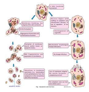
Apoptosis leads to several distinct cellular changes, including:
- Cell Shrinkage – The cell becomes smaller in size due to loss of cytoplasmic volume.
- Membrane Blebbing – The plasma membrane shows bubble-like protrusions without losing its integrity.
- Chromatin Condensation (Pyknosis) – The nucleus becomes dense due to condensation of chromatin.
- Nuclear Fragmentation (Karyorrhexis) – The condensed nucleus breaks into several fragments.
- Cytoplasmic Fragmentation – The cytoplasm is divided into smaller, membrane-bound fragments.
- Formation of Apoptotic Bodies – The cell is broken into small vesicles containing nuclear and cytoplasmic material, including intact organelles.
- Phagocytosis by Neighbouring Cells or Macrophages – Apoptotic bodies are rapidly cleared without inducing inflammation.

Cell death
Cell death is a normal process that has two functions: tissue remodelling and removal of damaged cells that harm the rest of the body. Integration of proliferation( cell division) and death is essential for normal homeostasis. There are two possible reasons for cell death:
* The cells may no longer be needed. When in a developing embryo, fingers are connected to each other by connective tissue, to initiate apoptosis connective tissue disappears and separate the fingers.
* Apoptosis may be a mechanism for preventing cancer when the cells have a genetic problem or damage that lead to cancer.
There are two types of cell death: apoptosis and necrosis. Apoptosis is a regulated process for numbers of cells but necrosis is dying cells by swelling and bursting. Unlikely necrosis, apoptosis is a regulated and controlled process. These cell death have different morphology and molecular features are the following:
Difference between apoptosis and necrosis
Apoptosis
Apoptosis is a highly regulated, energy-dependent form of programmed cell death that plays a crucial role in development, immune regulation, and removal of damaged or unwanted cells. It occurs in a controlled manner without causing harm to neighbouring cells.
Necrosis
Necrosis is an uncontrolled and accidental form of cell death that typically results from external injury, such as infection, toxins, trauma, or lack of blood supply (ischemia). It often leads to inflammation and damage to surrounding tissues.
| Feature | Apoptosis | Necrosis |
| Type of Process | Programmed cell death (regulated or Controlled) |
Accidental or pathological cell death (unregulated or uncontrolled) |
| Energy Requirement | Requires ATP (energy-dependent) | Passive process (does not require energy) |
| Trigger | Physiological stimuli (e.g., DNA damage, development) | External injury (e.g., trauma, toxins, infection, ischemia) |
| Cell Morphology | Cell shrinkage, membrane blebbing | Cell swelling, membrane rupture |
| Nuclear Changes | Chromatin condensation, fragmentation | Random degradation of nucleus |
| Plasma Membrane Integrity | Maintained until late stages | Lost early; membrane ruptures |
| Inflammation | No inflammation | Triggers inflammation |
| Formation of Apoptotic Bodies | Yes | No |
| Phagocytosis | Apoptotic bodies removed by phagocytes | Cell contents spill into surroundings |
| Physiological vs. Pathological | Often physiological (e.g., tissue remodeling) | Always pathological |
Removal of Dead Cells
Now LET’S TALK about how the apoptotic bodies are recognized by Macrophage. The removal (engulfment) of dead cells by phagocytic cells known as Phagocytosis. In a living cell, the phosphatidylserine is found on the cytosolic surface of the plasma membrane. The protein Flippase maintain the phosphatidylserine cytosolic surface of the plasma membrane. However, in the dead cell, its come out through Flip-flop movement and mark the cell for phagocytosis. The macrophages bind phosphatidylserine exposed only in apoptotic bodies through phosphatidylserine receptors. Tissue macrophages rapidly recognize and engulf apoptotic cells. These events require the display of so-called eat-me signals on the apoptotic cell surface, the most fundamental of which is phosphatidylserine (PtdSer). Eat-me signals also trigger macrophages to engulf virus-infected or metabolically traumatized, but still living, cells, and this ‘murder by phagocytosis’ may be a common phenomenon.
Molecular mechanisms of an apoptosis signalling pathway
Apoptosis can be initiated by various stimuli from outside or inside the cell, e.g. by a specific ligand of cell surface receptors, by DNA damage, failure of DNA repair mechanisms, certain cytotoxic drugs or irradiation of cell, by a lack of survival signals.
Caspases in Apoptosis
Now LET’S TALK about why they called as caspases and what is the role of caspases. Caspases are cysteine proteases. Caspases are endoproteases have an active site Cysteine (C) and which cleave at the C- terminal side of Aspartate residues ( asp ) and hence are known as caspases. Their catalytical activity depends on a critical cysteine-residue within a highly conserved active-site consist of pentapeptide Glutamine, Alanine, Cysteine, Arginine Glycine (QACRG). Caspases are essential enzymes that drive programmed cell death, or apoptosis, by cleaving cellular proteins and orchestrating the sequential events leading to cell demise. So far, 7 different caspases have been identified in Drosophila, and 14 in mammals, with caspase-11 and caspase–12 only identified in the mouse. These caspases form a cascade of proteases which are activated in this process. Caspases are expressed as zymogens (procaspases), which are activated by different apoptosis inducers.
Caspases is classified into three groups on the basis of their functions:
Caspases (Cysteine-aspartic proteases) are a family of protease enzymes that play essential roles in apoptosis (programmed cell death), necrosis, and inflammation. Based on their functions, caspases are classified into three main groups:
- Initiator Caspases
- Function: These caspases initiate the apoptosis signaling pathways.
- Examples: Caspase-2, Caspase-8, Caspase-9, and Caspase-10
- Characteristic: They have long pro-domains (such as CARD or DED) that allow them to interact with adaptor proteins in apoptotic complexes like the apoptosome (for caspase-9) or death-inducing signaling complex (DISC, for caspase-8/10).
- Executioner (Effector) Caspases
- Function: These caspases execute apoptosis by cleaving various cellular substrates.
- Examples: Caspase-3, Caspase-6, and Caspase-7
- Characteristic: They are activated by initiator caspases and carry out the degradation of cellular components leading to apoptosis.
- Inflammatory Caspases
- Function: These caspases are involved in cytokine maturation and inflammation rather than apoptosis.
- Examples: Caspase-1, Caspase-4, Caspase-5 (humans), and Caspase-11 (mice)
- Characteristic: Caspase-1 activates pro-inflammatory cytokines like IL-1β and IL-18 and is involved in pyroptosis, a type of inflammatory cell death.
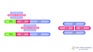
Caspase Activation Process
Caspases are synthesized as inactive zymogens (procaspases) and require proteolytic cleavage for activation. The activation involves multiple, highly specific hydrolysis steps, as follows:
- Cleavage of N-terminal Propeptide
- The first step involves hydrolysis at a specific site between the N-terminal propeptide and the large subunit of the procaspase.
- This removes the regulatory pro-domain.
- Cleavage Between Subunits
- A second hydrolysis occurs between the large (p20) and small (p10) catalytic subunits.
- This cleavage releases both subunits from the precursor form.
- Dimerization
- The large and small subunits form a heterodimer (p20/p10).
- Two heterodimers then associate to form the active caspase tetramer (p20/p10–p20/p10).
- Active Enzyme Formation
- The final active enzyme is a tetramer composed of two large and two small subunits, functioning as a protease to cleave specific substrates during apoptosis or inflammation.


44 Comments
Komal Sharma
May 4, 2025Wao sir kitna bdiya concept h ye website pe e Book amazing 😍
Suraj sir always best ✨✨✨
Seema
May 4, 2025Suraj sir ✨️✨️👌🫰🫰
Superb sir
Aditi
May 4, 2025Superb sir ✨✨ bahut achhe se explain kr rkha h concept ko
Riya
May 4, 2025Superb explanation 👍
Ekta yadav
May 4, 2025osm concept 🫶🏻👌🏼
Ekta yadav
May 4, 2025osm concept sir 🫶🏻👌🏼
Parul Verma
May 4, 2025Wow sir, ek ek chij ache se clear hogyi , lecture ke sth sth written concept bhi, thankyou sir
Parul Verma
May 4, 2025Wow sir, ek ek chij ache se clear hogyi , lecture ke sth sth written concept bhi thankyou sir
Beena Meena
May 4, 2025Thank you sir 🙂
Sachin Sharma
May 4, 2025The kind of content that a CSIR aspirant really needs🤩… LTA & Suraj sir always delivers✨
Suman bhakar
May 4, 2025Superb sir 👍 thanks sir 😊
Ujjwal
May 4, 2025Thank you sir for supporting us and providing all the content related to csir
Akshay mahawar
May 4, 2025Best explanation of apoptosis 👍👍
MOHIT AKHAND
May 4, 2025Excellent 👌🏻 sir……..and Thank you 😊 for this.
This type of concept a student deserves. 😍😊
Muskan meena
May 4, 2025Great sir thank u so much
It will be very helpful for us..
Muskan meena
May 4, 2025It will be very helpful thank u sir
Ashwini shekhawat
May 4, 2025Thank you for your efforts sir
You are incredible !!!
Vikram gahlot
May 4, 2025Best explanation of apoptosis. 👌
Vikram gahlot
May 4, 2025Best explanation of apoptosis 👌
Vikram gahlot
May 4, 2025Best explanation of apoptosis and necrosis
Rupali soni
May 4, 2025Outstanding … explanation sir 👍
yogesh sharma
May 4, 2025God is on the way to show an another miracle
Hat’s off to you sir 🙌
yogesh sharma
May 4, 2025Sir This concept will prove to be a boon for all the students who are not able to join your class. Salute to your struggle and lively thinking.
🙌🙌❣️🙏🙏
yogesh sharma
May 4, 2025You are really god of life sciences 🧬
Hemant verma
May 4, 2025Thank you suraj sir for this content…🙏😍 literally amazing for see all students 💖
Ashok
May 4, 2025Best explanation ✌
Priyanka yadav
May 4, 2025Best explanation of apoptosis 👌👍👍
Thanku so much sir 🙂☺😊
Prami Masih
May 4, 2025Thank you so much sir ji ❤️❤️
Pallavi gautam
May 4, 2025Thank you sir for your efforts
Understanding on this topic now much more clear.
Komal Pareek
May 4, 2025The content is well-organized and easy to understand.
Highly Recommended!!
Ritika Jangir
May 4, 2025Great explanation and easy to understand.
Thank you Sir 😊
Parul
May 4, 2025Thankyou so much sir for the great help. Your explanations are always exceptional. 💥💥
Mahima
May 4, 2025Very well explained sir🤍🌞
Mahima
May 4, 2025Very well explained sir in minimal words 🤍🌞
Mahima
May 4, 2025Great conceptual clarify in minimal words 🤍🌞
Mahima
May 4, 2025Great conceptual clarity
Chandan
May 4, 2025Good explanation…thank you sir ✨
Shreeji Charan
May 4, 2025Great explanation sir
SEETA CHOUDHARY
May 4, 2025This is amazing 🤩 Suraj Sir thank you so much for the best and great explanation 🤞💫❤️ this e book is very helpful
Thank you so much sir 😊
Kabeer Narwal
May 5, 2025Easily understandable 👍👍
Anish Choudhary
May 5, 2025SURAJ SIR IS KIND KING
Renu Saini
May 5, 2025Best book for life science
Yogita Choudhary
May 5, 2025Great Explanation Sir.
Krishma saini
May 7, 2025Outstanding sir mja aa gya 🤞🤞