67. In the avian embryo, the blastocoel-like fluid-filled cavity is formed between: (1) epiblast and hypoblast (2) hypoblast and yolk (3) primary hypoblast and secondary hypoblast (4) Koller's sickle and Posterior Marginal Zone 

Formation and Role of the Blastocoel-like Cavity in Avian Embryo Development

The early embryonic development in amphibians and aves serve as two different model plans of development. In the former the germ layer formation is initiated from a fluid-filled ball like blastula, while in the latter the germ layer formation is initiated on a flat blastodisc. Given below are some of the terms for amphibian embryo in column l and from avian embryo in column ll: Which of the following is the all correct match of the terms in Column l with that of Column ll? (1) A-iv, B-iii, C-ii, D-v, E-l (2) A-iv, B-ii, C-iii, D-v, E-i (3) A-v, B-i, C-ii, D-iv, E-iii (4) A-i, B-ii, C-iii, D-iv, E-v 

Comparative Terms in Amphibian and Avian Embryonic Development: Blastocoel, Primitive Streak, and Organizer Equivalents

65. The major structural characteristic of avian gastrulation is the primitive streak, which becomes the blastopore lips of amniotic embryos. Migration through the primitive streak is controlled by Fgf8. What would happen if the Fgf8 protein, which repels migrating cells away from the streak, is over expressed in the primitive streak? (1) The yolk sac will be deformed. (2) Wnt signalling will be activated and orientation of the primitive streak will change (3) Cells of the streak will not form the paraxial mesoderm. (4) Cells generate mesodermal portions of the embryo

Impact of Fgf8 Overexpression on Primitive Streak and Mesoderm Formation during Avian Gastrulation

Following statements were made about the events occurring during chick development. A. The fertilized chick egg undergoes discoidal meroblastic cleavage; however the cleavage does not extend into the yolk cytoplasm. B. Development of primary hypoblast is mediated by localized migration of a group of highly specified and connected cluster of 30-40 cells. C. By the stage XIII of chick embryogenesis and little prior to primitive streak formation, the formation of the hypoblast is just complete. D. Hansen's node of the chick embryo signifies a region at the anterior end of the primitive streak with regional thickening of cells. E. Inhibition of Wnt planar cell polarity pathway in the epiblast causes the mesoderm and endoderm to form centrally instead of peripherally. Which one of the following combinations represents all correct statements? (1) A, B and D (2) A, C and E (3) A, B and C (4) A, C and D 

Key Events in Chick Embryogenesis: Cleavage, Hypoblast Formation, Primitive Streak, and Hensen’s Node

Hensenis node is established as the avian equivalent of the amphibian dorsal blastopore lip. The following observations are presumed to be support of the same. A. It is the region whose cells induce and pattern a second embryonic axis when transplanted into other locations of the gastrula. B. It is equivalent in terms of tissue structure. C. It expresses the same marker genes as the Spemann's organizer in Amphibians. D. The same micro RNA can interfere with the formation of pre-chordal plate in both Hensen's node and Spemann's organizer. Choose the correct set among the following: (1) A and D (2) A and C (3) B and C (4) A and B 

Hensen’s Node as the Avian Equivalent of the Amphibian Spemann Organizer: Axis Induction and Genetic Markers

The part of the embryo from which the ectoderm, mesoderm and endoderm are formed in chick is known as (1) primitive streak (2) hypoblast (3) epiblast (4) cytotrophoblast 

Chick Embryo Germ Layer Formation: Role of the Epiblast and Primitive Streak in Early Development

Cells from an early frog blastula were remove from the animal pole and used to replace cells from the vegetal pole of the blastula. The following events may be expected. A. Transplanted cells would develop normally as part of the cells of the vegetal pole. B. Transplanted cells would develop as cells of the animal pole of the adult on the vegetal pole. C. Region of the animal pole from where the cells were removed would be missing in the adult. D. Remaining cells in the animal pole would compensate for the cells that were removed. Which of the following are true? (1) B, C and D (2) A, B and D (3) A, B and C (4) A, C and D 

Fate and Compensation of Frog Blastula Cells: Animal Pole to Vegetal Pole Transplantation Outcomes

In an experiment, activin-secreting beads were placed on unspecified cells from an early Xenopus embryo. The activin then diffused from the beads. If the beads contained 1nM of activin, it elicited expression of Xbra gene in cells near to the beads. If the beads contained 4 nM activin, the expression of Xbra was elicited in cells, but only at a distance of several cell diameters away from the beads. In the latter case, expression of goosecoid gene was observed near the source bead. Beads with no activin did not elicit the expression of the two genes. Following statements were made regarding the observations and the role of activin in determining cell fate. A. High concentration of activin activates goosecoid, whereas lower concentrations activate Xbra. B. Lower concentrations of activin help specify the dorsal-most structures of the frog's embryo C. Concentrations of activin that do not lead to expression of the two genes specifies the cell to become blood vessels and heart Which of the above statement(s) are correct? (1) A only (2) C only (3) A and C (4) B and C 

Activin Morphogen Gradient in Xenopus: Concentration-Dependent Gene Activation and Cell Fate Specification

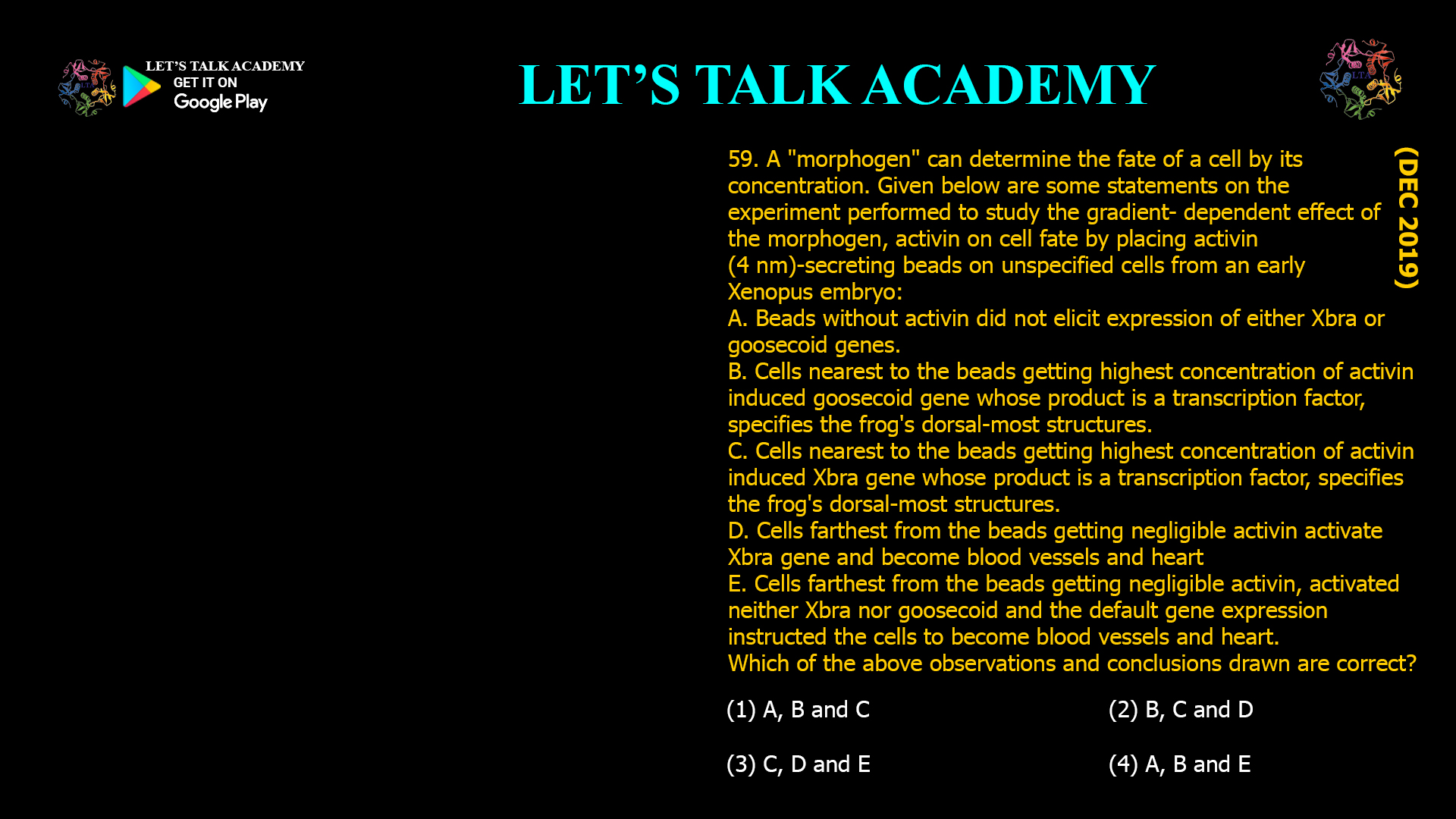
59. A "morphogen" can determine the fate of a cell by its concentration. Given below are some statements on the experiment performed to study the gradient- dependent effect of the morphogen, activin on cell fate by placing activin (4 nm)-secreting beads on unspecified cells from an early Xenopus embryo: A. Beads without activin did not elicit expression of either Xbra or goosecoid genes. B. Cells nearest to the beads getting highest concentration of activin induced goosecoid gene whose product is a transcription factor, specifies the frog's dorsal-most structures. C. Cells nearest to the beads getting highest concentration of activin induced Xbra gene whose product is a transcription factor, specifies the frog's dorsal-most structures. D. Cells farthest from the beads getting negligible activin activate Xbra gene and become blood vessels and heart E. Cells farthest from the beads getting negligible activin, activated neither Xbra nor goosecoid and the default gene expression instructed the cells to become blood vessels and heart. Which of the above observations and conclusions drawn are correct? (1) A, B and C                                                          (2) B, C and D (3) C, D and E                                                          (4) A, B and E

Activin Morphogen Gradient in Xenopus: Differential Gene Activation and Cell Fate Determination

In Xenopus, the Noggin protein, accomplishes two major functions of the organizer: it induces dorsal ectoderm to form neural tissues, and it dorsalizes mesoderm cells. Which one of the following observations is correct with respect to Noggin? (1) If a plasmid clone expressing Noggin protein is microinjected into a lithium chloride treated Xenopus gastrula, it should rescue the abnormalities induced by lithium chloride treatment. (2) If a plasmid clone expressing Noggin protein is microinjected into UV-treated embryo which does not give rise to neural tube, it will rescue the abnormality. (3) RNA in situ hybridization of noggin cDNA on Xenopus embryo will show its presence in all regions except the dorsal blastopore lip. (4) Microinjection of noggin mRNA into the embryo region fated to make the ventral part will promote its ventralization. 

Noggin in Xenopus Embryos: Rescuing Dorsal Structures and Neural Tube Formation after UV Treatment

Injection of Noggin mRNA in cells that will become the future ventral side of a frog embryo mimics the effect of an organizer graft to the ventral side. This experiment demonstrates that A. Noggin is a transcription factor B. Noggin induces ventral fates C. Noggin is involved in organizer fate D. Noggin is required to induce a secondary axis Which one of the following options represents correct combination of statement/s? (1) A and C (2) C and D (3) A and B (4) B and C 

Noggin mRNA Injection in Frog Embryos: Mimicking Organizer Function and Inducing Secondary Axis Formation

56. The following statements are made regarding the amphibian early-embryonic development: A. The Nieuwkoop center cells are mesodermal in origin. B. Chordin, Noggin and Goosecoid are secreted by the Organizer. C. The default fate of the ectoderm is to become neural tissue. D. BMP levels are high in the presumptive dorsal mesoderm.

Understanding Amphibian Early Embryonic Development: Nieuwkoop Center, Organizer Secretions, Neural Induction, and BMP Signaling

Following statements are made regarding amphibian development: A. Fibronectin plays an important role in enabling the mesodermal cells to migrate into the embryo. B. Organizer secrete proteins that block the BMP signal, which allows the ectodermal cells to become epidermis. C. Wnt signalling causes a gradient of B-catenin along the anterior-posterior axis of the neural plate, which appears to specify the regionalization of the neural tube. D. The more ventral blastomeres in the endoderm have high expression of nodal-related proteins. Which one of the following options represents the combination of all correct statements? (1) A and B (2) A and C (3) B and C (4) C and D 

Roles of Fibronectin, BMP Inhibition, Wnt Signaling, and Nodal Proteins in Amphibian Development: Migration, Neural Induction, and Patterning

Which one of the following statements with respect to amphibian development is correct? (1) The organizer is itself induced by the Nieuwkoop Centre located in the dorsal most mesodermal cells. (2) The organizer functions by secreting proteins like Noggin, Chordin and Follistatin that blocks BMP signal that would otherwise dorsalize the (3) In the presence of BMP activators the ectodermal cells form neural tissue. (4) Wnt signalling causes a gradient of β-catenin along the anterior-posterior axis of the neural tube that appears to specify the regionalization of the neural tube.

Key Principles of Amphibian Development: The Nieuwkoop Centre, Organizer Function, BMP Signaling, and Wnt in Neural Patterning

52. In Xenopus embryos, β-catenin plays an important role in the Dorsal/Ventral axis development. What would you expect if the endogenous glycogen synthase kinase 3 (GSK3) is knocked out by a dominant negative form of GSK3 in the ventral cells of the early embryos? (1) Blocking of GSK3 on the ventral side has no effect. A normal embryo will form. (2) The resulting embryo will only have ventral sides (3)A second axis will form (4) The dorsal fate is suppressed. 

Dominant Negative GSK3 in Xenopus Ventral Cells: Induction of Secondary Axis and Role in Dorsal-Ventral Axis Formation

51. The presence of β-catenin in the nuclei of blastomeres in the dorsal portion of the amphibian embryo is one of the determinants for laying down the dorso-ventral axis. What will be the outcome of expressing a dominant negative form of GSK3 in the ventral cells of early embryo ? (1) The dorsal cells will be ventralized (2) A second axis will be formed (3) The primary organizer will not be formed (4) The embryo will develop normally 

Dominant-Negative GSK3 Expression in Ventral Amphibian Embryos: Mechanism, Outcome, and Axis Duplication

In amphibian oocyte, the germplasm which gets segregated during cleavage to give rise to primordial germ cells (PGC's) is normally (1) distributed evenly throughout the oocyte. (2) localized at animal pole. (3) localized at vegetal pole. (4) aggregated in central part of oocyte. 

Vegetal Pole Localization of Germplasm in Amphibian Oocytes: Key to Primordial Germ Cell Segregation

49. The dorsal-most vegetal cells of the amphibian embryo that is capable of inducing the organizer is called as Nieuwkoop centre and is marked by the presence of (1) Chordin (2) β-catenin (3) Goosecoid (4) Nanos

Nieuwkoop Center in Amphibian Embryo: Function and Molecular Markers

Which one of the following statements regarding amphibian development is correct? (1) The Nieuwkoop centre is formed on the dorsal side of the embryo due to accumulation of β-catenin which helps activate the siamois and twin genes (2) The entodermal cells form neural tissues in the presence of BMP molecules. (3) Brain formation requires the activation of both Wnt and BMP pathway. (4) There is a gradient of Nodal-related protein across the endoderm with low concentration on the dorsal side of the embryo 

Understanding Amphibian Development: The Role of the Nieuwkoop Centre and β-Catenin

In the table below Column I lists terms related to development and Column II contains their descriptions not in a sequential manner. Select the option with all correct matches between Column I and Column II. (1) A-i, B-iii, C-ii, D-iv (2) A-ii, B-i, C-iv, D-iii (3) A-iii, B-i, C-ii, D-iv (4) A-iv, B-ii, C-i, D-iii

Spemann organizer: dorsal blastopore lip mesoderm cells organizing embryo

46. The group of cells of amphibian blastula capable of inducing the organizer is called as (1) Hensen's node                                                      (2) Nieuwkoop centre (3) Dorsal blastopore lip                                  (4) Hypoblast 

Nieuwkoop Center: The Inducer of the Amphibian Organizer in Blastula Embryos

45. Which protein secreted by the amphibian organizer induces neural tissue formation by inhibiting Bone Morphogenetic Protein? (1) β-catenin.                                                 (2) Noggin. (3) Dickkopf.                                                 (4) Dishevelled.

Noggin: The Key Protein from the Amphibian Organizer that Induces Neural Tissue by Inhibiting BMP

44. In case of is Xenopus levis which cells make up the Knewkoop center and Spemann's organizer? (1) endodermal and mesodermal, respectively (2) mesodermal and endodermal, respectively (3) endodermal and ectodermal, respectively (4) ectodermal and endodermal, respectively 

Cells Comprising the Nieuwkoop Center and Spemann’s Organizer in Xenopus laevis: Endoderm and Mesoderm Explained

43. The blastopore region of amphibian embryo that secretes BMP inhibitors and dorsalizes the surrounding tissue is known as (1) Brachet's cleft                                            (2) Nieuwkoop center (3) Hensen's node                                           (4) Spemann's organizer 

Role of Spemann’s Organizer in Amphibian Embryos: BMP Inhibition and Dorsalization Explained

42. The initial dorsal-ventral axis in amphibian embryos is determined by (1) the point of sperm entry. (2) gravity. (3) the point of contact with the uterus. (4) genetic differences in the cells. 

How the Initial Dorsal-Ventral Axis Is Determined in Amphibian Embryos: The Crucial Role of Sperm Entry

The lateral separation of amphibian embryo at two celled stage will result in (1) Identical twins (2) Two embryos joined at belly region (3) Single embryo (4) Two embryos missing various organs 

What Happens When an Amphibian Embryo is Laterally Separated at the Two-Celled Stage? Identical Twins Explainedv

40. The grafting of the dorsal lip of the blastopore from an early Xenopus gastrula onto the ventral side of an early embryo will result in two complete embryos. Thus dorsal can be designated as (1) Primary organizer (2) Cytoplasmic determinant (3) Morphogen (4) Commitment

 The Role of the Dorsal Lip as the Primary Organizer in Xenopus Embryonic Development

Dorsal lip of amphibian is equivalent to chick's (1) Hensen's node (2) Primitive grove (3) Animal pole (4) Vegetal pole 

Understanding the Equivalent of the Amphibian Dorsal Lip in Chick Embryos: Hensen’s Node

38. The specification of sea urchin micromeres involves the activation of a repressor protein Pmar1, which represses the expression of hesC, which also encodes a repressor protein. One of the genes controlled by HesC is Delta, whose expression is used as a marker for micromere lineage. The image below represents a sea urchin embryo on which whole mount in situ hybridization (WMISH) was performed using delta probe, indicated by the area 'A'. The rest of the embryo is labeled 'B'. The table below summarizes a set of experiments (column A) and the area in which hybridization is observed (column B) Which one of the following options is a correct match between columns A and B? (1) A-ii, B-iii, C-l                                              (2) A-ii, B-iii, C-iv (3) A-i, B-ii, C-iv                                              (4) A-i, B-ii, C-iii

 Gene Regulatory Network and Delta Expression in Sea Urchin Micromere Specification

37. In sea urchins, a group of cells at the vegetal pole become specified as the large micromere cells. These cells are determined to become skeletogenic mesenchyme cells that will leave the blastula epithelium to ingress into the blastocoel. This specification is controlled by the expression of Pmar1 which is a repressor of HesC. HesC represses the genes encoding transcription factors activating skeleton forming genes. The gene regulatory network is given below. Below, column I lists the experiments carried with mRNA/antisense RNA of different genes injected into single-celled sea urchin embryo while column II lists the developmental outcomes: Match the following: Which of the following combinations is correct? (1) A-2, B-1, C-1, D-2                                                (2) A-1, B-1, C-2, D-2 (3) A-1, B-2, C-2, D-1                                                (4) A-2, B-2, C-2, D-2

Gene Regulatory Network Controlling Micromere Specification in Sea Urchin Embryos

Given below are few statements regarding the role of Disheveled (Dsh) and β-catenin (β-cat) in the development of sea urchin. A. Dsh is localized in the vegetal cortex of the oocyte before fertilization and in the region of the 16-cell embryo about to become the micromeres. B. Dsh is localized in the cytosol of the oocyte during oogenesis and in the micromere forming blastomeres of a 16- cell embryo. C. β-cat accumulates predominantly in the micromeres and somewhat in the veg2 tier cells. D. Treatment of embryos with lithium chloride does not allow the accumulation of β-cat in the nuclei of all blastula cells, and the animal cells thus become specified as endoderm and mesoderm. E. When β-cat is prevented from entering the nucleus, the embryo develops as a ciliated ectodermal ball. Which one of the following options represents a combination of correct statements? (1) B, C and E (2) A, C and D (3) A, C and E                                                          (4) B, D and E 

Understanding the Role of Disheveled and β-Catenin in Early Sea Urchin Embryonic Development

Following statements with respect to development in sea urchin were put forth: A. The cell fates are determined both by autonomous and conditional modes of specification. B. Large micromeres are conditionally specified. C. Large micromeres produce paracrine and juxtacrine factors that specify the fates of their neighbours. D. B-catenin is not required for the specification of the micromeres. Which one of the following options represents the combination of all correct statements? (1) A and B (2) A and C (3) C and D (4) B and D 

 Autonomous and Conditional Cell Fate Specification in Sea Urchin Embryos

Impact of GSK-3 Inhibition on β-Catenin Nuclear Accumulation and Cell Fate in Sea Urchin Embryos

33. In a given experiment, transplantation o micromeres from the vegetal pole of a 16-cell sea urchin embryo onto the animal pole of a host 16-cell sea Urchin embryo would initiate: (1) The transplanted micromeres to invaginate into the blastocoel to create a new set of skeletogenic mesenchyme cells (2) The transplanted micromeres to ingress into the blastocoel to create a new set of skeletogenic mesenchymal cells (3) The transplanted micromeres will mingle with the host micromeres to ingress into the blastocoel to create skeletogenic mesenchyme cells. (4) The transplanted micromeres will form the secondary archenteron 

Sea Urchin Micromere Transplantation: Formation of Skeletogenic Mesenchyme Cells Through Ingression

When the 4 blastomere pairs of the 8-cell stage tunicate embryo are dissociated, each forms most of the structures it would have formed had it remained in the embryo. However, the notochord and nervous system get specified only if different blastomeres get the chance to interact. Given below are certain interpretations formulated from the above results: A. Each pair of blastomeres forming respective structures indicate autonomous specification B. Each pair of blastomeres forming respective structures indicate conditional specification C. The notochord and nervous system development indicate autonomous specification D. The notochord and nervous system development indicate conditional specification. Which combination of interpretations is most appropriate? (1) A and C (2) B and D (3) A and D (4) B and C 

 Autonomous and Conditional Specification in Tunicate Embryonic Development

31. Which one of the following about development of sea urchin embryos is TRUE? (1) Each blastomere of a 4-cell stage possesses a portion of the original animal-vegetal axis and if isolated and allowed to develop will form a complete but smaller size larva. (2) Each blastomere of a 8-cell stage has the capacity to form a complete embryo but by the 16-cell stage, blastomeres will develop according to their presumptive fate. (3) Any blastomere isolated till the pluteus larva formation will regulate to go on and develop into a full sized embryo. (4) After an intricate recombination at the 16 cell stage, the resulting embryo looses its ability to form a complete larva. 

Understanding Sea Urchin Embryo Development: Cellular Potency and Axis Formation

Dreisch performed the "pressure plate experiment" to alter the distribution of nuclei in a 8-cell sea urchin embryo. He obtained normal larvae from these embryos. Following possible conclusions could be drawn: A. Prospective potency of the blastomeres is less than the actual prospective fate. B. Sea urchin embryo is a "harmonious equipotential system" implying that cell interaction is critical for normal development. C. Prospective potency of the blastomere is greater than the actual prospective fate. D. Prospective potency of the blastomere is equal to the prospective fate. Which one of the following combinations of statements represents the correct inference from the experiment? (1) A and B (2) Band C (3) B only (4) D only 

 Driesch’s Pressure Plate Experiment Reveals Equipotentiality and Potency in Sea Urchin Embryos

29. Driesch performed famous 'pressure plate" experiments involving intricate recombination with 8- celled Sea urchin embryo. This procedure reshuffled the nuclei that normally would have been in the region destined to form endoderm into the presumptive ectoderm region. If segregation of nuclear determinants had occurred, resulting embryo should have been disordered. However, Driesch obtained normal larvae form these embryos possible interpretations regarding the 8-celled sea urchin embryo are: A. The prospective potency of an isolated blastomere is greater than its actual prospective fate B. The prospective potency and prospective fate of blastomere were identical C. Sea-urchin embryo is a "harmoniously equipotential system" because all of its potentially independent parts interacted together to form single embryo. D. Regulative development occurs where location of a cell in the embryo determines its fate. Which of the interpretation(s) is/are true? (1) only A                                                      (2) only D (3) only A and B                                             (4) A, C and D 

Driesch’s Pressure Plate Experiment: Insights into Regulative Development in Sea Urchins

28. With respect to the extra embryonic structures formed in the mammals, the possible functional attributes have been designated: A. Allantoin stores urinary waste and helps mediate gas exchange. It is derived from splanchnopleure at the caudal end of the primitive streak. B. Amnion is a water sac and protects the embryo and its surrounding amniotic fluid. This epithelium is derived from somatopleure. C. Chorion is essential for gas exchange in amniote embryos. It is generated from the splanchnopleure. Yolk sac is the last embryonic membrane to form and is derived from somatopleure. Which of the above statements are correct? (1) A and B (2) A and C (3) B and C (4) A and D

Mammalian Extraembryonic Structures: Origins and Functional Roles

Which one of the following mRNAs is a BMP inhibitor and can rescue the dorsal structures of ventralized Xenopus embryo when injected into it? (1) beta-catenin (2) Noggin (3) Disheveled (4) Siamos 

Noggin: A Key BMP Inhibitor That Rescues Dorsal Structures in Ventralized Xenopus Embryos

Following statements are made regarding animal development: A. The cell is first specified towards a given fate, suggesting that it would develop into this cell type, even in a neutral environment. B. Holoblastic rotational cleavage is observed in tunicates. C. Infolding of sheet of cells is called ingression. D. Conditional specification can be observed in sea urchin embryos. Which one of the following options represents the combination of all correct statements? (1) A and B (2) B and C (3) A and D (4) C and D 

Understanding Cell Fate Specification and Cleavage Patterns in Animal Development

Which one of the following statements with respect to development in amphibians is correct? (1) Gastrulation begins with the invagination of bottle cells, followed by coordinated involution of the mesodermal precursors and the epiboly of the prospective ectoderm (2) The organizer induces the Nieuwkoop centre (3) The organizer is formed by the accumulation of B- catenin (4) In the absence of BMP inhibitors ectodermal cells form neural tube BMP 

Key Steps in Amphibian Gastrulation: Invagination, Involution, and Epiboly Explained

23. Which one of the following developmental processes in animals is more dependent on cellular movements? (1) pattern formation                                                (2) morphogenesis (3) cell differentiation                                       (4) growth 

Morphogenesis: The Crucial Role of Cellular Movements in Animal Development

Movement of epithelial sheet spreading as a unit to enclose deeper layers of the embryo is termed as (1) Epiboly (2) Emboly (3) Involution (4) Ingression 

 Epiboly: The Coordinated Spreading of Epithelial Sheets During Embryonic Development

Which one of the following is NOT a typical movement pattern observed during gastrulation? (1) Involution (2) Epiboly (3) Furrowing (4) Delamination 

Understanding Typical and Atypical Cell Movements During Gastrulation

The splitting or migration or one sheet of cells into two sheets as seen during hypoblast formation in bird embryogenesis is termed as (1) delamination (2) ingression (3) involution                                                 (4) invagination

Delamination: The Splitting of Cell Sheets During Hypoblast Formation in Bird Embryos

Inward movement of an expanding outer layer spreading over the internal surface during gastrulation is termed as (1) invagination (2) Ingression (3) involution (4) delamination 

 Understanding Involution: A Key Morphogenetic Movement During Gastrulation

Bones of vertebrates embryonic are derived from (1) ectoderm (2) epiderm (3) mesoderm (4) endoderm

Understanding the Embryonic Origin of Vertebrate Bones: The Role of the Mesoderm

14. Human chorionic gonadotropin (hCG) is known to facilitate attachment of blastocyst to uterus. In women with mutation in hCG gene, biologically inactive hCG was formed but implantation occurred. When hcG was immune-neutralized in the uterus of normal woman, implantation failed. This suggests that for implantation in humans: (1) biologically active circulating hCG is not required. (2) blastocyst can produce the required hCG, which helps locally in uterine attachment. (3) trophoblastic cells do not require hCG for the invasion of uterus. (4) extra-embryonic tissue is not responsible for the attachment of embryo to uterus 

Why Local hCG Production by the Blastocyst Is Essential for Human Embryo Implantation

Compaction during early embryonic development involves activation of actin filaments. Which one of the following inhibitors would prevent formation of the blastula from the morula? (1) An inhibitor that would block the action of Glin. (2) A specific inhibitor for stabilizing the function of Axin. (3) A specific inhibitor for GSK3B (4) An inhibitor which would block Smad4

The Essential Role of Actin Filaments and Cofilin1 in Embryonic Compaction and Blastula Formation

The figure above represents a late zebrafish gastrula. The following concepts may be proposed during further development of the embryo. A. The concentration of FGF decreases from the yolk towards the epidermis, along with the increase of BMP activity from the dorsal to the ventral axis. B. Increase in FGF activity in the epidermis with concomitant decrease in BMP activity towards the ventral axis. C. Neural induction in zebrafish is independent of the organizer and depends on activation of BMP signaling. D. In comparison, both Xenopus and chick embryos require activation of FGF for neural induction to occur in addition to BMP inhibition. Which of the above statements are true? (1) A and C (2) B and C (3) A and D (4) C and D 

Morphogen Gradients and Neural Induction in Zebrafish and Comparison to Other Vertebrates

11. Gradients of morphogens determine the future dorsal- ventral and anterior-posterior axes of the developing embryo in many organisms. How is the anterior- posterior axis developed in C. elegans? (1) Sperm entry leads to reorganization of the cytoskeleton and redistribution of naturally packed PAR proteins, which in turn determine the anterior- posterior axis. (2) After fertilization β-catenin gets localized into the nucleus of the future anterior cells. (3) The P-granules are localized in a way consistent with a role as a morphogenetic determinant and they act through translational regulation to initiate anterior-posterior axis. (4) The localization of the maternally expressed polypeptide SKN-1 is responsible for the

How Sperm Entry and PAR Proteins Establish the Anterior-Posterior Axis in C. elegans

10. Given below are fate map of two organisms and the pattern by which embryos undergo cleavage. Which of the following is/are the right combination(s)? (1) B only                                            (2) B and A (3) A and C                                          (4) B and D

Matching Fate Maps and Cleavage Patterns: Finding the Right Combination in Embryology

The table below lists cleavage pattern and names of species. Match the cleavage patterns with the species. (1) A- i; B- ii; C- iii; D- iv (2) A- ii; B- iv; C- i; D- iii (3) A- iv; B- i; C- iii; D- ii (4) A- iii; B- i; C- iv; D- ii 

 Matching Embryonic Cleavage Patterns with Representative Species: A Comprehensive Guide

8. Match the following cleavage patterns with the species in which they occur. (1) A-iv, B-iii, C-i, D-ii                                       (2) A-iii, B-i, C-iv, D-ii (3) A-ii, B-iii, C-i, D-iv                                       (4) A-ii, B-iv, C-iii, D-i

 A Detailed Guide to Embryonic Cleavage Patterns and Their Species Associations

The pattern of embryonic cleavage specific to a species is determined by two major parameters. (A) The amount and distribution of yolk protein within the cytoplasm. (B) The factors in the cytoplasm that influence the angle mitotic spindles and the timings of its formation. Which of the following statements are true? (1) Species having teloecithal egg follow a holoblastic cleavage. (2) Species having isolecithal egg follow a holoblastic cleavage. (3) Species having centrolecithal egg follow a holoblastic cleavage. (4) Species having isolecithal egg follow a meroblastic cleavage.

How Yolk Distribution and Cytoplasmic Factors Determine Embryonic Cleavage Patterns

Centrolecithal eggs show (1) superficial cleavage (2) displaced radial cleavage (3) bilateral cleavage (4) discoidal cleavage 

Understanding Cleavage in Centrolecithal Eggs: The Superficial Cleavage Pattern

5. Amphibian zygote will generally undergo (1) Holoblastic radial cleavage. (2) Holoblastic rotational cleavage. (3) meroblastic bilateral cleavage. (4) meroblastic rotational cleavage. 

Understanding Amphibian Embryonic Development: Holoblastic Radial Cleavage Explained

What is the pattern of cleavage observed in mammals? (1) Radial (2) Spiral (3) Rotational (4) Bilateral

 The Rotational Cleavage Pattern in Mammalian Embryonic Development

Which kind of cleavage is shown in mammals? (1) Holoblastic rotational (2) Meroblastic rotational (3) Holobastic radial (4) Meroblastic radial 

Understanding Mammalian Embryonic Cleavage: The Holoblastic Rotational Pattern

If an embryo undergoes 13 cleavage divisions during embryogenesis, then the size of the embryo compared to zygote (1) increases 13 times. (2) increases only 6-7 times. (3) increases in an exponential fashion. (4) Remains almost the same.

Why Embryo Size Remains Constant During Early Cleavage Divisions

Which is true for amount of yolk and cleavage in egg of amphibian? (1) Mesolecithal and holoblastic cleavage (2) Isolecithal and holoblastic cleavage (3) Mesolecithal and meroblastic cleavage (4) Microlecithal and meroblastic cleavage 

Understanding Amphibian Eggs: Mesolecithal Yolk Amount and Holoblastic Cleavage

Latest Courses